. 2015 Nov 9:13:93.
doi: 10.1186/s12915-015-0203-8.

Molecular insights into the surface-specific arrangement of complement C5 convertase enzymes

Affiliations

Molecular insights into the surface-specific arrangement of complement C5 convertase enzymes

Evelien T M Berends et al. BMC Biol. .

Abstract

Background: Complement is a large protein network in plasma that is crucial for human immune defenses and a major cause of aberrant inflammatory reactions. The C5 convertase is a multi-molecular protease complex that catalyses the cleavage of native C5 into its biologically important products. So far, it has been difficult to study the exact molecular arrangement of C5 convertases, because their non-catalytic subunits (C3b) are covalently linked to biological surfaces through a reactive thioester. Through development of a highly purified model system for C5 convertases, we here aim to provide insights into the surface-specific nature of these important protease complexes.

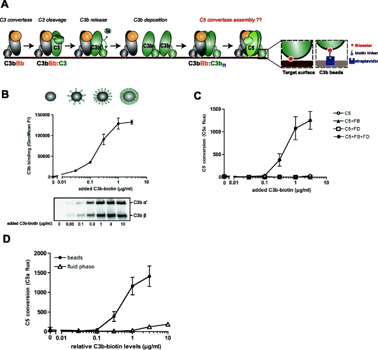
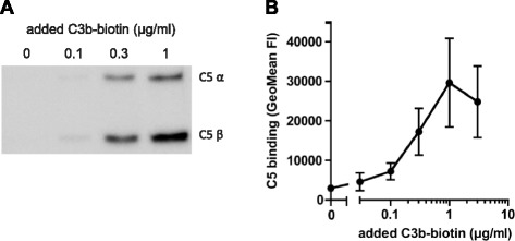
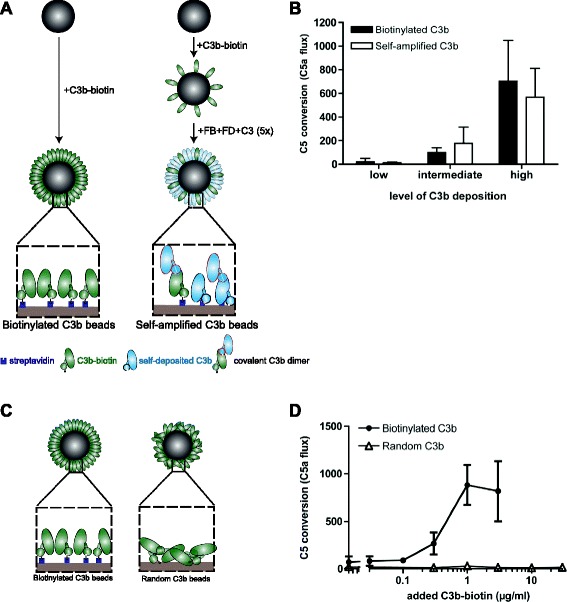
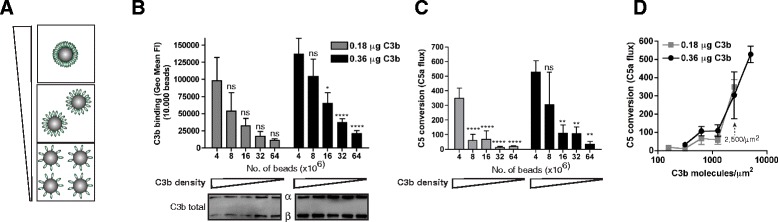
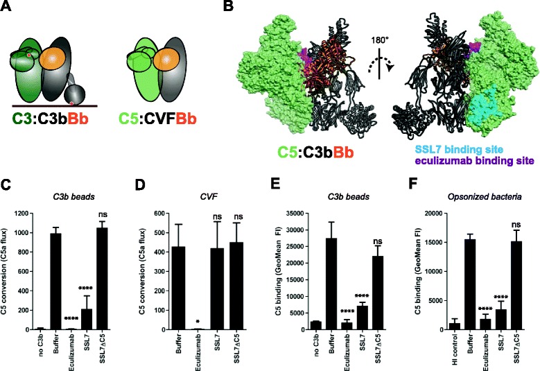
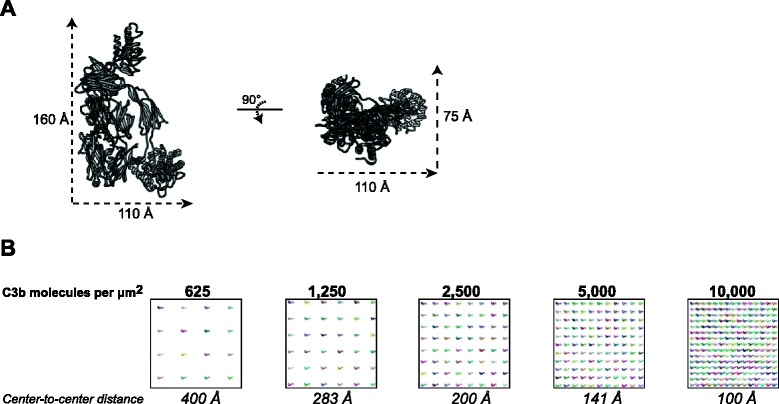
Results: Alternative pathway (AP) C5 convertases were generated on small streptavidin beads that were coated with purified C3b molecules. Site-specific biotinylation of C3b via the thioester allowed binding of C3b in the natural orientation on the surface. In the presence of factor B and factor D, these C3b beads could effectively convert C5. Conversion rates of surface-bound C3b were more than 100-fold higher than fluid-phase C3b, confirming the requirement of a surface. We determine that high surface densities of C3b, and its attachment via the thioester, are essential for C5 convertase formation. Combining our results with molecular modeling explains how high C3b densities may facilitate intermolecular interactions that only occur on target surfaces. Finally, we define two interfaces on C5 important for its recognition by surface-bound C5 convertases.

Conclusions: We establish a highly purified model that mimics the natural arrangement of C5 convertases on a surface. The developed model and molecular insights are essential to understand the molecular basis of deregulated complement activity in human disease and will facilitate future design of therapeutic interventions against these critical enzymes in inflammation.


Figures

Fig. 1
Fig. 1
A novel bead-based assay model for purified alternative pathway (AP) C5 convertases. a Proposed model for assembly of C5 convertases in the AP. Surface-bound C3 convertase (C3bBb) cleaves multiple C3 molecules into C3b that covalently binds to target surfaces via the reactive thioester (red dot). Association of deposited C3b molecules with the existing C3 convertase gives rise to multimeric complexes (C3b-C3bn) that, together with Bb, can convert C5. The precise arrangement of surface-specific C5 convertases is currently unknown. In the novel C5 convertase assay model described in this study, C3b molecules are site-specifically biotinylated via the thioester and loaded on bacteria-sized streptavidin beads (2.8 μm) to mimic their natural density and orientation on target surfaces. b Loading of streptavidin beads with biotinylated C3b was analyzed by flow cytometry or immunoblotting (below). c C5 convertase activity of C3b-coated beads that were incubated with factor B (FB), factor D (FD) (together needed to form Bb) and C5. Conversion of C5 was determined by measuring release of C5a in the supernatant using a calcium mobilization assay with U937-C5aR cells. Values represent absolute C5a flux (mean fluorescence of stimulated cells subtracted by the mean fluorescence before stimulus). d C5 convertase activity of C3b molecules on beads versus C3b molecules in solution. The amount of C3b molecules in solution was adjusted to the levels of C3b loaded onto the beads (relative C3b-biotin levels) and both were incubated with FB, FD and C5. bd Data of three independent experiments, presented as means ± standard deviation (SD). Immunoblot is a representative of three independent experiments
Fig. 2
Fig. 2
C5 binds to C3b-coated beads. C3b-coated streptavidin beads were incubated with C5 (in the absence of FB and FD). Binding of C5 to C3b beads was determined by a Western blotting or b flow cytometry. a is a representative gel of three independent experiments; b shows data of three independent experiments, presented as means ± standard deviation (SD)
Fig. 3
Fig. 3
Attachment of C3b with the thioester toward the surface is critical for C5 convertase activity. a Left, streptavidin beads with site-specifically biotinylated C3b molecules. Right, self-amplified C3b beads were generated by coating streptavidin beads with a low concentration of C3b-biotin after which FB, FD and C3 were added for five repeating incubations to allow natural deposition of C3b and formation of covalently associated C3b multimers (outlined in red). b C5 convertase activity on self-amplified and biotinylated C3b beads. Beads (containing equal levels of C3b) were incubated with FB, FD and C5 and C5a release was determined by calcium mobilization. c Random C3b beads were generated by coupling C3b-biotin onto tosyl-activated beads. d C5 convertase activity on random and biotinylated C3b beads. (b, d) Data of three independent experiments, presented as means ± standard deviation (SD)
Fig. 4
Fig. 4
High surface density of C3b is critical for C5 convertase activity. a Preparation of beads with different densities of C3b. b Mixing a fixed amount of C3b molecules with increasing numbers of streptavidin beads results in lower C3b densities per bead (top, flow cytometry) while total levels of C3b per sample are equal (below, immunoblot). c C5 convertase activity on streptavidin beads with different C3b densities. d C5 convertase activity plotted against the absolute number of C3b molecules per μm2 (calculated from the results in c). bd Data of three independent experiments, presented as means ± standard deviation (SD). Immunoblot graphs are representative of three independent experiments. Measures of statistical significance were determined by one-way ANOVA for the different amounts of beads versus 4 × 10⁶ beads and displayed as: ns; *P <0.05; **P <0.01; ***P <0.005; and ****P <0.001
Fig. 5
Fig. 5
Inhibitors reveal two important interaction sites for C5 with surface-bound C3b. a Left, schematic representation of the proposed interaction between substrate C3 and the alternative pathway (AP) C3 convertase (based on crystal structure [8]). Right, binding of C5 to CVFBb (based on the CVF:C5 crystal structure [24]). CVF is a potent C3b homologue that lacks the thioester domain and forms stable C5 convertases in solution. b Structural model of the previously proposed AP C5 convertase. The C3/C5 convertase (C3bBb) is shown in ribbon representation, with C3b in gray and Bb in orange, respectively. C5 (green) is shown as a molecular surface, with residue involved in eculizumab (magenta) and SSL7 (cyan) binding colored on the surface. The left and right representations represent the same complex rotated 180° about the vertical axis. c C5 conversion on C3b-coated beads in the absence or presence of 20 μg/ml C5 inhibitors (SSL7, SSL7ΔC5, eculizumab) as determined by calcium mobilization of U937-C5aR cells. d C5 conversion by soluble CVFBb in the absence or presence of 20 μg/ml C5 inhibitors. e C5 binding to C3b-coated beads (loaded with 1 μg/ml C3b-biotin) in absence or presence of 20 μg/ml C5 inhibitors, determined by flow cytometry. f C5 binding to pre-opsonized bacteria in absence or presence of 20 μg/ml C5 inhibitors (flow cytometry). cf Data of three independent experiments, presented as means ± standard deviation (SD). Measures of statistical significance were determined by one-way ANOVA for the various inhibitors versus buffer control alone and displayed as: ns; *P <0.05; **P <0.01; ***P <0.005; and ****P <0.001
Fig. 6
Fig. 6
Molecular model for C3b density. a The crystallographic structure of C3b [PDB:2I07]. b Model for C3b density on a 150 × 150 nm area representing a portion of the bead surface. For each C3b density, the number of molecules per 150 × 150 nm surface area was calculated, and intermolecular distance was calculated assuming a uniform distribution of molecules. C3b molecules were then placed such that the center-to-center distance was equal to the calculated intermolecular distance. C3b is oriented such that the long axis is perpendicular to the surface and the thioester (and in turn the biotin linker) was in contact with the surface

Similar articles

Cited by

References

    1. Walport MJ. Complement. First of two parts. N Engl J Med. 2001;344:1058–66. doi: 10.1056/NEJM200104053441406. - DOI - PubMed
    1. Ricklin D, Hajishengallis G, Yang K, Lambris JD. Complement: a key system for immune surveillance and homeostasis. Nat Immunol. 2010;11:785–97. doi: 10.1038/ni.1923. - DOI - PMC - PubMed
    1. Müller-Eberhard HJ. The membrane attack complex of complement. Annu Rev Immunol. 1986;4:503–28. doi: 10.1146/annurev.iy.04.040186.002443. - DOI - PubMed
    1. de Jong HK, van der Poll T, Wiersinga WJ. The systemic pro-inflammatory response in sepsis. J Innate Immun. 2010;2:422–30. doi: 10.1159/000316286. - DOI - PubMed
    1. Mollnes TE, Kirschfink M. Strategies of therapeutic complement inhibition. Mol Immunol. 2006;43:107–21. doi: 10.1016/j.molimm.2005.06.014. - DOI - PubMed

Publication types

Substances

LinkOut - more resources