

Coarctation of the aorta with left pulmonary artery stenosis: a rare association diagnosed with ECG-gated multislice dual-source CT angiography
- PMID: 26552877
- PMCID: PMC4654156
- DOI: 10.1136/bcr-2015-210897
Coarctation of the aorta with left pulmonary artery stenosis: a rare association diagnosed with ECG-gated multislice dual-source CT angiography
Abstract
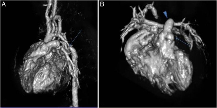
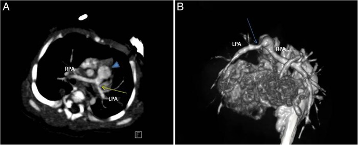
We report an extremely rare case of coarctation of the aorta with associated left pulmonary artery stenosis. This violates the traditional fetal flow pattern theory. Two-dimensional echocardiography, although being the initial imaging modality for diagnosing coarctation of the aorta, is limited in the demonstration of branch pulmonary arteries. Retrospective ECG-gated multidetector CT angiography is a non-invasive imaging technique for comprehensive assessment of the site, length and severity of the stenosed aortic segment. It is also helpful in differentiation of an interrupted aortic arch from severe coarctation. Associated pulmonary arterial and venous system anomalies can be evaluated, which dictates the management of aortic coarctation.
2015 BMJ Publishing Group Ltd.
Figures
Similar articles
-
[Clinical value of ECG-gated dual-source computed tomography and angiography in assessing coarctation of aorta].Sheng Wu Yi Xue Gong Cheng Xue Za Zhi. 2013 Feb;30(1):89-94. Sheng Wu Yi Xue Gong Cheng Xue Za Zhi. 2013. PMID: 23488145 Chinese.
-
Anomalous left coronary artery arising from the right pulmonary artery in association with coarctation of the aorta.Cardiol Young. 2016 Apr;26(4):802-4. doi: 10.1017/S104795111500195X. Epub 2015 Sep 14. Cardiol Young. 2016. PMID: 26365716
-
[A rare association of double discordance with aortic arch anomalies: value of multislice CT scan].Arch Mal Coeur Vaiss. 2006 May;99(5):523-5. Arch Mal Coeur Vaiss. 2006. PMID: 16802748 French.
-
Coarctation of aorta--intervention from neonates to adult life.Indian Heart J. 2008 Nov-Dec;60(4 Suppl D):D22-33. Indian Heart J. 2008. PMID: 19845083 Review.
-
Echocardiography of coarctation of the aorta, aortic arch hypoplasia, and arch interruption: strategies for evaluation of the aortic arch.Cardiol Young. 2016 Dec;26(8):1553-1562. doi: 10.1017/S1047951116001670. Cardiol Young. 2016. PMID: 28148317 Review.
References
-
- Ruhela M, Randhawa H, Bagarhatta P et al. . Coarctation of aorta with supravalvular pulmonary stenosis in an adult patient: a rare exception of the fetal flow pattern theory. Am J Med Case Rep 2015;3:53–8.
-
- Beekman RH. Coarctation of the aorta. In: Allen HG, Gutgessell HP, Clark EB et al., eds Moss and Adams’ heart disease in infants, children, and adolescents including the fetus and young adults. 6th edn Philadelphia: Lippincott Williams and Wilkins, 2001:988–1010.
Publication types
MeSH terms
LinkOut - more resources
Full Text Sources
Other Literature Sources
Medical