

Endometrial stem/progenitor cells: the first 10 years
- PMID: 26552890
- PMCID: PMC4755439
- DOI: 10.1093/humupd/dmv051
Endometrial stem/progenitor cells: the first 10 years
Abstract
Background: The existence of stem/progenitor cells in the endometrium was postulated many years ago, but the first functional evidence was only published in 2004. The identification of rare epithelial and stromal populations of clonogenic cells in human endometrium has opened an active area of research on endometrial stem/progenitor cells in the subsequent 10 years.
Methods: The published literature was searched using the PubMed database with the search terms 'endometrial stem cells and menstrual blood stem cells' until December 2014.
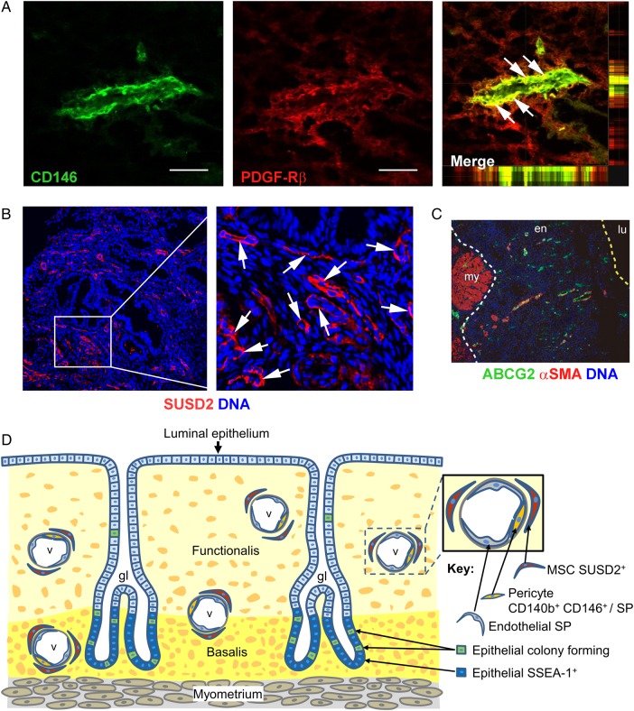
Results: Endometrial epithelial stem/progenitor cells have been identified as clonogenic cells in human and as label-retaining or CD44(+) cells in mouse endometrium, but their characterization has been modest. In contrast, endometrial mesenchymal stem/stromal cells (MSCs) have been well characterized and show similar properties to bone marrow MSCs. Specific markers for their enrichment have been identified, CD146(+)PDGFRβ(+) (platelet-derived growth factor receptor beta) and SUSD2(+) (sushi domain containing-2), which detected their perivascular location and likely pericyte identity in endometrial basalis and functionalis vessels. Transcriptomics and secretomics of SUSD2(+) cells confirm their perivascular phenotype. Stromal fibroblasts cultured from endometrial tissue or menstrual blood also have some MSC characteristics and demonstrate broad multilineage differentiation potential for mesodermal, endodermal and ectodermal lineages, indicating their plasticity. Side population (SP) cells are a mixed population, although predominantly vascular cells, which exhibit adult stem cell properties, including tissue reconstitution. There is some evidence that bone marrow cells contribute a small population of endometrial epithelial and stromal cells. The discovery of specific markers for endometrial stem/progenitor cells has enabled the examination of their role in endometrial proliferative disorders, including endometriosis, adenomyosis and Asherman's syndrome. Endometrial MSCs (eMSCs) and menstrual blood stromal fibroblasts are an attractive source of MSCs for regenerative medicine because of their relative ease of acquisition with minimal morbidity. Their homologous and non-homologous use as autologous and allogeneic cells for therapeutic purposes is currently being assessed in preclinical animal models of pelvic organ prolapse and phase I/II clinical trials for cardiac failure. eMSCs and stromal fibroblasts also exhibit non-stem cell-associated immunomodulatory and anti-inflammatory properties, further emphasizing their desirable properties for cell-based therapies.
Conclusions: Much has been learnt about endometrial stem/progenitor cells in the 10 years since their discovery, although several unresolved issues remain. These include rationalizing the terminology and diagnostic characteristics used for distinguishing perivascular stem/progenitor cells from stromal fibroblasts, which also have considerable differentiation potential. The hierarchical relationship between clonogenic epithelial progenitor cells, endometrial and decidual SP cells, CD146(+)PDGFR-β(+) and SUSD2(+) cells and menstrual blood stromal fibroblasts still needs to be resolved. Developing more genetic animal models for investigating the role of endometrial stem/progenitor cells in endometrial disorders is required, as well as elucidating which bone marrow cells contribute to endometrial tissue. Deep sequencing and epigenetic profiling of enriched populations of endometrial stem/progenitor cells and their differentiated progeny at the population and single-cell level will shed new light on the regulation and function of endometrial stem/progenitor cells.
Keywords: adenomyosis; endometrial stem cells; endometriosis; endometrium; epithelial progenitor cells; immunomodulation; menstrual blood; mesenchymal stem cells; regenerative medicine; sushi domain containing-2.
© The Author 2015. Published by Oxford University Press on behalf of the European Society of Human Reproduction and Embryology.
Figures
Comment in
-
An update on endometrial stem cells and progenitors.Hum Reprod Update. 2016 Jun;22(4):529-30. doi: 10.1093/humupd/dmw010. Epub 2016 Mar 24. Hum Reprod Update. 2016. PMID: 27016288 No abstract available.
-
Reply: An update on endometrial stem cells and progenitors by Deepa Bhartiya.Hum Reprod Update. 2016 Jun;22(4):530-1. doi: 10.1093/humupd/dmw011. Epub 2016 Mar 24. Hum Reprod Update. 2016. PMID: 27016290 No abstract available.
References
-
- Atala A. Engineering organs. Curr Opin Biotechnol 2009;20:575–592. - PubMed
-
- Azedi F, Kazemnejad S, Zarnani AH, Behzadi G, Vasei M, Khanmohammadi M, Khanjani S, Edalatkhah H, Lakpour N. Differentiation potential of menstrual blood versus bone marrow-stem cells into glial-like cells. Cell Biol Int 2014;38:615–624. - PubMed
Publication types
MeSH terms
Substances
LinkOut - more resources
Full Text Sources
Other Literature Sources
Medical
Miscellaneous
