. 2015 Dec 15;195(12):5582-91.
doi: 10.4049/jimmunol.1501092. Epub 2015 Nov 9.

Structural and Functional Changes of the Invariant NKT Clonal Repertoire in Early Rheumatoid Arthritis

Affiliations

Structural and Functional Changes of the Invariant NKT Clonal Repertoire in Early Rheumatoid Arthritis

Salah Mansour et al. J Immunol. .

Abstract

Invariant NKT cells (iNKT) are potent immunoregulatory T cells that recognize CD1d via a semi-invariant TCR (iNKT-TCR). Despite the knowledge of a defective iNKT pool in several autoimmune conditions, including rheumatoid arthritis (RA), a clear understanding of the intrinsic mechanisms, including qualitative and structural changes of the human iNKT repertoire at the earlier stages of autoimmune disease, is lacking. In this study, we compared the structure and function of the iNKT repertoire in early RA patients with age- and gender-matched controls. We analyzed the phenotype and function of the ex vivo iNKT repertoire as well as CD1d Ag presentation, combined with analyses of a large panel of ex vivo sorted iNKT clones. We show that circulating iNKTs were reduced in early RA, and their frequency was inversely correlated to disease activity score 28. Proliferative iNKT responses were defective in early RA, independent of CD1d function. Functional iNKT alterations were associated with a skewed iNKT-TCR repertoire with a selective reduction of high-affinity iNKT clones in early RA. Furthermore, high-affinity iNKTs in early RA exhibited an altered functional Th profile with Th1- or Th2-like phenotype, in treatment-naive and treated patients, respectively, compared with Th0-like Th profiles exhibited by high-affinity iNKTs in controls. To our knowledge, this is the first study to provide a mechanism for the intrinsic qualitative defects of the circulating iNKT clonal repertoire in early RA, demonstrating defects of iNKTs bearing high-affinity TCRs. These defects may contribute to immune dysregulation, and our findings could be exploited for future therapeutic intervention.


Figures

Figure 1
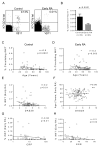
Figure 1. iNKT cell frequency in early RA patients and controls
(A) Ex vivo flow cytometry staining of live CD3+ iNKT cells (KRN7000-CD1dtet+Vβ11+) from the peripheral blood of a representative control individual and an early RA patient. (B) Cumulative staining data of iNKT frequency in early RA and controls demonstrates lower iNKT frequency in early RA patients. (C) and (D) Spearman’s correlation of ex vivo iNKT frequency with age in controls (C) and early RA (D). In both groups, iNKT frequency declines with age. (E) and (F) Spearman’s correlation of DAS28 with ex vivo iNKT frequency (E) and age (F) in early RA, demonstrating an inverse correlation between DAS28 score and iNKT frequency in early RA patients, but no correlation with age. (G) Spearman’s correlation of ex vivo iNKT frequency with CRP in treatment naïve early RA patients. (H) Spearman’s correlation of ex vivo iNKT frequency with ESR in treatment naïve early RA patients. iNKT frequency inversely correlates with both ESR and CRP in treatment naïve early RA patients.
Figure 2
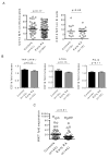
Figure 2. In vitro antigen-induced iNKT expansion in early RA patients and controls
Live CD3+KRN7000-CD1dtet+Vβ11+ iNKT cells were stained on day 0 “ex vivo” and again on day 14 after in vitro culture with KRN7000 pulsed CD1d+ monocytes. Plots show percentage of iNKT cells, ex vivo and after in vitro culture, in a control (A) and an early RA patient (B). (C) Peripheral blood T cells were stimulated with monocytes pulsed with either KRN7000 or OCH. Data showing fold expansion (ratio of in vitro grown iNKT/ex vivo frequency) of iNKT cells in response to KRN7000- (C) or OCH-(D) pulsed CD1d+monocytes in early RA patients and controls. In early RA, expansion in response to both stimuli is reduced.
Figure 3
Figure 3. Analysis of CD1d expression in monocytes and B cells from early RA patients and controls
(A) CD14+ monocytes and CD19+ B cells were purified from the peripheral blood of early RA patients and controls. Monocytes and B cells were stained with anti-CD1d and an isotype control. CD1d MFI (ratio of anti-CD1d/isotype) on the cell surface of monocytes and B cells from early RA patients and controls is presented. No difference in CD1d expression was observed. (B) CD14+ monocytes were co-cultured overnight with either PBS, TNF-α/IFN-γ, ATRA, or PGJ2. Plots show CD1d expression fold increase, in response to inflammatory stimuli, on monocytes from early RA patients and controls. No difference between controls and early RA patients was observed. (C) CD14−CD19− peripheral blood T cells were stimulated with KRN7000-CD1d MACSibeads. Live CD3+KRN7000-CD1dtet+Vβ11+ iNKT cells were stained after 14 days of in vitro iNKT expansion. Data show iNKT fold expansion (ratio of in vitro grown iNKT/ex vivo frequency) in early RA patients and controls. iNKT expansion is reduced in early RA patients.
Figure 4
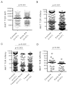
Figure 4. Clonal distribution of iNKT-TCR expression and binding properties in early RA patients and controls
(A) Single Vα24+/Vβ11+ iNKT clones were sorted and subsequently expanded with PHA. TCR expression was measured by KRN7000-CD1d-tetramers or with anti-Vα24 antibody. No difference in iNKT-TCR expression (anti-Vα24 MFI) in clones from early RA patients (175 clones) and controls (121 clones) was demonstrated. (B) iNKT clones were stained with KRN7000-CD1d− and OCH-CD1d− tetramers and iNKT-TCR affinity was calculated as the ratio of (OCH-CD1d MFI/KRN7000-CD1d MFI). iNKT-TCR affinity was significantly lower in clones from early RA patients (175 clones) compared to controls (121 clones). (C) iNKT-TCR affinity is lower in clones from both treatment-naïve (64 clones) and treated (111 clones) early RA patients relative to controls (121 clones). (D) iNKT clones of the highest iNKT-TCR affinity from early RA patients (27 clones) is lower than that from controls (21 clones).
Figure 5
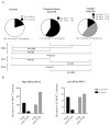
Figure 5. T-Helper phenotype of iNKT cells in early RA patients and controls
iNKT cells were co-cultured overnight with KRN7000 pulsed (200μg/ml) T2-CD1d. Culture supernatants were collected and IFN-γ, TNF-α, IL-4 and IL-10 were measured by Luminex array. (A) The observed proportions of Th1, Th2, and Th0 cytokine patterns in iNKT clones from treatment-naïve and treated early RA patients, and controls significantly differ. (B) The number of high-affinity iNKT clones and their Th0 vs nonTh0 proportions are significantly (p=0.0004) skewed towards nonTh0 in early RA patients (n=21) compared to controls (n=11) (left panel). The number of low-affinity iNKT clones and their Th0 vs nonTh0 proportions were not significantly different in early RA patients (n=11) compared to controls (n=6) (right panel). Fisher’s exact test was used to determine p values of differences between proportions.

References

    1. Brennan PJ, Brigl M, Brenner MB. Invariant natural killer T cells: an innate activation scheme linked to diverse effector functions. Nature reviews. Immunology. 2013;13:101–117. - PubMed
    1. Novak J, Lehuen A. Mechanism of regulation of autoimmunity by iNKT cells. Cytokine. 2011;53:263–270. - PubMed
    1. Wu L, Van Kaer L. Natural killer T cells and autoimmune disease. Curr Mol Med. 2009;9:4–14. - PubMed
    1. Jung S, Park YK, Shin JH, Lee H, Kim SY, Lee GR, Park SH. The requirement of natural killer T-cells in tolerogenic APCs-mediated suppression of collagen-induced arthritis. Exp Mol Med. 2010;42:547–554. - PMC - PubMed
    1. Coppieters K, Dewint P, Van Beneden K, Jacques P, Seeuws S, Verbruggen G, Deforce D, Elewaut D. NKT cells: manipulable managers of joint inflammation. Rheumatology (Oxford) 2007;46:565–571. - PubMed

Publication types