. 2015 Dec 15;195(12):5592-601.
doi: 10.4049/jimmunol.1501929. Epub 2015 Nov 9.

The Bacterial Enzyme IdeS Cleaves the IgG-Type of B Cell Receptor (BCR), Abolishes BCR-Mediated Cell Signaling, and Inhibits Memory B Cell Activation

Affiliations

The Bacterial Enzyme IdeS Cleaves the IgG-Type of B Cell Receptor (BCR), Abolishes BCR-Mediated Cell Signaling, and Inhibits Memory B Cell Activation

Sofia Järnum et al. J Immunol. .

Abstract

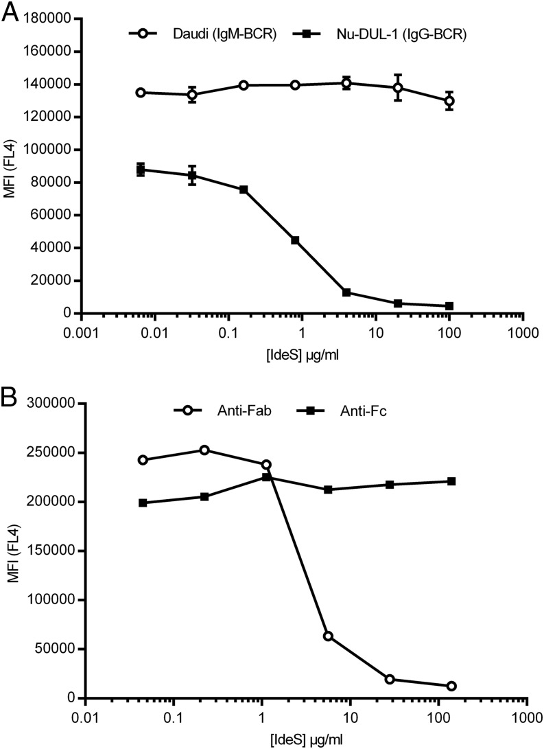
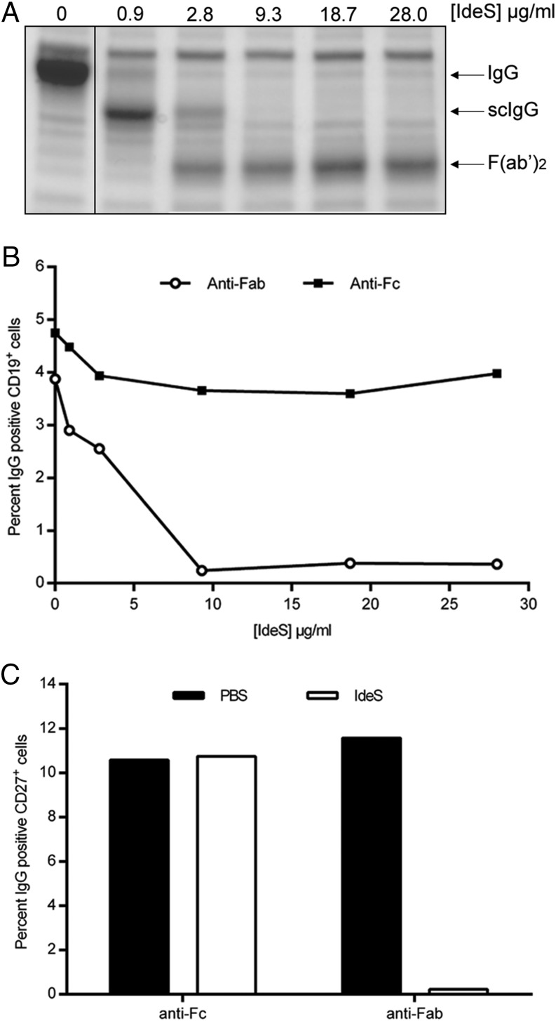
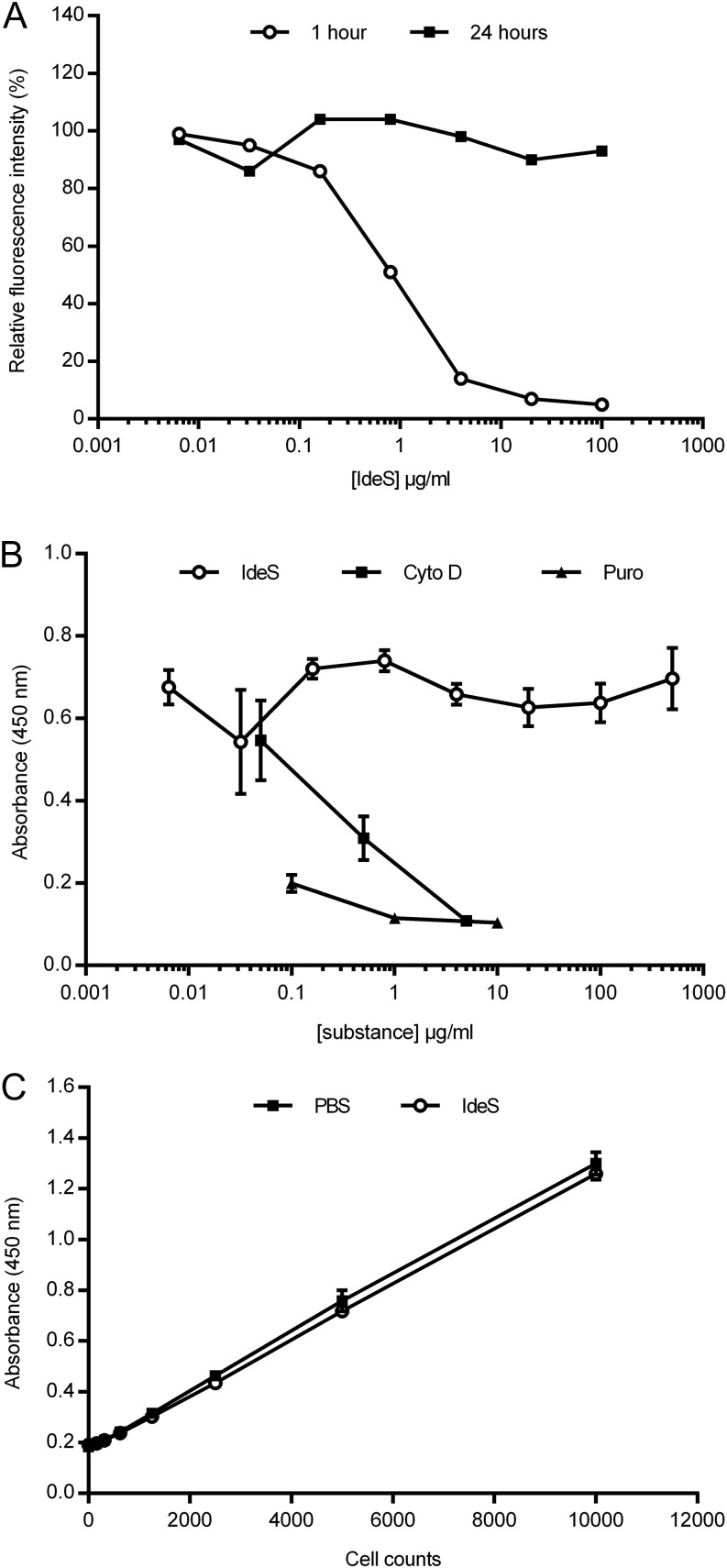
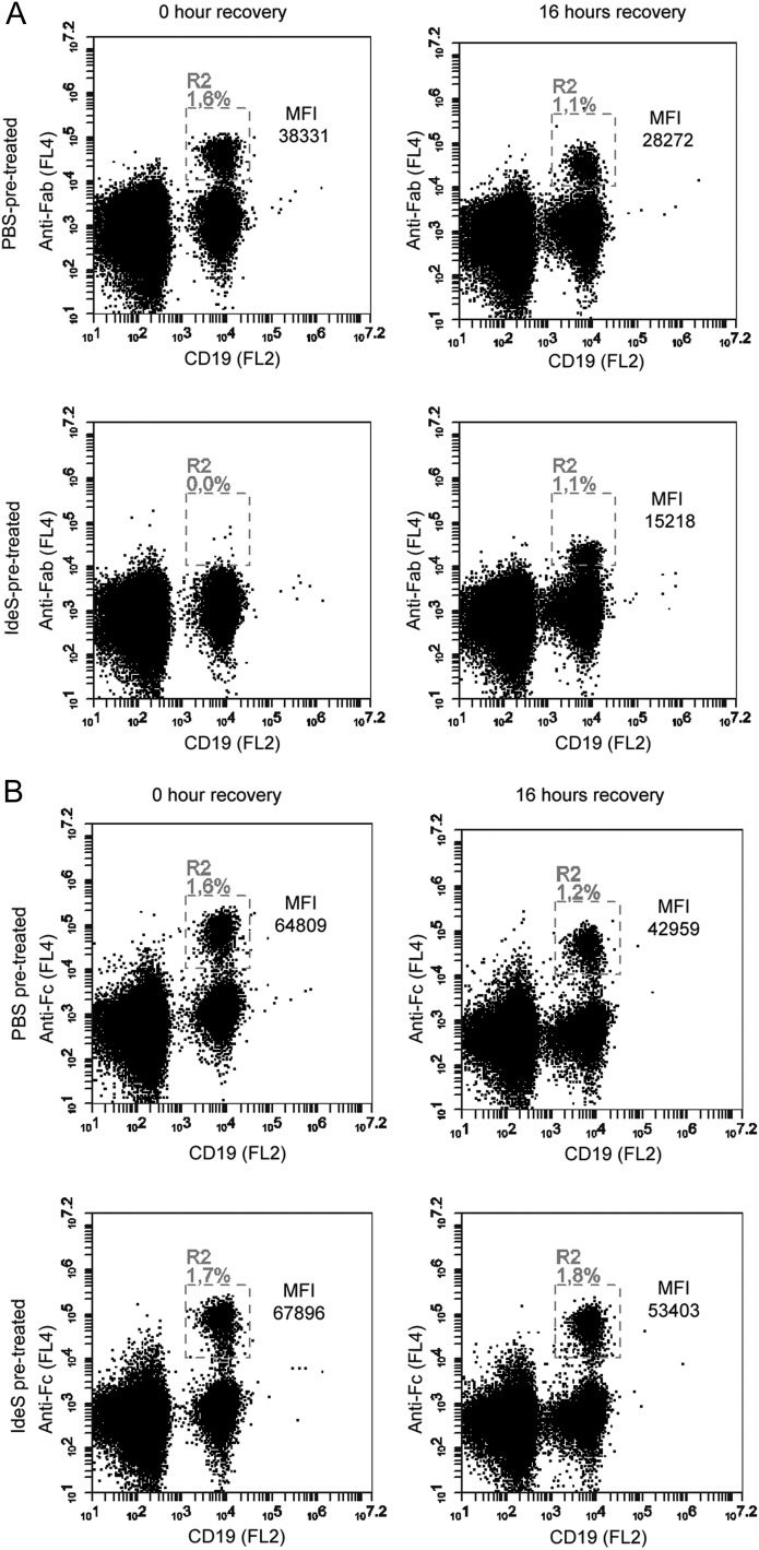
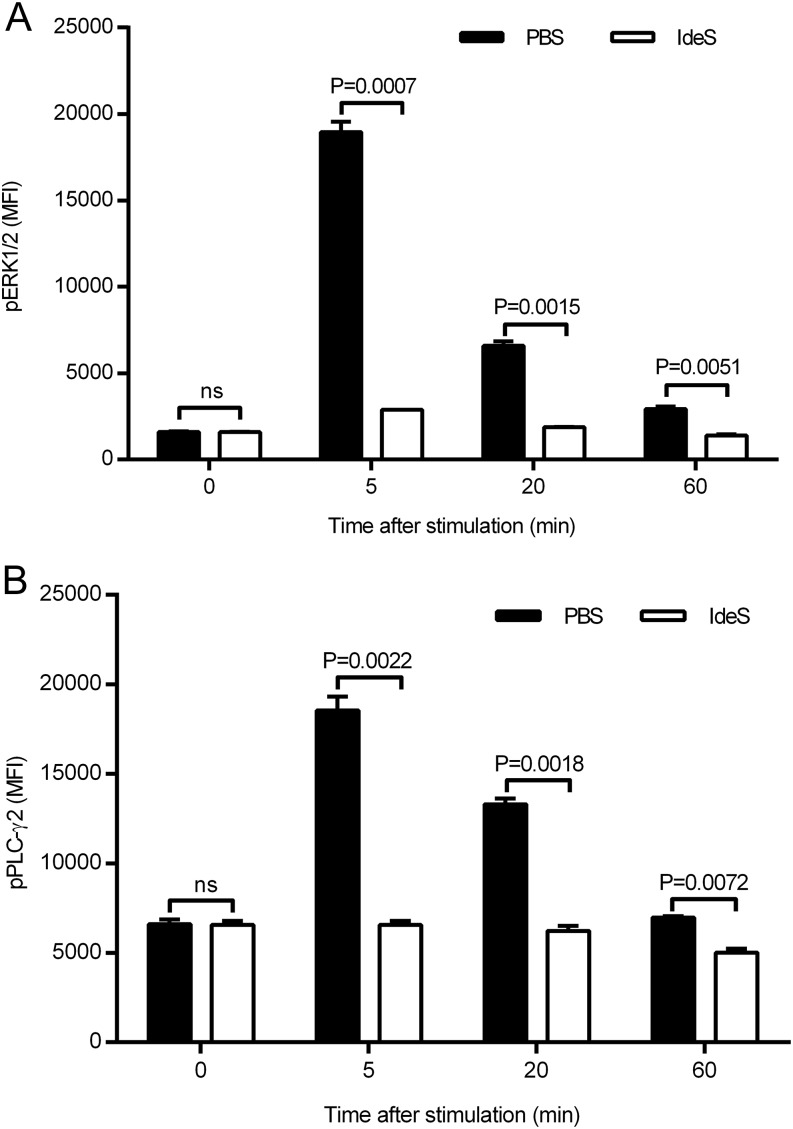
Ag binding to the BCR is a critical step in B cell development and activation, initiating a cascade of signaling events ultimately leading to proliferation, differentiation, or cell death. A bacterial enzyme, IgG-degrading enzyme of Streptococcus pyogenes (IdeS), was shown to specifically cleave IgG molecules below the hinge region of soluble IgG and when IgG is bound to Ag, resulting in one F(ab')2 molecule and one homodimeric Fc fragment. Whether IdeS could also cleave the IgG molecule when it is present in the BCR attached to the B cell membrane in a complex with CD79a and CD79b is unknown. In this article, we present human in vitro and ex vivo data showing that IdeS cleaves the IgG present in the BCR complex and very efficiently blocks Ag binding to the BCR. As a consequence of IdeS cleaving the BCR, signaling cascades downstream of the BCR are blocked, and memory B cells are temporarily silenced, preventing them from responding to antigenic stimulation and their transition into Ab-producing cells.


Figures

FIGURE 1.
FIGURE 1.
IdeS cleaves IgG-type, but not IgM-type, of BCR on B cells. (A) Flow cytometry analysis of cells stained with biotinylated anti-Fab Ab, followed by streptavidin-allophycocyanin, after treatment of Nu-DUL-1 cells (IgG-type) and Daudi cells (IgM-type) with the indicated concentrations of IdeS. The y-axis shows MFI of duplicate samples in FL4, and vertical lines represent SD. The data are representative of two independent experiments. (B) Flow cytometry analysis of Nu-DUL-1 cells (IgG-type) stained with either biotin-conjugated anti-Fab or anti-Fc, followed by streptavidin-allophycocyanin, after treatment with the indicated concentrations of IdeS. The y-axis shows MFI in FL4. The data are representative of two independent experiments.
FIGURE 2.
FIGURE 2.
IdeS cleaves the IgG-type of BCR with similar efficacy as soluble IgG. (A) Heparinized peripheral blood was treated with PBS or different concentrations of IdeS. After an incubation period, plasma was isolated and separated by SDS-PAGE. Intact IgG, scIgG, and F(ab′)2 fragments are indicated to the right. (B) PBMCs purified from the same PBS- or IdeS-treated blood were double stained for CD19+ (PE-coupled Ab) and anti-Fc or anti-Fab (biotin-conjugated Abs, followed by streptavidin-allophycocyanin) and analyzed by flow cytometry. Lymphocytes were gated using forward scatter/side scatter, and CD19+ cells within the lymphocyte gate were monitored for anti-Fab and anti-Fc signal. The data are representative of two independent experiments. (C) B cells were negatively selected (RosetteSep) after PBS or IdeS treatment (30 μg/ml). This CD19+-enriched population was double stained for CD27+ (PE-conjugated Ab) and anti-Fab or anti-Fc (biotin-conjugated Abs, followed by streptavidin-allophycocyanin) and analyzed by flow cytometry. The y-axis shows the percentage of CD27+ and IgG+ double-positive cells. The data are representative of two independent experiments.
FIGURE 3.
FIGURE 3.
Recovery of surface BCR expression on B cells after IdeS treatment. (A) Flow cytometry analysis of the presence of F(ab′)2 fragments on the surface of Nu-DUL-1 cells after treatment with different amounts of IdeS. IdeS was removed, and cells were cultured and analyzed after 1 and 24 h. Cells were stained with biotin-conjugated anti-Fab Ab, followed by streptavidin-allophycocyanin. The y-axis shows relative fluorescent intensity for which non-IdeS–treated signals were assigned 100%. The data are representative of two independent experiments. (B) Nu-DUL-1 cells were treated with different amounts of IdeS or antiproliferative control substances (cytochalasin D and puromycin) and cultured for 24 h prior to a 6-h BrdU incorporation. Data are mean ± SD of triplicate samples. The data are representative of two independent experiments. (C) Nu-DUL-1 cells were treated with PBS or IdeS (30 μg/ml) for 24 h before an intracellular hydrogenase activity–based viability assay (CCK-8) was used as read-out. The data are representative of two independent experiments.
FIGURE 4.
FIGURE 4.
Recovery of IgG-type BCR expression on ex vivo IdeS-treated PBMCs. PBMCs were treated with PBS or IdeS (30 μg/ml) prior to the start of staining. Lymphocytes were gated using forward scatter/side scatter (data not shown), and CD19+ cells (PE-conjugated Ab) within the lymphocyte gate were monitored for anti-Fab or anti-Fc signal using biotin-conjugated fragment-specific Abs, followed by streptavidin-allophycocyanin. (A) Flow cytometry analysis of anti-Fab signal on CD19+ cells immediately after PBS or IdeS treatment and after 16 h of IdeS-free culturing. Double-positive cells are found in R2 and expressed as the percentage of cells in the lymphocyte gate. MFI (anti-Fab) in R2 is shown. (B) Flow cytometry analysis of anti-Fc signal on CD19+ cells immediately after PBS or IdeS treatment and after 16 h of IdeS-free culturing. Double-positive cells are found in R2 and expressed as the percentage of cells in the lymphocyte gate. MFI (anti-Fc) in R2 is shown. The data are representative of two independent experiments.
FIGURE 5.
FIGURE 5.
IdeS treatment inhibits BCR signaling. Nu-DUL-1 cells were treated with PBS or IdeS (30 μg/ml) prior to cross-linking using a F(ab′)2-specific Ab. (A) ERK1/2 phosphorylation was followed at different time points after stimulation using a phospho-specific Ab in flow cytometry. MFI of duplicate samples (mean ± SD) is shown for each time point. (B) PLC-γ2 phosphorylation was followed at different time points after stimulation using a phospho-specific Ab in flow cytometry. MFI of duplicate samples (mean ± SD) is shown for each time point. The p values were calculated using GraphPad Prism and an unpaired, two-tailed t test. All data are representative of three independent experiments. ns, not significant.
FIGURE 6.
FIGURE 6.
IdeS specifically blocks B cell maturation of IgG-producing cells. PBMCs were treated with PBS or IdeS and stimulated with rIL-2 and R848 to activate memory B cells and differentiate them into Ig-producing cells. ELISPOT filter plates were evaluated for the number of IgG-producing cells. (A) The filter plate was seeded with 50,000 or 100,000 cells that were treated or not with rIL-2/R848 on day 0 and with/without IdeS (30 μg/ml) on day 0 or 3. (B) Number of IgA-, IgM-, and IgG-producing cells after stimulation with rIL-2/R848 in the presence or absence of 30 μg/ml IdeS for 96 h. Data are mean (± SD) of four to six wells. The p value was calculated using GraphPad Prism and an unpaired, two-tailed t test. (C) Number of IgG-producing cells after stimulation with rIL-2/R848 in the presence or absence of 0.3–30 μg/ml IdeS for 72 h. Data are mean (± SD) of two to four wells. (D) Number of IgG-producing cells after pretreating cells for 1 h with 0.3–30 μg/ml IdeS prior to removing IdeS and subjecting cells to 72 h of stimulation with rIL-2/R848. Data are mean (± SD) of two to four wells. (E) Graphical illustration of the ELISPOT experiments in which IdeS was added from the start or at the end of the culturing period. Data in (B)–(D) are representative of two independent experiments. ns, not significant.
FIGURE 7.
FIGURE 7.
Recovery of IgG-type BCR expression on PBMCs collected after a single i.v. dose of 0.24 mg/kg BW IdeS in a human healthy subject. Flow cytometry analysis of CD19+/IgG+ cells at different time points after a single dose of IdeS. PBMCs were purified, and lymphocytes gated using forward scatter/side scatter (P1). B cells (CD19+) were monitored in FL2 (PE), and IgG was monitored in FL4 (allophycocyanin). Double-positive cells for CD19 (PE) and the Fc portion of IgG (FL4) predose and up to 96 h postdosing (upper panels). Double-positive cells for CD19 (FL2) and the Fab portion of IgG (FL4) predose and up to 96 h postdosing (lower panels). MFI for double-positive cells are given for R1 (CD19/anti-Fc) and R2 (CD19/anti-Fab). The data are representative of two staining experiments.
FIGURE 8.
FIGURE 8.
IdeS cleaves the IgG-type of BCR in vivo in humans. Healthy human subjects were dosed with 0.24 mg/kg BW IdeS, and PBMCs were collected at different time points after dosing. The percentage of cells double positive for CD19 (PE) and F(ab′)2 (allophycocyanin) was analyzed using flow cytometry. Hours postdosing are shown on the x-axes and MFI × cell frequency is shown on the y-axes. Data are the mean (± SD) of two independent flow cytometry experiments for each patient.

References

    1. von Pawel-Rammingen U., Johansson B. P., Björck L. 2002. IdeS, a novel streptococcal cysteine proteinase with unique specificity for immunoglobulin G. EMBO J. 21: 1607–1615. - PMC - PubMed
    1. Su Y. F., Chuang W. J., Wang S. M., Chen W. Y., Chiang-Ni C., Lin Y. S., Wu J. J., Liu C. C. 2011. The deficient cleavage of M protein-bound IgG by IdeS: insight into the escape of Streptococcus pyogenes from antibody-mediated immunity. Mol. Immunol. 49: 134–142. - PubMed
    1. Reth M. 1989. Antigen receptor tail clue. Nature 338: 383–384. - PubMed
    1. Reth M., Wienands J. 1997. Initiation and processing of signals from the B cell antigen receptor. Annu. Rev. Immunol. 15: 453–479. - PubMed
    1. Kurosaki T. 2002. Regulation of B-cell signal transduction by adaptor proteins. Nat. Rev. Immunol. 2: 354–363. - PubMed