

RNA sequencing of Sleeping Beauty transposon-induced tumors detects transposon-RNA fusions in forward genetic cancer screens
- PMID: 26553456
- PMCID: PMC4691744
- DOI: 10.1101/gr.188649.114
RNA sequencing of Sleeping Beauty transposon-induced tumors detects transposon-RNA fusions in forward genetic cancer screens
Abstract
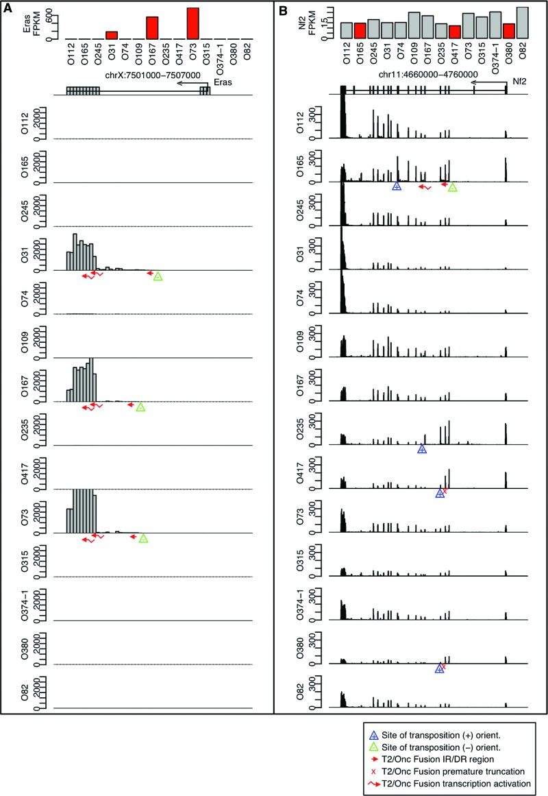
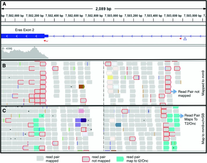
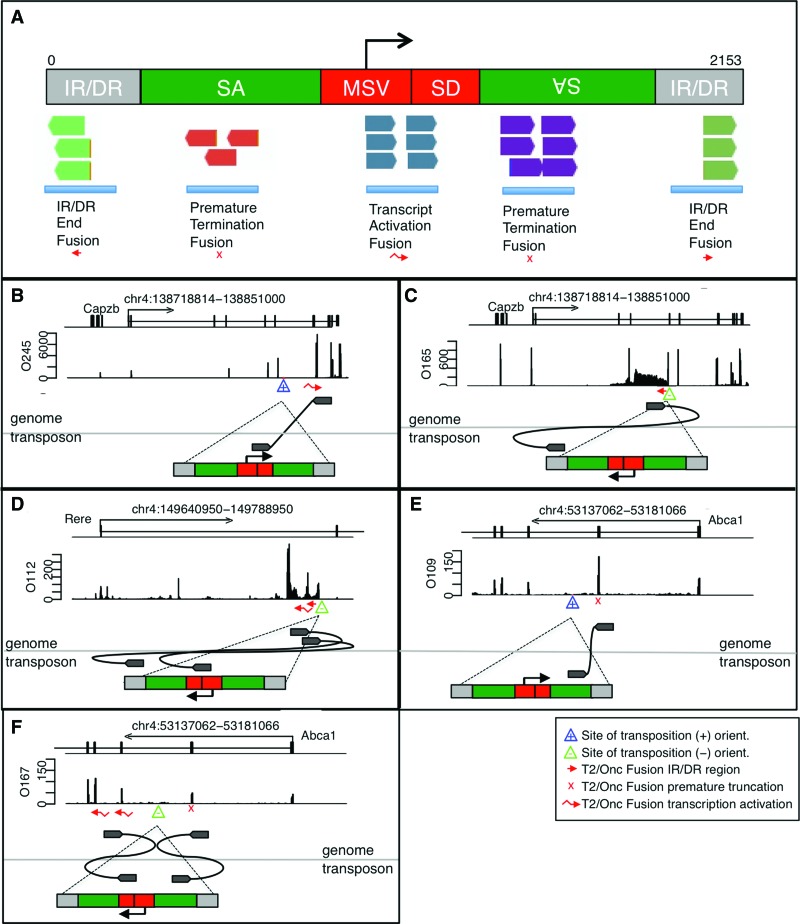
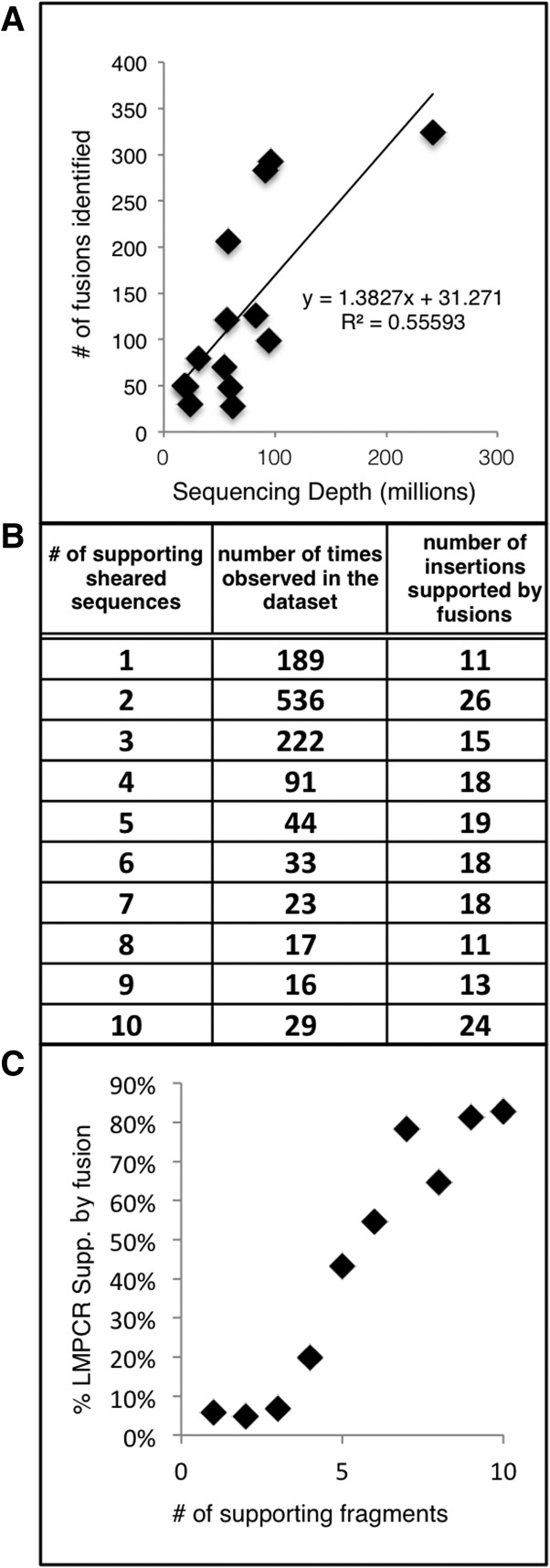
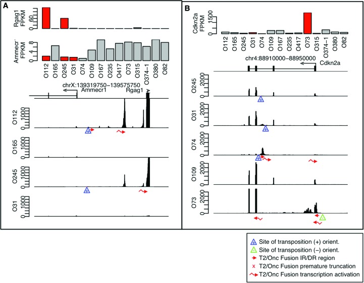
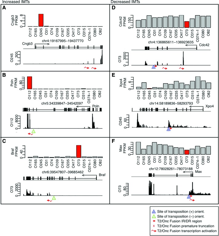
Forward genetic screens using Sleeping Beauty (SB)-mobilized T2/Onc transposons have been used to identify common insertion sites (CISs) associated with tumor formation. Recurrent sites of transposon insertion are commonly identified using ligation-mediated PCR (LM-PCR). Here, we use RNA sequencing (RNA-seq) data to directly identify transcriptional events mediated by T2/Onc. Surprisingly, the majority (∼80%) of LM-PCR identified junction fragments do not lead to observable changes in RNA transcripts. However, in CIS regions, direct transcriptional effects of transposon insertions are observed. We developed an automated method to systematically identify T2/Onc-genome RNA fusion sequences in RNA-seq data. RNA fusion-based CISs were identified corresponding to both DNA-based CISs (Cdkn2a, Mycl1, Nf2, Pten, Sema6d, and Rere) and additional regions strongly associated with cancer that were not observed by LM-PCR (Myc, Akt1, Pth, Csf1r, Fgfr2, Wisp1, Map3k5, and Map4k3). In addition to calculating recurrent CISs, we also present complementary methods to identify potential driver events via determination of strongly supported fusions and fusions with large transcript level changes in the absence of multitumor recurrence. These methods independently identify CIS regions and also point to cancer-associated genes like Braf. We anticipate RNA-seq analyses of tumors from forward genetic screens will become an efficient tool to identify causal events.
© 2016 Temiz et al.; Published by Cold Spring Harbor Laboratory Press.
Figures
Similar articles
-
Advances in functional genetic screening with transposons and CRISPR/Cas9 to illuminate cancer biology.Curr Opin Genet Dev. 2018 Apr;49:85-94. doi: 10.1016/j.gde.2018.03.006. Epub 2018 Mar 26. Curr Opin Genet Dev. 2018. PMID: 29587177 Free PMC article. Review.
-
Identification of Cancer Genes Based on De Novo Transposon Insertion Site Analysis Using RNA and DNA Sequencing.Methods Mol Biol. 2019;1907:73-79. doi: 10.1007/978-1-4939-8967-6_5. Methods Mol Biol. 2019. PMID: 30542991
-
Identification of Sleeping Beauty transposon insertions in solid tumors using linker-mediated PCR.J Vis Exp. 2013 Feb 1;(72):e50156. doi: 10.3791/50156. J Vis Exp. 2013. PMID: 23407503 Free PMC article.
-
TAPDANCE: an automated tool to identify and annotate transposon insertion CISs and associations between CISs from next generation sequence data.BMC Bioinformatics. 2012 Jun 29;13:154. doi: 10.1186/1471-2105-13-154. BMC Bioinformatics. 2012. PMID: 22748055 Free PMC article.
-
Identification of cancer driver genes using Sleeping Beauty transposon mutagenesis.Cancer Sci. 2021 Jun;112(6):2089-2096. doi: 10.1111/cas.14901. Epub 2021 May 1. Cancer Sci. 2021. PMID: 33783919 Free PMC article. Review.
Cited by
-
Development and characterization of the novel human osteosarcoma cell line COS-33 with sustained activation of the mTOR pathway.Oncotarget. 2020 Jul 7;11(27):2597-2610. doi: 10.18632/oncotarget.27611. eCollection 2020 Jul 7. Oncotarget. 2020. PMID: 32676162 Free PMC article.
-
Advances in functional genetic screening with transposons and CRISPR/Cas9 to illuminate cancer biology.Curr Opin Genet Dev. 2018 Apr;49:85-94. doi: 10.1016/j.gde.2018.03.006. Epub 2018 Mar 26. Curr Opin Genet Dev. 2018. PMID: 29587177 Free PMC article. Review.
-
Pharmacologic inhibition of CSF-1R suppresses intrinsic tumor cell growth in osteosarcoma with CSF-1R overexpression.J Transl Med. 2025 Aug 12;23(1):900. doi: 10.1186/s12967-025-06920-6. J Transl Med. 2025. PMID: 40797330 Free PMC article.
-
CRISPR and transposon in vivo screens for cancer drivers and therapeutic targets.Genome Biol. 2020 Aug 19;21(1):204. doi: 10.1186/s13059-020-02118-9. Genome Biol. 2020. PMID: 32811551 Free PMC article. Review.
-
Mouse models in the era of large human tumour sequencing studies.Open Biol. 2018 Aug;8(8):180080. doi: 10.1098/rsob.180080. Open Biol. 2018. PMID: 30111589 Free PMC article. Review.
References
-
- Collier LS, Carlson CM, Ravimohan S, Dupuy AJ, Largaespada DA. 2005. Cancer gene discovery in solid tumours using transposon-based somatic mutagenesis in the mouse. Nature 436: 272–276. - PubMed
-
- Dromard M, Bompard G, Glondu-Lassis M, Puech C, Chalbos D, Freiss G. 2007. The putative tumor suppressor gene PTPN13/PTPL1 induces apoptosis through insulin receptor substrate-1 dephosphorylation. Cancer Res 67: 6806–6813. - PubMed
-
- Egas-Bejar D, Anderson PM, Agarwal R, Corrales-Medina F, Devarajan E, Huh WW, Brown RE, Subbiah V. 2014. Theranostic profiling for actionable aberrations in advanced high risk osteosarcoma with aggressive biology reveals high molecular diversity: the human fingerprint hypothesis. Oncoscience 1: 167–179. - PMC - PubMed
-
- Ivics Z, Hackett PB, Plasterk RH, Izsvák Z. 1997. Molecular reconstruction of Sleeping Beauty, a Tc1-like transposon from fish, and its transposition in human cells. Cell 91: 501–510. - PubMed
Publication types
MeSH terms
Substances
Grants and funding
LinkOut - more resources
Full Text Sources
Other Literature Sources
Research Materials
Miscellaneous