. 2015 Nov 9;7(6):1759091415610761.
doi: 10.1177/1759091415610761. Print 2015 Nov-Dec.

The Circadian Clock Gene Period1 Connects the Molecular Clock to Neural Activity in the Suprachiasmatic Nucleus

Affiliations

The Circadian Clock Gene Period1 Connects the Molecular Clock to Neural Activity in the Suprachiasmatic Nucleus

Takashi Kudo et al. ASN Neuro. .

Abstract

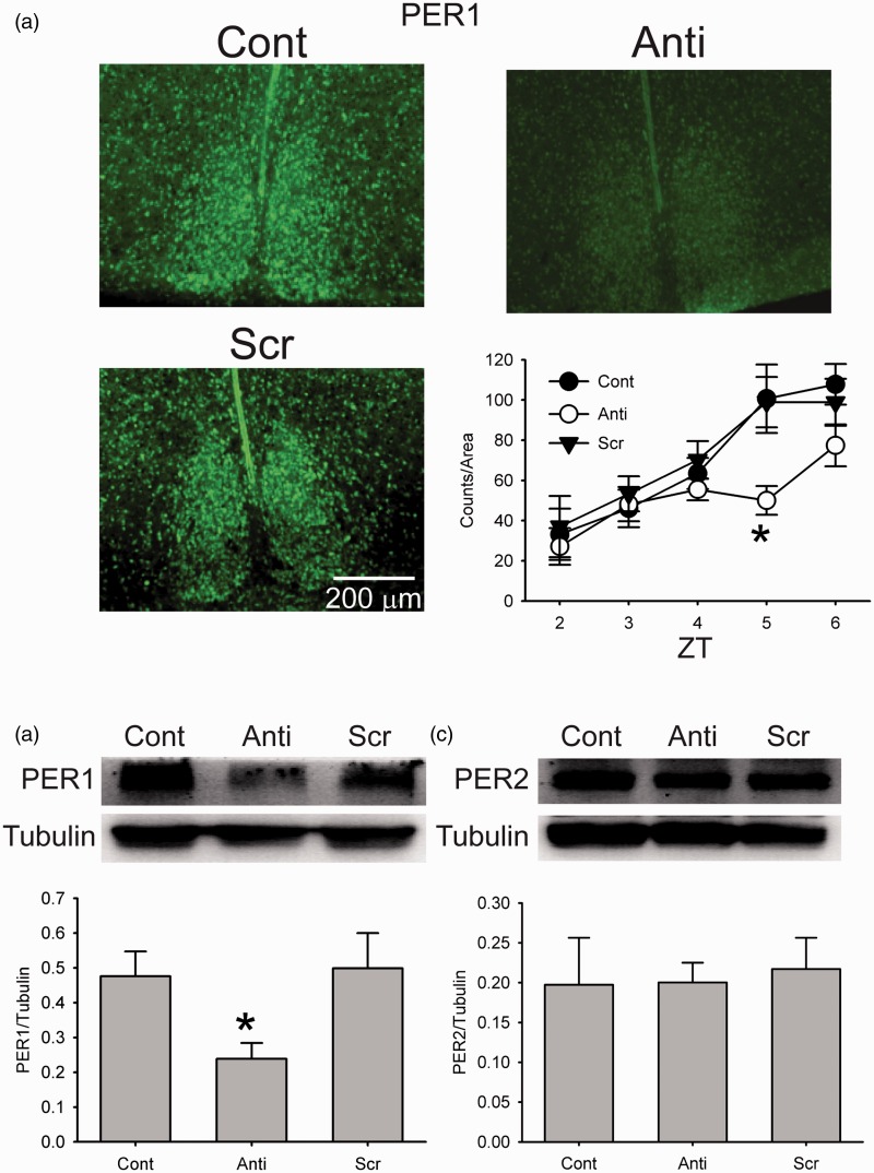
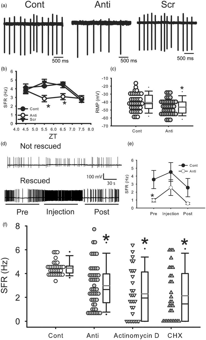
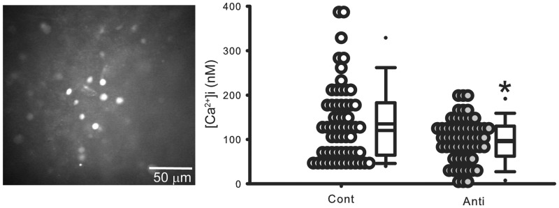
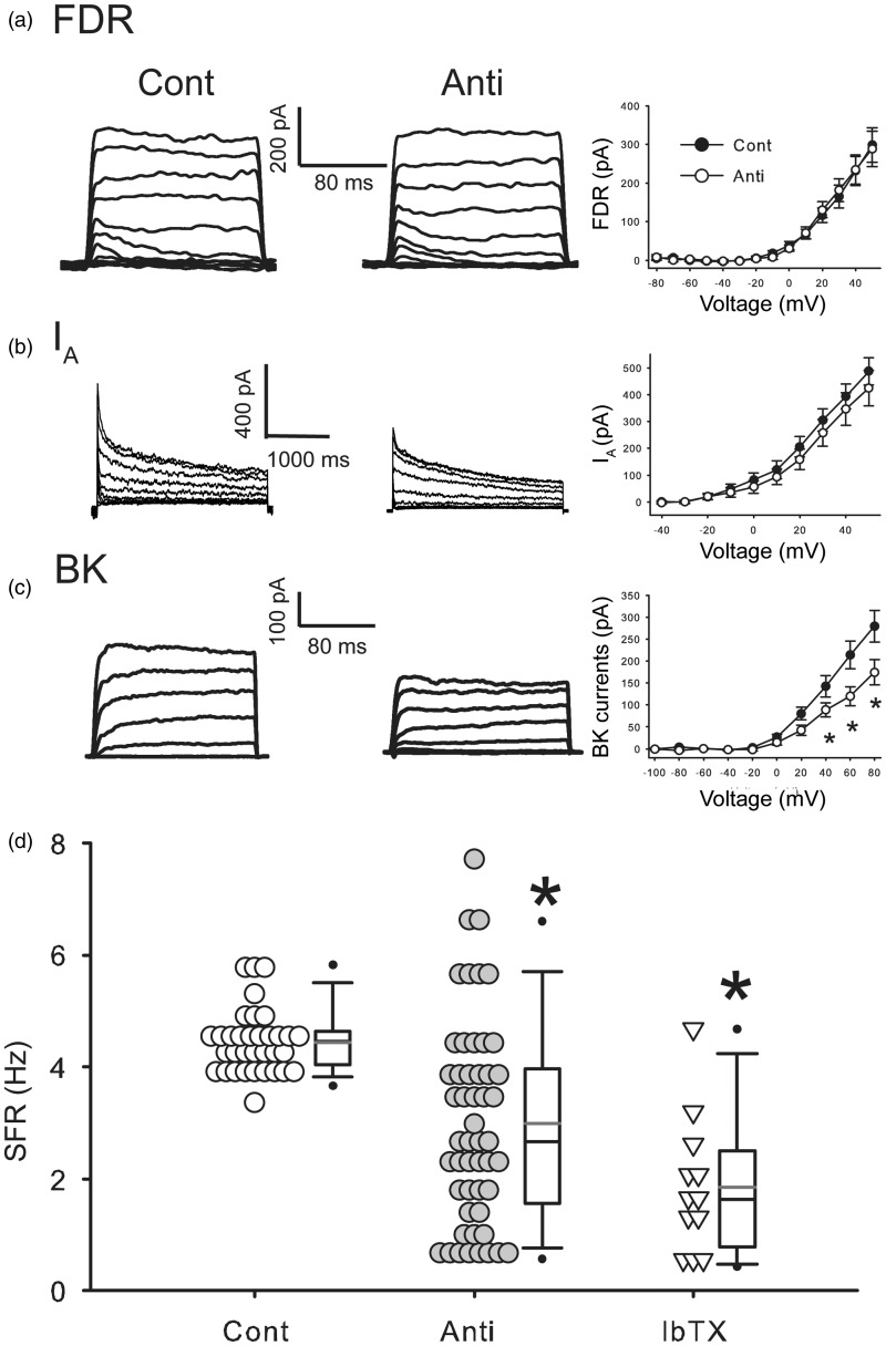
The neural activity patterns of suprachiasmatic nucleus (SCN) neurons are dynamically regulated throughout the circadian cycle with highest levels of spontaneous action potentials during the day. These rhythms in electrical activity are critical for the function of the circadian timing system and yet the mechanisms by which the molecular clockwork drives changes in the membrane are not well understood. In this study, we sought to examine how the clock gene Period1 (Per1) regulates the electrical activity in the mouse SCN by transiently and selectively decreasing levels of PER1 through use of an antisense oligodeoxynucleotide. We found that this treatment effectively reduced SCN neural activity. Direct current injection to restore the normal membrane potential partially, but not completely, returned firing rate to normal levels. The antisense treatment also reduced baseline [Ca(2+)]i levels as measured by Fura2 imaging technique. Whole cell patch clamp recording techniques were used to examine which specific potassium currents were altered by the treatment. These recordings revealed that the large conductance [Ca(2+)]i-activated potassium currents were reduced in antisense-treated neurons and that blocking this current mimicked the effects of the anti-sense on SCN firing rate. These results indicate that the circadian clock gene Per1 alters firing rate in SCN neurons and raise the possibility that the large conductance [Ca(2+)]i-activated channel is one of the targets.

Keywords: BK currents; Per1; calcium; circadian.


Figures

Figure 1.
Figure 1.
Antisense ODN for Per1 reduced PER1 expression in the SCN. (a) Photomicrographs illustrate that the application of antisense ODN for Per1 (10 µM) reduces PER1 expression as measured by immunohistochemistry (IHC). Cont: control, Anti: antisense ODN for Per1, and Scr: scrambled ODN. Bottom right, bar graphs show PER1 positive cell counts for Cont, Anti, and Scr (±SE, n = 3 for each time point). *Significant difference (p < .05) analyzed by two-way ANOVA followed by Holm-Sidak method for multiple comparisons (vs. Cont). ZT = Zeitgeber time. (b) Example of Western blots measuring PER1 expression in the SCN tissue. Tubulin protein expression was measured as a control for loading. Bottom: Levels of PER1 protein expression (+SE, normalized to tubulin, n = 3 for each group). *Significant difference (p < .05) analyzed by one-way ANOVA followed by Holm-Sidak method for multiple comparisons (vs. Cont). (c) Example of Western blots measuring PER2 expression in the SCN tissue. Tubulin protein expression was measured as a control for loading. Bottom: Levels of PER2 protein expression (+SE, normalized to tubulin, n = 3 for each group).
Figure 2.
Figure 2.
Antisense ODN for Per1 reduced firing rate in dorsal SCN neurons during the day. (a) Representative examples illustrating the reduced firing rates by the antisense ODN for Per1 (10 µM). Cont: control, Anti: antisense ODN for Per1, and Scr: scrambled ODN. (b) Average firing rate for each group ( ± SE). *Significant difference (p < .05) analyzed by two-way ANOVA followed by Holm-Sidak method for multiple comparisons (vs. Cont). For each group n = 17–28 cells from 4 mice at each time point (in total, 271 cells from 48 mice). (c) Average resting membrane potential for each group ( ± SE). *Significant difference (p < .05) analyzed by unpaired Student’s t-test. For each group, n = 40 cells from 30 to 37 mice. (d) Representative examples illustrating the effects of current injections on SFR under antisense ODN for Per1. (e) Average firing rate for each group (±SE). *Significant difference (p < .05) analyzed by two-way repeated measures ANOVA followed by Holm-Sidak method for multiple comparisons (vs. Cont). For each group, n = 10 cells from 7 to 9 mice. (f) Effects of actinomycin D and cycloheximide (CHX) on SFR in the SCN during daytime. Actinomycin D solution (0.8 µM final concentration), CHX (final 36 µM), or water (as a control) was added to the culture medium 3 h before the start of recordings. The SFR was measured by loose-patch method. Average firing rate for each group (±SE). *Significant difference (p < .05) analyzed by one-way ANOVA followed by Dunn’s method for multiple comparisons (vs. Cont). For each group, n = 30–51 cells from 4 to 8 mice. Cont and Anti data are the same as (b).
Figure 3.
Figure 3.
Antisense ODN for Per1 reduced [Ca2+]i levels in cells in the SCN during daytime. In these experiments, resting [Ca2+]i levels were estimated in SCN neurons in brain slices with antisense ODN for Per1 and compared with data obtained from the control group. Animals were sacrificed at ZT 0. Each cell is sampled only once. Left: Representative pictures in Fura2 imaging in the SCN. Right: Dot density plot showing that [Ca2+]i levels in SCN cells were reduced by antisense ODN for Per1. Cont: control, Anti: antisense ODN for Per1. *Significant difference (p < .05) analyzed by unpaired Student’s t-test. Bottom: Histograms illustrating the distribution of [Ca2+]I levels. For each group (±SE), n = 59 cells from four mice.
Figure 4.
Figure 4.
Antisense ODN for Per1 reduced BK currents in the SCN during daytime. (a) Left, representative examples showing that application of antisense ODN for Per1 (10 µM) did not decrease the magnitude of fast delayed rectifier (FDR) currents in the SCN neurons. Right, current-voltage (I–V) relationship of FDR currents in SCN neurons. Cont: control, Anti: antisense ODN for Per1. For each group (±SE), n = 20 cells from 10 mice. (b) Left, representative examples showing the IA currents. Right, I–V curve of IA measured in SCN neurons. For each group (±SE), n = 11–15 cells from 4 to 13 mice. (c) Left, representative examples showing BK currents. Right, I–V curve of BK current measured in SCN neurons. *Significant difference (p < .05) analyzed by two-way repeated measures ANOVA followed by Holm-Sidak method for multiple comparisons (vs. Cont). For each group (±SE), n = 20–21 cells from 15 to 18 mice. (d) Average firing rate of control, antisense ODN for Per1, and iberiotoxin (IbTX). *Significant difference (p < .05) analyzed by one-way ANOVA followed by Dunn’s method for multiple comparisons (vs. Cont). For each group n = 12–51 cells from 6 to 8 mice. Cont and Anti data are the same as Figure 2(b). The currents were normalized by capacitance.

Similar articles

Cited by

References

    1. Akiyama M., Kouzu Y., Takahashi S., Wakamatsu H., Moriya T., Maetani M., Shibata S. (1999) Inhibition of light- or glutamate-induced mPer1 expression represses the phase shifts into the mouse circadian locomotor and suprachiasmatic firing rhythms. The Journal of Neuroscience: The Official Journal of the Society for Neuroscience 19: 1115–1121. - PMC - PubMed
    1. Albus H., Bonnefont X., Chaves I., Yasui A., Doczy J., van der Horst G. T., Meijer J. H. (2002) Cryptochrome-deficient mice lack circadian electrical activity in the suprachiasmatic nuclei. Current Biology: CB 12: 1130–1133. - PubMed
    1. Belle M. D., Diekman C. O., Forger D. B., Piggins H. D. (2009) Daily electrical silencing in the mammalian circadian clock. Science 326: 281–284. - PubMed
    1. Brancaccio M., Maywood E. S., Chesham J. E., Loudon A. S., Hastings M. H. (2013) A Gq-Ca2+ axis controls circuit-level encoding of circadian time in the suprachiasmatic nucleus. Neuron 78: 714–728. - PMC - PubMed
    1. Brown S. A., Kowalska E., Dallmann R. (2012) (Re)inventing the circadian feedback loop. Developmental Cell 22: 477–487. - PubMed

Publication types

MeSH terms