. 2016 Apr 1;22(7):1744-56.
doi: 10.1158/1078-0432.CCR-15-1606. Epub 2015 Nov 9.

Dysregulation of miR-212 Promotes Castration Resistance through hnRNPH1-Mediated Regulation of AR and AR-V7: Implications for Racial Disparity of Prostate Cancer

Affiliations

Dysregulation of miR-212 Promotes Castration Resistance through hnRNPH1-Mediated Regulation of AR and AR-V7: Implications for Racial Disparity of Prostate Cancer

Yijun Yang et al. Clin Cancer Res. .

Abstract

Purpose: The causes of disproportionate incidence and mortality of prostate cancer among African Americans (AA) remain elusive. The purpose of this study was to investigate the mechanistic role and assess clinical utility of the splicing factor heterogeneous nuclear ribonucleoprotein H1 (hnRNP H1) in prostate cancer progression among AA men.

Experimental design: We employed an unbiased functional genomics approach coupled with suppressive subtractive hybridization (SSH) and custom cDNA microarrays to identify differentially expressed genes in microdissected tumors procured from age- and tumor grade-matched AA and Caucasian American (CA) men. Validation analysis was performed in independent cohorts and tissue microarrays. The underlying mechanisms of hnRNPH1 regulation and its impact on androgen receptor (AR) expression and tumor progression were explored.

Results: Aberrant coexpression of AR and hnRNPH1 and downregulation of miR-212 were detected in prostate tumors and correlate with disease progression in AA men compared with CA men. Ectopic expression of miR-212 mimics downregulated hnRNPH1 transcripts, which in turn reduced expression of AR and its splice variant AR-V7 (or AR3) in prostate cancer cells. hnRNPH1 physically interacts with AR and steroid receptor coactivator-3 (SRC-3) and primes activation of androgen-regulated genes in a ligand-dependent and independent manner. siRNA silencing of hnRNPH1 sensitized prostate cancer cells to bicalutamide and inhibited prostate tumorigenesis in vivo

Conclusions: Our findings define novel roles for hnRNPH1 as a putative oncogene, splicing factor, and an auxiliary AR coregulator. Targeted disruption of the hnRNPH1-AR axis may have therapeutic implications to improve clinical outcomes in patients with advanced prostate cancer, especially among AA men.


Conflict of interest statement

Disclosure of Potential Conflicts of Interest

No potential conflicts of interest were disclosed.

Figures
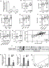
Figure 1.
Figure 1.
Selective expression and correlation of hnRNPH1 with AR expression, prostate cancer progression, and recurrence in AA men. A, qRT-PCR analysis of hnRNPH1 gene expression relative to the β-actin in LCM-procured normal prostate epithelium (NE) and tumor cells (T) of an independent cohort of AA and CA (n = 12/group). B, analysis of gene expression of Affymetrix GSE17386 dataset (NCBI Geo; Oncomine) denotes a significant expression of hnRNPH1 in prostate tumors in AA men in comparison with CA men. C, analysis of the second dataset reveals significant elevation of hnRNPH1 transcripts levels in prostate tumor cells (T) in comparison with the normal prostate epithelium (NE). D, analysis of second database indicates significant hnRNPH1 gene expression in prostate tumor cells compared with the normal epithelium in both groups, but to a greater extent in AA men than CA men. E–G, aberrant hnRNPH1 gene expression correlates with extraprostatic extension (EPE) and high Gleason sum score. H, hnRNPH1 gene expression increases in 5-year recurrent tumors in comparison with nonrecurrent tumors. I and J, positive correlation between hnRNPH1 and AR gene expression in prostate tumors in comparison with the normal epithelium. Affymetrix Human Genome U133 Plus 2.0 array datasets (26, 27) AR and hnRNPH1 expression data were extracted as log2 median and compared using Pearson correlation. K and L, qRT-PCR analysis demonstrates coexpression of hnRNPH1 and AR transcripts, respectively, in microdissected tumor cells relative to their matched normal epithelium in an independent cohort of AA men (n = 13) than CA men (n = 17). Gene expression is significant (P < 0.05) relative to normal adjacent tissue (a) and to CA men (b). M, regression analysis depicts a positive correlation (r = 0.8323; P < 0.005) between AR and hnRNPH1 transcript levels shown in K and L.
Figure 2.
Figure 2.
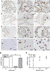
Ethnicity-based TMA-4 analysis reveals prostate tumor cell nuclear immunoreactivity and correlation with disease progression in AA men. A–C, a representative prostate adenocarcinoma core depicting intense nuclear immunoreactivity to hnRNPH1 (arrow) in comparison with stroma (arrowhead). D–I, representative cores of benign prostatic hyperplasia (d–f) and normal prostate epithelium (g–i) demonstrating weak nuclear immunoreactivity (arrow) in the epithelial cells in comparison with the adjacent stroma (arrowhead). J, validation of the hnRNPH1 antibody specificity with negative staining of prostate tumor cores in the absence of primary antibody. K and L, weak and intense immunostaining of hnRNPH1 in cell cores of AR-naïve PC-3 and AR-expressing LNCaP, respectively. M, histoscore of hnRNPH1 in tumors (T) relative to the normal epithelium (NE) in AA (n = 148) and CA men (n = 152) expressed as mean ± SEM. N, hnRNPH1 histoscore stratified by Gleason scores. *, denotes significant difference at P < 0.05 in comparison with controls.
Figure 3.
Figure 3.
Expression and functional significance of miR-212 and miR-22 in the regulation of hnRNPH1 and AR and its splice variants in prostate cancer cells. A and B, in silico analysis of different miRNA predicting targets programs (TargetScan, miRNA and Microcosm Targets) predicted hnRNPH1 as a target for miR-22 and miR-212 among others. qRT-PCR analysis of hnRNPH1-associated miR-22 and miR-212 expression in prostate tumors derived from AA (n = 13) and CA men (n = 17) relative to the normal epithelium in both groups. The data are expressed as fold change relative to U6 as an internal control. *, significance at P < 0.05 relative to controls. #, a significant difference in hnRNPH1 levels in prostate tumor cells procured from CA men compared with AA men. C and D, transfection efficiencies of miR-22 and/or miR-212 in C4–2B cells, respectively, as measured by qRT-PCR relative to cells transfected with nontargeting control miRNA (control miR). Ectopic expression of either miR-22 and/or miR-212 mimics significantly reduced expression of hnRNPH1 and AR-FL and all N-terminal containing AR variants (AR-nt) in C4–2B cells compared with control miR–transfected cells, as measured by qRT-PCR (E and F) and conventional PCR analyses (G). In addition to reduction of hnRNPH1 and AR-FL transcripts, the ectopic expression of miR-212 mimics significantly inhibited the AR splice variant AR-V7 mRNA in C4–2B cells when measured by qRT-PCR (H), conventional PCR (I) and Western blot (J) analyses. hnRNPH1 shRNA silencing causes reduction of AR-FL and AR-V7 mRNAs compared with control shRNA-transfected C4–2B cells as measured by qRT-PCR (K) and immunoblot (L) assay, as quantified relative to GAPDH by densitometric analysis (M).
Figure 4.
Figure 4.
hnRNPH1 confers growth stimulation and hormone resistance through activation of AR in prostate cancer cells. A, qRT-PCR analysis of hnRNPH1 transcripts in AR-expressing (C4–2B and MDA-PCa-2b) and AR-null PC-3 cells. B, immunocytochemical analysis of hnRNPH1 protein expression in MDA-PCa-2b cells in presence of complete medium. C and D, optimization of siRNA silencing and transfection efficiencies in prostate cancer cells by GFP and siGLO Lamin A/C, respectively. E and F, endogenous mRNA and proteins levels of hnRNPH1, respectively, at 24 hours following siRNA transfection. G, assessment of growth inhibitory effects by a cell counting assay kit in hnRNPH1 siRNA-silenced MDA-PCa-2b cells cultured in complete medium for up to 120 hours. H and I, cell growth of MDA-PCa-2b and C4–2B cells, respectively, pretransfected with siControl or hnRNPH1 siRNA and cultured in RPMI containing charcoal-stripped serum and various concentrations of BIC with (+) or without (−) DHT for 24 hours (n = 3). J, immunoblot analysis of C4–2B cells stably transfected with control shRNA or hnRNPH1 shRNA plasmids. K and L, body weight and tumor volume in animals transplanted with C4–2B cell stable clones, respectively. M, resected tumor sizes of siControl-, or siRNPH1-transfected C4–2B cells. N and O, COS-7 and CV-1 cells, respectively, were cultured in charcoal-stripped FBS medium in the absence (ethanol) or presence of DHT and cotransfected with hnRNPH1, pCMV-AR, and psPSA-Luc plasmids. P, LNCaP cells cotransfected with hnRNPH1 and psPSA-Luc plasmids and cultured with or without DHT. Q, C4–2B cells cotransfected with non-target siRNA (siControl) or HnRNPHI siRNA (siRNPH1) and psPSA-Luc reporter and cultured with or without DHT. For normalization, all cells were cotransfected with five ng pRL-SV40. Activity was measured with the dual luciferase system, and the results were expressed as fold change of relative light units (RLU). * and **, significant difference at P < 0.05 and P < 0.01, respectively, in comparison with controls (n = 3).
Figure 5.
Figure 5.
AR–hnRNPH1 interaction and transcriptional regulation of AR and PSA in prostate cancer cells. A, prostate cancer cell lysates cultured in complete medium were subjected to immunoprecipitation (IP) using anti-AR or anti-hnRNPH1 antibody, followed by immunoblotting (IB) with the indicated antibodies in a reversed order as shown. B, lysates of prostate cancer cells cultured in charcoal-stripped medium with or without DHT were analyzed for AR–hnRNPH1 interaction by co-immunoprecipitation (co-IP) analysis as shown above (n = 3). C, representative deconvolution photomicrographs (Leica DMRXA) depicting endogenous expression and colocalization of AR and hnRNPH1 in prostate cancer cells under DHT-treated and hormone-deprived conditions for 2 hours. Cells were fixed and stained with DAPI nuclear counterstain (blue) and then reacted with hnRNPH1- or AR-specific antibody followed by a secondary antibody conjugated with Alexa Fluor 488 (green) or Alexa Fluor 568 (red). Inset depicts hnRNPH1 not localized (white arrow) or weakly colocalizes with AR (yellow arrow) in the nucleus in the absence of DHT. In merged photomicrographs, DHT increases both expression and nuclear colocalization of hnRNPH1 and AR (green arrow) in prostate cancer cells. Scale bar, 10 μm. D and E, immunoblot analysis demonstrating weak cytosolic and strong nuclear coprecipitation of SRC-3 with AR or hnRNPH1 antibodies in C4–2B cells, respectively. The purity of nuclear and cytoplasmic fractions was assessed by TATA-binding protein (TBP), α-tubulin, GAPDH, and lamin B, whereas actin was used as a loading control (n = 3). F and G, qRT-PCR analysis of hnRNPH1 expression in MDA-PCa-2b and C4–2B cells, respectively, cultured in phenol red–free, charcoal-stripped media and transfected with siControl (nontarget siRNA) or hnRNPH1 siRNA (siRNPH1) with or without DHT. H and I, immunoblot analysis of PSA, AR, and hnRNPH1 in cytoplasmic and nuclear lysates of siRNPH1-silenced or siControl-transfected MDA-PCa-2b and C4–2B cells, respectively, with or without DHT. J–M, qRT-PCR analysis of AR and PSA transcripts in MDA-PCa-2b and C4–2B cells, respectively, cultured in phenol red–free, charcoal-stripped media and transfected with siControl or siRNPH1 with or without DHT. * and **, statistically significant difference at P < 0.05 and P < 0.01, respectively (n = 3).
Figure 6.
Figure 6.
hnRNPH1 mediates hormone-dependent and independent AR binding to AREs in prostate cancer cells. A, schematic representation of three AREs (underlined) encompassing the proximal ARE-I (−170), ARE-II (−394) and the enhancer element ARE-III (−4258) on the PSA promoter. B, nuclear extract of MDA-PCa-2b cells cultured in the complete medium was used for EMSA analysis with labeled DIG-labeled oligonucleotides corresponding to PSA AREs (Supplementary Table S4) in the presence or absence of hnRNPH1 antibody. Specific AR-DNA binding was observed in all AREs (arrowhead), which was reduced by the molar excess of cognate unlabeled ARE oligo. Binding of hnRNPH1 to ARE complex was evident by supershift (arrow) upon addition of a specific hnRNPH1 antibody (n = 2). C, EMSA analysis of hnRNPH1 binding to PSA enhancer ARE-III domain in MDA-PCa-2b cells under DHT treated or deprived conditions. Note the addition of hnRNPH1 antibody markedly inhibited ARE-III binding under both hormone-naïve and induced conditions (n = 3). D, siRNA silencing of hnRNPH1 caused potent reduction of both hormone-naïve and induced ARE-III binding in MDA-PCa-2b cells. E, ChIP assay performed using anti-hnRNPH1 and PCR amplification of sequences flanking AREs of PSA gene (Supplementary Table S5) in presence or absence of DHT (n = 3). F, PCR-amplified exon B, in the DNA-binding domain (DBD), and exons D, E (containing ARE-1 and 2, respectively), and H in the hormone-binding domain (HBD) of AR gene. G, ChIP analysis of hnRNPH1 binding to exons B, D, E, and H of AR gene as influenced by DHT in prostate cancer cells. Input DNA and rabbit control IgG were used as controls (n = 3).

Similar articles

Cited by

References

    1. Chen CD, Welsbie DS, Tran C, Baek SH, Chen R, Vessella R, et al. Molecular determinants of resistance to antiandrogen therapy. Nat Med 2014;10:33–9. - PubMed
    1. Heemers HV, Tindall DJ. Androgen receptor (AR) coregulators: a diversity of functions converging on and regulating the AR transcriptional complex. Endocr Rev 2007;28:778–808. - PubMed
    1. Siegel RL, Miller KD, Jemal A. Cancer Statistics. CA Cancer J Clin 2015;65: 5–29. - PubMed
    1. Powell IJ, Banerjee M, Novallo M, Sakr W, Grignon D, Wood DP, et al. Prostate cancer biochemical recurrence stage for stage is more frequent among African American than white men with locally advanced but not organ-confined disease. Urology 2000;55:246–51. - PubMed
    1. Powell IJ, Bollig-Fischer A. Minireview: the molecular and genomic basis for prostate cancer health disparities. Mol Endocrinol 2013;27:879–91. - PMC - PubMed

Publication types

MeSH terms