. 2016 Jan 15;291(3):1251-66.
doi: 10.1074/jbc.M115.662171. Epub 2015 Nov 9.

Exosomes from HIV-1-infected Cells Stimulate Production of Pro-inflammatory Cytokines through Trans-activating Response (TAR) RNA

Affiliations

Exosomes from HIV-1-infected Cells Stimulate Production of Pro-inflammatory Cytokines through Trans-activating Response (TAR) RNA

Gavin C Sampey et al. J Biol Chem. .

Abstract

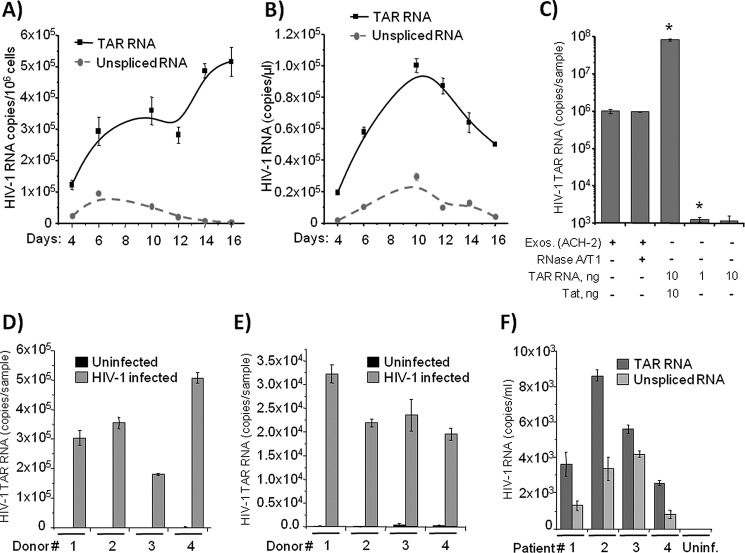
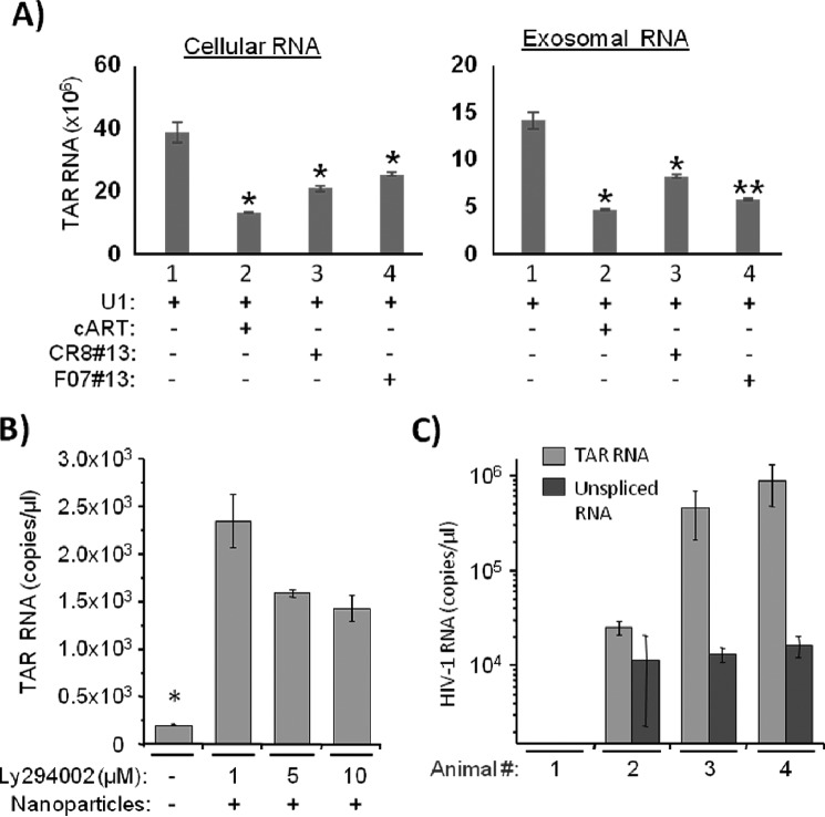
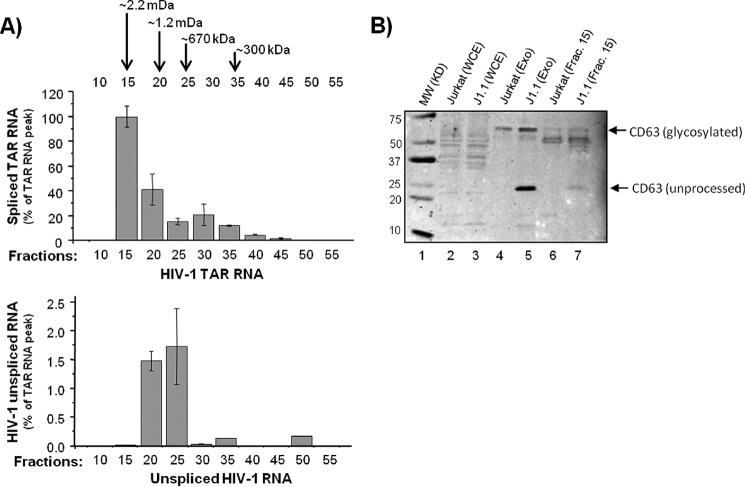
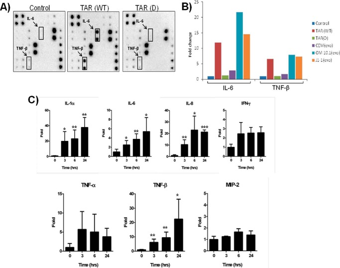
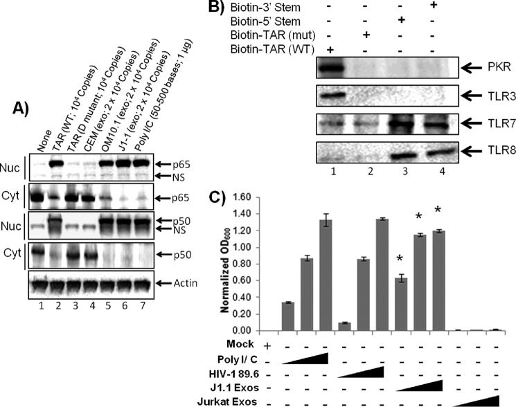
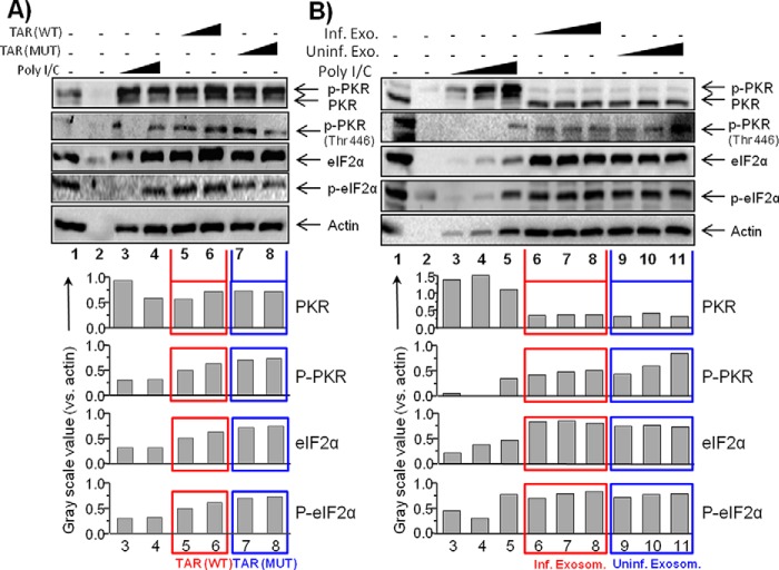
HIV-1 infection results in a chronic illness because long-term highly active antiretroviral therapy can lower viral titers to an undetectable level. However, discontinuation of therapy rapidly increases virus burden. Moreover, patients under highly active antiretroviral therapy frequently develop various metabolic disorders, neurocognitive abnormalities, and cardiovascular diseases. We have previously shown that exosomes containing trans-activating response (TAR) element RNA enhance susceptibility of undifferentiated naive cells to HIV-1 infection. This study indicates that exosomes from HIV-1-infected primary cells are highly abundant with TAR RNA as detected by RT-real time PCR. Interestingly, up to a million copies of TAR RNA/μl were also detected in the serum from HIV-1-infected humanized mice suggesting that TAR RNA may be stable in vivo. Incubation of exosomes from HIV-1-infected cells with primary macrophages resulted in a dramatic increase of proinflammatory cytokines, IL-6 and TNF-β, indicating that exosomes containing TAR RNA could play a direct role in control of cytokine gene expression. The intact TAR molecule was able to bind to PKR and TLR3 effectively, whereas the 5' and 3' stems (TAR microRNAs) bound best to TLR7 and -8 and none to PKR. Binding of TAR to PKR did not result in its phosphorylation, and therefore, TAR may be a dominant negative decoy molecule in cells. The TLR binding through either TAR RNA or TAR microRNA potentially can activate the NF-κB pathway and regulate cytokine expression. Collectively, these results imply that exosomes containing TAR RNA could directly affect the proinflammatory cytokine gene expression and may explain a possible mechanism of inflammation observed in HIV-1-infected patients under cART.

Keywords: TAR RNA; cytokinesis; exosome (vesicle); human immunodeficiency virus (HIV); macrophage; protein kinase RNA-activated (PKR); toll-like receptor (TLR).


Figures

FIGURE 1.
FIGURE 1.
TAR RNA is abundant in the exosomes from HIV-1-infected primary cells. A, purified T cells from healthy donors were stimulated with PHA/IL-2 and then infected with HIV-1 NL4-3. Cells were lysed on 4, 6, 10, 12, 14, and 16 days post-infection; total RNA was isolated and subjected to RT reaction and then to SYBR Green real time PCR with the primers specific for HIV-1 TAR and gag to quantitate numbers of copies of TAR and unspliced HIV-1 RNA. Error bars indicate ± S.D. of three independent measurements. B, cell supernatants from the HIV-1-infected T cells used in A were harvested at 4, 6, 10, 12, 14, and 16 days post-infection and then incubated with a mixture of Nanotrap particles NT080 and NT082 (to trap exosomes) for 1 h at 4 °C. The exosomes bound by the Nanotrap particles were lysed; total RNA was isolated and subjected to qRT-PCR using specific primers as in A. Error bars indicate ±S.D. of three independent measurements. C, trapping of exosomes and RNP complex by nanoparticles. One milliliter of infected ACH2 supernatants was incubated with/without RNase A/T1 mix (RNase A 800 ng/ml and RNase T1 2500 units/ml; Pharmingen) for 1 h at 37 °C. Samples were then treated with 25 μl of 30% slurry of NT080 + 82 overnight at 4 °C. Ten nanograms of purified Tat (76, 77) and purified TAR were incubated with beads overnight (total of 100 μl) at 4 °C. Also, purified TAR RNA (1 and 10 ng) was incubated with beads overnight at 4 °C. All samples were washed the next day with PBS (two times), and RNA was isolated for TAR RT-qPCR. Error bars indicate ±S.D. of three independent measurements. Asterisks indicate p ≤ 0.01 between indicated samples and exosomal samples. D, PBLs were separated from human PBMCs from four donors using incubation with PHA (1 μg/ml) for 24 h. After 48 h of incubation with 25 units/ml IL-2, the PBLs were infected with HIV-1 strain 89.6 and then cultured for 1 week with ART mixture (lamivudine/emitricitabine, indinavir, and tenofovir; 10 μm of each). Then the cells were cultivated with IL-7 (1 ng/ml) to transfer T cells to the quiescent phase. Culture supernatants were then harvested, and exosomes were separated with NT080/NT082 Nanotrap particle mixture as described in B. Total RNA was extracted, and TAR and unspliced viral RNA were quantitated as described in A. Error bars indicate ±S.D. of three independent measurements. E, human monocytes obtained from four donors were cultured for 1 week with 10 ng/ml M-CSF for differentiation of MDM. The MDM were infected with HIV-1 dual-tropic strain 89.6 and then cultured for 2 weeks (for the 1st week with ART mixture as described in D). The exosomes were then captured from culture media as described in D; RNA was extracted and HIV-1 TAR and unspliced RNA were quantitated using qRT-PCR. Error bars indicate ±S.D. of three independent measurements. F, serum specimens from four HIV-1-infected patients who had undetectable plasma HIV-1 RNA on cART were obtained from The Washington D. C. Metropolitan Site of Women's Interagency HIV Study. The exosomes were captured from 1.0 ml of serum as described in C; RNA was extracted and HIV-1 TAR, and unspliced RNA were quantitated using RT-qPCR. Error bars indicate ±S.D. of three independent measurements.
FIGURE 2.
FIGURE 2.
Exosome packaging of TAR RNA is dependent on its expression level in HIV-1-infected cells. A, promonocytic chronically HIV-1-infected U1 cells were treated with cART mixture (described in Fig. 1) for 5 days and subsequently treated with two different transcription inhibitors, F07 number 13 and CR8 number 13 (1 μm each), for 24 h. Exosomes were then isolated using NT080 and NT082 nanoparticles overnight at 4 °C and then tested for quantity of TAR RNA by qRT-PCR. Total RNA was also isolated from treated cells for TAR RNA analysis. Error bars indicate ±S.D. of three independent measurements. Asterisk indicates p ≤ 0.05. B, chronically HIV-1-infected T cells J1.1 were treated with three different concentrations (1, 5, and 10 μm) of PI3K inhibitor LY294002, and the supernatants (500 μl) were tested for TAR RNA by qRT-PCR following pulldown with a mixture of Nanotrap particles NT080 and NT082 as described in Fig. 1A. Error bars indicate ±S.D. of three independent measurements. Asterisk indicates p ≤ 0.05. C, three groups (2–4) of three humanized mice were subcutaneously infected with the dual tropic HIV-1 89.6 (100 μl). Total RNA was isolated from the blood plasma of infected mice (25–50 μl) after 14 days of infection. The exosomes were captured using Nanotrap particle mixture NT080/NT082. Copy numbers of both TAR RNA and unspliced HIV-1 RNA were measured using qRT-PCR as described in Fig. 1A. Results are presented as mean ± S.D. of at least three independent measurements.
FIGURE 3.
FIGURE 3.
TAR RNA is a part of the endogenous fractions of pre-released exosomes. A, whole cell extracts from uninfected Jurkat and HIV-1-infected J1.1 cells were prepared and then separated at 2.5 mg of total protein run on a Superose 6 size-exclusion chromatography column in the presence of 500 mm salt buffer. No detergent was used during fractionation. A total of 55 fractions (500 μl each) from each cell extract was collected. Every 5th fraction (from fraction 10 to 55) from the J1.1 extract was subjected to isolation of total RNA followed RT reaction and real time PCR of cDNA with the primers specific for either TAR RNA (upper panel) or for unspliced HIV-1 RNA (gag-specific primers; lower panel). B, 500 μl of the chromatography fraction 15 from Jurkat (lane 6) and J1.1 (lane 7) cell extracts were incubated for 30 min with a mixture of Nanotrap particles NT080 and NT082. Exosomes were separated by centrifugation and then tested for the presence of CD63 (an exosome marker) by Western blot. Exosomes were similarly isolated from the Jurkat (lane 4) and J1.1 (lane 5) supernatants using the Nanotrap particles and tested for the CD63 marker protein by Western blot. The whole cell extracts from Jurkat (lane 2) and J1.1 (lane 3) were also separated onto a 4–20% Tris-glycine SDS-polyacrylamide gel and immunoblotted with anti-CD63 antibody.
FIGURE 4.
FIGURE 4.
Exposure to TAR RNA alters cytokine profiles in primary monocyte-derived macrophages. A, primary human MDMs were differentiated by incubating in 10 ng/ml M-CSF and phorbol 12-myristate 13-acetate for 1 week before incubating 106 cells with 104 copies of either TAR WT or mutated TAR D RNA packaged into DOTAP liposomal transfection reagent or with DOTAP only (control). After 48 h, supernatants were analyzed for the presence of 23 cytokine proteins using human cytokine antibody array I. Cytokines that are highly up-regulated after treatment with TAR are indicated by enclosed rectangles. B, exosomes were first isolated from CEM, OM10.1, and J1.1 cell supernatants using Nanotrap particles as described in Fig. 1B; total RNA was purified and then packaged into DOTAP reagent as in A. The DOTAP (liposomes) containing extracellular RNA, as well as TAR (WT) and mutated TAR (D) RNA, were incubated with MDM for 48 h, and then supernatants were analyzed for 23 cytokines (data not shown) as in A. Only IL-6 and TNF data are shown as bar graphs. MDMs were incubated with DOTAP alone as a control. C, primary macrophages were treated with either DOTAP alone (control) or DOTAP/TAR wild type (104 copies/10 μl/experimental), and supernatants were assayed for the presence of cytokines after 0, 3, 6, and 24 h. Results are presented as mean ± S.D. of at least three independent measurements.
FIGURE 5.
FIGURE 5.
Effects of exosomal TAR RNA on NF-κB and TLR pathways. A, exosomes were prepared from CEM (uninfected), OM10.1 (HIV-1-infected), and J1.1 (HIV-1-infected) cell culture supernatants using Nanotrap particles. The total exosomal RNAs were purified and then mixed with DOTAP transfection reagent before incubating with MDM (7-day cultures). DOTAP liposome-packaged TAR WT or mutant TAR D RNAs were also incubated with macrophages; poly(I-C) was used as positive control. Nuclear and cytoplasmic extracts were prepared 24 h post-treatment, run on a 4–20% SDS-polyacrylamide gel, and then Western blotted for NF-κB components p65 and p50 both from cytoplasm and nuclear extracts. Actin antibody served as a control. B, biotin-labeled WT TAR, TAR D mutant, and TAR miRNAs (5′ stem or 3′ stem) were incubated with total MDM (5 × 10⁵) extracts. Three hundred micrograms of extracts were incubated with labeled RNA (1 μg) overnight at 4 °C, and the next day 30% slurry of streptavidin-Sepharose beads were added. Samples were washed once with TNE 50 + 0.1% Nonidet P-40, then once with RIPA buffer, and then one final time with TNE50 + 0.1% Nonidet P-40 buffer. Bound samples were separated on 4–20% SDS-polyacrylamide gel and then Western-blotted for PKR, TLR3, TLR7, and TLR8. C, HEK-Blue hTLR3 cells containing a TLR3 activation-inducible SEAP reporter gene were incubated in a SEAP detection medium with HIV-1 89.6 virus (1, 10, and 100 ng of p24/well), exosomes from HIV-1-infected J1.1 cells (0.1, 1, and 10 μg/well), or exosomes from uninfected Jurkat cells (0.1, 1, and 10 μg/well). Similarly, cells were incubated with poly(I-C) (10, 50, and 250 ng/ml) as a positive control. After 18 h of incubation, absorbance (600 nm) of each sample was measured and normalized to PBS controls. Error bars indicate ±S.D. of three independent measurements. Asterisk indicates p ≤ 0.01.
FIGURE 6.
FIGURE 6.
Effect of TAR on NFκB pathway. A, IKK-β complex components are altered in HIV-1-infected cells. Extracts from uninfected Jurkat and infected J1.1 cells were fractionated in a Superose 6 size-exclusion column (AKTA). A total of 70 fractions were obtained, and every fifth fraction was analyzed for IKK-α, -β, and -γ complexes and for β-actin. B, Jurkat cells were transfected with either an HIV-Luc (TAR+; 20 μg) or CMV-Luc (TAR−; 20 μg), and cell lysates were prepared for chromatography and Western blots. C, fractions 19 (large complex) and 34 (small complex) were used (500 μl) for immunoprecipitation with α-IKK-β antibody (10 μg), bound to A + G beads, washed, and used for kinase reaction, which included GST-IKBα (0.5 μg) or histone H1 (0.5 μg) as substrates and [γ-32P]ATP. Samples were run on SDS-polyacrylamide gel, dried, and exposed to a cassette.
FIGURE 7.
FIGURE 7.
Effect of exosomal TAR RNA on PKR. A, uninfected CEM T cells (7.5 × 10⁵) were treated with DOTAP TAR or DOTAP TAR mutant at varying concentrations (5 × 10⁴/1 μg and 10 × 104/10 μg) for 24 h. Total cell extracts were isolated and used for Western blot using antibodies against PKR, p-PKR, eIF2α, p-eIF2α, and actin as a reference protein. Grayscale values of each Western blot band were quantified using ImageJ software, and results are presented as a ratio of analyzed protein to actin in the same sample. B, similar to A, CEM cells were treated with the exosomes (1, 5, or 10 μg) from either CEM (uninfected) or ACH2 (HIV-1-infected) cells for 24 h. Total cell extracts were isolated for Western blot with PKR, p-PKR, eIF2α, p-eIF2α, and actin antibodies. The grayscale values of each Western blot band were measured as described in A.
FIGURE 8.
FIGURE 8.
Inhibition of exosome release from HIV-1-infected cells make naive recipient cells less susceptible to HIV-1 infection. A, chronically HIV-1-infected J1.1 cells were treated with 1 μm MVB pathway inhibitors manumycin A or brefeldin A and tested for exosome production by Western blot using antibodies specific for exosomal proteins, CD63 and HSP70. Antibody for actin and cytochrome c were used as control. B, supernatants from inhibitor-treated or untreated J1.1 cells used in A were utilized in RT assay for quantification of virus. Results are presented as mean ± S.D. of at least three independent measurements. C, naive CEM cells were pre-treated with exosomes (10 μg) from either manumycin-treated, GW4869-treated (100 nm), or untreated infected J1.1 cells prior to addition of HIV-1 89.6 and scored for p24 activity. D, humanized NSG mice were divided into two groups and treated with either DMSO or manumycin A (5 mg/kg) and then infected with 100 μl (5 ng/μl of p24) of HIV-1 89.6. Animals were then euthanized, and blood, brain, and other tissues were collected. Brains were harvested, and white matter (midbrain) from each animal (1 mm) were diced and digested with trypsin. Half of the samples were processed for qRT-PCR using LTR-specific (upper panel) and Nef-specific (lower panel) primers. E, other half-samples of midbrain were co-cultured with CEM cells (5 × 10⁵) for 9 days and processed for the quantitation of virus using RT assay from the supernatants. Results are presented as mean ± S.D. of three independent measurements.

References

    1. UNAIDS (2012) UNAIDS Report on the Global AIDS Epidemic 2012. http://www.unaids.org/sites/default/files/media_asset/20121120_UNAIDS_Gl...
    1. Prejean J., Song R., Hernandez A., Ziebell R., Green T., Walker F., Lin L. S., An Q., Mermin J., Lansky A., Hall H. I., and HIV Incidence Surveillance Group (2011) Estimated HIV incidence in the United States, 2006–2009. PLoS ONE 6, e17502. - PMC - PubMed
    1. Laird G. M., Eisele E. E., Rabi S. A., Lai J., Chioma S., Blankson J. N., Siliciano J. D., and Siliciano R. F. (2013) Rapid quantification of the latent reservoir for HIV-1 using a viral outgrowth assay. PLoS Pathog. 9, e1003398. - PMC - PubMed
    1. van der Sluis R. M., Jeeninga R. E., and Berkhout B. (2013) Establishment and molecular mechanisms of HIV-1 latency in T cells. Curr. Opin. Virol. 3, 700–706 - PubMed
    1. Kiage J. N., Heimburger D. C., Nyirenda C. K., Wellons M. F., Bagchi S., Chi B. H., Koethe J. R., Arnett D. K., and Kabagambe E. K. (2013) Cardiometabolic risk factors among HIV patients on antiretroviral therapy. Lipids Health Dis. 12, 50. - PMC - PubMed

Publication types

MeSH terms

LinkOut - more resources