. 2016 Jan 1;291(1):103-9.
doi: 10.1074/jbc.C115.700492. Epub 2015 Nov 9.

A Genome-wide CRISPR (Clustered Regularly Interspaced Short Palindromic Repeats) Screen Identifies NEK7 as an Essential Component of NLRP3 Inflammasome Activation

Affiliations

A Genome-wide CRISPR (Clustered Regularly Interspaced Short Palindromic Repeats) Screen Identifies NEK7 as an Essential Component of NLRP3 Inflammasome Activation

Jonathan L Schmid-Burgk et al. J Biol Chem. .

Abstract

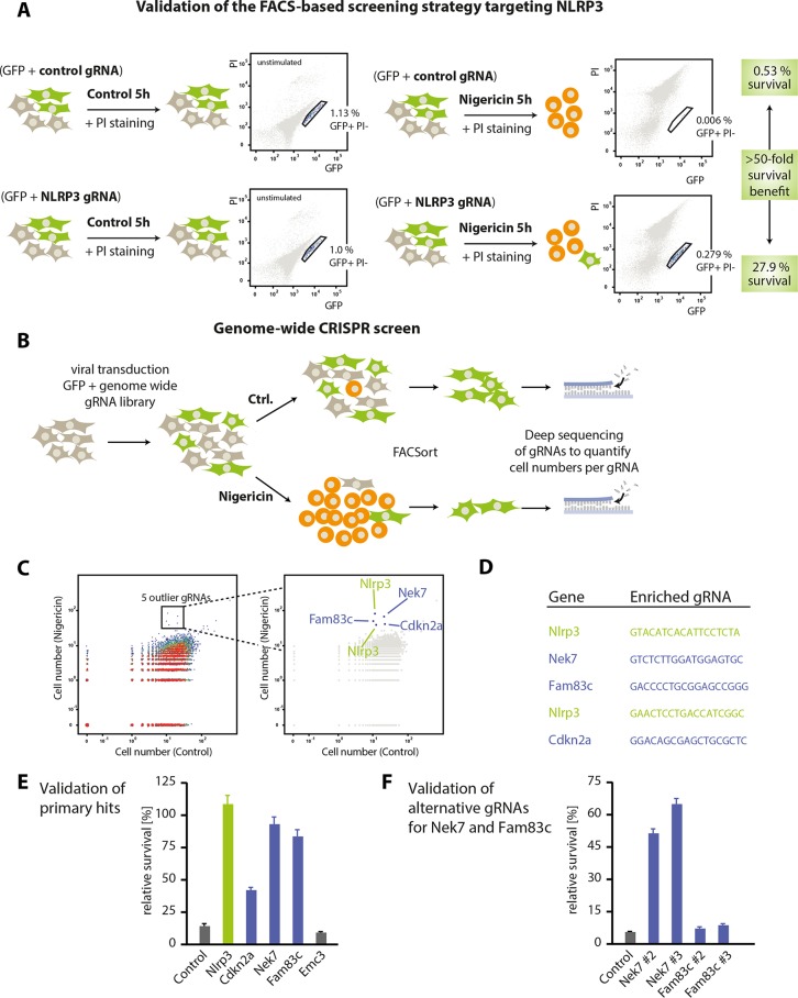
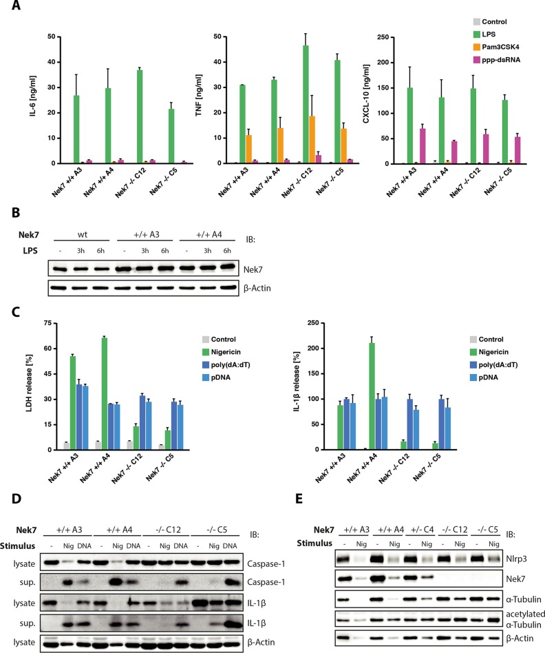
Inflammasomes are high molecular weight protein complexes that assemble in the cytosol upon pathogen encounter. This results in caspase-1-dependent pro-inflammatory cytokine maturation, as well as a special type of cell death, known as pyroptosis. The Nlrp3 inflammasome plays a pivotal role in pathogen defense, but at the same time, its activity has also been implicated in many common sterile inflammatory conditions. To this effect, several studies have identified Nlrp3 inflammasome engagement in a number of common human diseases such as atherosclerosis, type 2 diabetes, Alzheimer disease, or gout. Although it has been shown that known Nlrp3 stimuli converge on potassium ion efflux upstream of Nlrp3 activation, the exact molecular mechanism of Nlrp3 activation remains elusive. Here, we describe a genome-wide CRISPR/Cas9 screen in immortalized mouse macrophages aiming at the unbiased identification of gene products involved in Nlrp3 inflammasome activation. We employed a FACS-based screen for Nlrp3-dependent cell death, using the ionophoric compound nigericin as a potassium efflux-inducing stimulus. Using a genome-wide guide RNA (gRNA) library, we found that targeting Nek7 rescued macrophages from nigericin-induced lethality. Subsequent studies revealed that murine macrophages deficient in Nek7 displayed a largely blunted Nlrp3 inflammasome response, whereas Aim2-mediated inflammasome activation proved to be fully intact. Although the mechanism of Nek7 functioning upstream of Nlrp3 yet remains elusive, these studies provide a first genetic handle of a component that specifically functions upstream of Nlrp3.

Keywords: NEK7; NLRP3; caspase 1 (CASP1); cell death; genetic screen; inflammasome; inflammation; pyroptosis.


Figures

FIGURE 1.
FIGURE 1.
A CRISPR loss-of-function screen identifies Nek7 as a component involved in Nlrp3 signaling. A, Nlrp3-Cas9 macrophages were transduced with a control gRNA or a gRNA targeting Nlrp3, which additionally encoded for GFP at an MOI of 0.01. Following stable transduction, macrophages were either stimulated with nigericin for 5 h or left untreated. Subsequently, cells were labeled with PI and subjected to FACS analysis. Depicted are schematic views of the transduction and stimulation modalities as well as FACS plots of a representative result. Highlighted are the frequencies of cells in the stringent live gate that contains GFP-positive and PI-negative cells. Data are representative of three independent experiments with comparable results. B, schematic view of the genome-wide screening approach that was undertaken to identify factors that confer resistance to nigericin-induced cell death. Please see “Experimental Procedures” for details. Ctrl., control. C, dot plot representation of the screening results. Each dot represents the frequency of cells carrying a specific gRNA that were found in the live gate of mock-treated cells (x axis) or of nigericin-treated cells (y axis). Highlighted are five outlier gRNAs targeting Nlrp3 (x2), Nek7, Fam83c, or Cdkn2a. D, depicted are the gRNA sequences found in the outlier population. E, Nlrp3-Cas9 macrophages were transduced with the same gRNAs identified in the screen and a control gRNA targeting Emc3. Subsequently, cells were subjected to nigericin stimulation, and cell survival was analyzed by FACS. The relative survival benefit was calculated by dividing the gated cell fraction from the stimulated condition by that from the unstimulated condition and is depicted as a mean value ± S.E. of three independent experiments. F, two alternative gRNAs targeting Nek7 and Fam83c were used to transduce Nlrp3-Cas9 macrophages. Nigericin stimulation and subsequent cell survival were analyzed as in E.
FIGURE 2.
FIGURE 2.
Nek7 functions specifically upstream of the Nlrp3 inflammasome. A, Nek7-deficient Nlrp3-Cas9 macrophages or wild-type cells were stimulated with Pam3CSK4, LPS, or 5′-triphosphate dsRNA. 16 h later, supernatants were collected and analyzed for TNF, IL-6, and CXCL10 production. Data are presented as mean values ± S.E. of two independent experiments. B, wild-type macrophages or Nlrp3-Cas9 macrophages of the indicated genotype were stimulated with 200 ng/ml LPS for the durations indicated. Nek7 protein expression was analyzed by immunoblot (IB), whereas β-actin served as a loading control. Representative data from one experiment out of two independent experiments are depicted. C, macrophages of the indicated genotypes were primed with LPS and subsequently stimulated with nigericin, poly(dA:dT), or plasmid DNA (pDNA). 6 h after stimulation, supernatants were analyzed for LDH release (left panel) or IL-1β production (right panel). IL-1β data were normalized to the poly(dA:dT) condition. D, lysates or supernatants (sup.) of cells stimulated with nigericin (Nig) for 2 h or poly(dA:dT) for 6 h were analyzed for IL-1β, caspase-1, or β-actin by immunoblot. Representative data from one experiment out of two independent experiments are depicted. E, cells of the indicated genotypes were stimulated with nigericin or left untreated (note that one heterozygote Nek7 cell clone was included in the analysis). 6 h after stimulation, cells were analyzed for Nlrp3, Nek7, α-tubulin, acetylated α-tubulin, and β-actin expression by immunoblot. Representative data from one experiment out of two independent experiments are depicted.

References

    1. Medzhitov R. (2007) Recognition of microorganisms and activation of the immune response. Nature 449, 819–826 - PubMed
    1. Chen G. Y., and Nuñez G. (2010) Sterile inflammation: sensing and reacting to damage. Nat. Rev. Immunol. 10, 826–837 - PMC - PubMed
    1. Gross O., Thomas C. J., Guarda G., and Tschopp J. (2011) The inflammasome: an integrated view. Immunol. Rev. 243, 136–151 - PubMed
    1. Kayagaki N., Stowe I. B., Lee B. L., O'Rourke K., Anderson K., Warming S., Cuellar T., Haley B., Roose-Girma M., Phung Q. T., Liu P. S., Lill J. R., Li H., Wu J., Kummerfeld S., Zhang J., Lee W. P., Snipas S. J., Salvesen G. S., Morris L. X., Fitzgerald L., Zhang Y., Bertram E. M., Goodnow C. C., and Dixit V. M. (2015) Caspase-11 cleaves gasdermin D for non-canonical inflammasome signaling. Nature 526, 666–671 - PubMed
    1. Shi J., Zhao Y., Wang K., Shi X., Wang Y., Huang H., Zhuang Y., Cai T., Wang F., and Shao F. (2015) Cleavage of GSDMD by inflammatory caspases determines pyroptotic cell death. Nature 526, 660–665 - PubMed

Publication types