. 2016 Jan 15;291(3):1411-26.
doi: 10.1074/jbc.M115.662874. Epub 2015 Nov 9.

Unexpected Allosteric Network Contributes to LRH-1 Co-regulator Selectivity

Affiliations

Unexpected Allosteric Network Contributes to LRH-1 Co-regulator Selectivity

Paul M Musille et al. J Biol Chem. .

Abstract

Phospholipids (PLs) are unusual signaling hormones sensed by the nuclear receptor liver receptor homolog-1 (LRH-1), which has evolved a novel allosteric pathway to support appropriate interaction with co-regulators depending on ligand status. LRH-1 plays an important role in controlling lipid and cholesterol homeostasis and is a potential target for the treatment of metabolic and neoplastic diseases. Although the prospect of modulating LRH-1 via small molecules is exciting, the molecular mechanism linking PL structure to transcriptional co-regulator preference is unknown. Previous studies showed that binding to an activating PL ligand, such as dilauroylphosphatidylcholine, favors LRH-1's interaction with transcriptional co-activators to up-regulate gene expression. Both crystallographic and solution-based structural studies showed that dilauroylphosphatidylcholine binding drives unanticipated structural fluctuations outside of the canonical activation surface in an alternate activation function (AF) region, encompassing the β-sheet-H6 region of the protein. However, the mechanism by which dynamics in the alternate AF influences co-regulator selectivity remains elusive. Here, we pair x-ray crystallography with molecular modeling to identify an unexpected allosteric network that traverses the protein ligand binding pocket and links these two elements to dictate selectivity. We show that communication between the alternate AF region and classical AF2 is correlated with the strength of the co-regulator interaction. This work offers the first glimpse into the conformational dynamics that drive this unusual PL-mediated nuclear hormone receptor activation.

Keywords: allosteric regulation; diabetes; lipids; molecular dynamics; nuclear receptor; phosphatidylcholine.


Figures

FIGURE 1.
FIGURE 1.
Structure of the apo-LRH-1 LBD·TIF complex. A, ribbon diagram of apo-LRH-LBD (α-helices, teal; β-strands, yellow) with the TIF2 NR box 3 peptide (orange). The surface-bound CHAPS is depicted as sticks (C, pink; O, red; S, yellow; N, blue). The AF-2 surface is defined by H3, H4b and H12. B, ribbon diagram of apo-LRH·SHP NRBox2 complex (PDB code 4DOR) with the unobserved alternate AF region (defined by β1–2 and H6) represented by a dashed line. C, close-up view of the bound CHAPS molecules included in the crystallization buffer.
FIGURE 2.
FIGURE 2.
Structure of the LRH-1 LBD·E. coli PL·TIF2 complex. A, ribbon diagram of E. coli PL-bound LRH-LBD (α-helices, teal; β-strands, yellow) with the TIF2 NR box 3 peptide (green). The bound E. coli PL is depicted as sticks (C, green; O, red; P, magenta) The surface-bound CHAPS is depicted as sticks (C, yellow; O, red; S, yellow; N, blue). B, 2FoFc electron density (contoured at 1 σ) for the bound E. coli PL observed in this structure, along with side chains lining the ligand binding pocket of hLRH-1 that contact this ligand. C, close-up view of the bound CHAPS molecules included in the crystallization buffer along H3 and H4 in close proximity to the bound PL. Residues within 4.2 Å are depicted as sticks. D, close-up view of the bound CHAPS along H9, which interact with a crystallographic symmetry mate and in a position overlapping the CHAPS site in the apo-LRH-1·TIF2 complex. Residues within 4.2 Å of CHAPS are depicted as sticks.
FIGURE 3.
FIGURE 3.
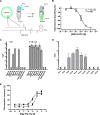
LRH-1 in vitro lipid binding profile. Binding affinities of LRH-1 to PLs of differing headgroup and tail compositions. A, PL binding was measured relative to probe ligand NBD-DLPE via FRET quenching of DCIA-labeled LRH-1. B, binding affinity of LRH-1 to NBD-DLPE probe. C, relative binding affinities of competing PLs of differing headgroups; 5 mm β-cyclodextrin added as indicated. D, relative binding affinities of competing saturated PCs of differing tail lengths. Data are reported are the means ± S.E. of three independent experiments. The presence of an × instead of a bar indicates that no binding was observed. E, example of an individual competitive binding curves for NBD-DLPE displacement. Solid line represents the inclusion of 5 mm β-cyclodextrin, although the dashed line is without 5 mm β-cyclodextrin as described under “Experimental Procedures.”
FIGURE 4.
FIGURE 4.
AF-2 charge clamp engagement is dictated by ligand-co-regulator combination. Ligand binding pocket entrance measurements and analysis of Glu-534-peptide charge clamp engagement for the apo-LRH-1·TIF2 complex (A), LRH-1·E. coli PL·TIF2 complex (B), LRH-1·GSK8470·TIF2 complex (PDB code 3PLZ) (C), LRH-1·DLPC·TIF2 complex (PDB code 4DOS) (D), apo-LRH-1·SHP complex (PDB code 4DOR) (E), and LRH-1·E. coli PL·SHP complex (PDB code 1YUC) (F) are shown.
FIGURE 5.
FIGURE 5.
ProSMART Procrustes central residue analysis of LRH-1 complexes. ProSMART analysis of LRH-1 with differentially bound ligands and co-regulator peptides is shown. Models were colored by the Procrustes score of the central residue of an aligned fragment pair according to the legend at top right. Areas colored white were omitted from the analysis. The following pairwise comparisons were made: A, apo-SHP (PDB code 4DOR) versus E. coli PL·SHP (PDB code 1YUC); B, apo-SHP versus apo-TIF2; C, apo-SHP versus E. coli PL·TIF2; D, apo-SHP versus DLPC-TIF2 (PDB code 4DOS); E, apo-TIF2 versus E. coli PL·SHP; F, apo-TIF2 versus E. coli PL·TIF2; G, E. coli PL·SHP versus E. coli PL·TIF2; H, apo-TIF2 versus DLPC-TIF2; I, E. coli PL·SHP versus DLPC-TIF2; J, E. coli PL·TIF2 versus DLPC-TIF2.
FIGURE 6.
FIGURE 6.
Correlated motion in LRH-1-PL co-regulator systems. Cross-correlation matrices showing correlated and anti-correlated motion over the 200-ns MD simulation for apo-LRH-1·TIF2 complex (A), apo-LRH-1·SHP complex (B), LRH-1·DLPC·TIF2 complex (C), LRH-1·DLPC·SHP complex (D), and LRH-1·E. coli PL·TIF2 complex (E). Cross-correlation between protein residues and the lipid headgroup phosphorus atom is mapped to the protein structure in LRH-1·DLPC·TIF2 complex (F), LRH-1·DLPC·SHP complex (G), and LRH-1·E. coli PL·TIF2 complex (H).
FIGURE 7.
FIGURE 7.
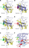
Allosteric paths from binding pocket to co-regulator. Allosteric communication pathways between the β-sheet-H6 and co-regulator binding regions of the LRH-1 LBD in the apo-LRH-1·TIF2 (A), LRH-1·DLPC·TIF2 (B), LRH-1·E. coli PL·TIF2 (C), apo-LRH-1·SHP (D), and LRH-1·DLPC·SHP (E) complexes. Schematic loop view of LRH-1 showing thick loops (yellow, LRH-1; green, TIF2; red, SHP) for regions of the protein identified along the allosteric path. F, LRH-1 mutations that alter PL binding or co-regulator recruitment lie on or adjacent to the allosteric pathway. Known mutations of mouse (m) or human (h) LRH-1 LBD are shown as C-α spheres on the LRH-1 protein backbone. Mutations shown in green enhance the degree of LRH-1 activation in response to co-activator binding; mutations shown in red selectively decrease LRH-1 sensitivity to SHP without affecting overall activation; mutations shown in brown decrease overall LRH-1 activity without affecting PL binding; mutations shown in blue decrease PL binding and overall activity.
FIGURE 8.
FIGURE 8.
Biologically relevant principal modes identified from the projections of the MD trajectories on PC1 versus PC2. An outward swing of helix 9 contributes to PC1 (A), whereas opening motions at the mouth of the lipid binding pocket results in translation along PC2 (B). Projections of snapshots were taken from MD onto PC1 and PC2 in apo-LRH-1·SHP (C), LRH-1·DLPC·SHP (D), apo-LRH-1·TIF2 (E), LRH-1·DLPC·TIF2 (F), and LRH-1·E. coli PL·TIF2 (G) complexes. Higher densities indicate more populated regions of the conformational subspace. Scale bar indicates how many snapshots (out of 10,000) were collected within a contour. Green and red rings indicate activating and repressing regions of the subspace, respectively.
FIGURE 9.
FIGURE 9.
Subtle disruption of residues on or near the allosteric pathway reduces LRH-1 activation. A, close-up view of the PL binding pocket of DLPC- (beige/orange) and E. coli PL-bound LRH-1 (blue). Helices are shown as cylinders, and helix 3 has been hidden. Residues within 4.2 Å of the phospholipid are depicted as sticks. B, junction of helices 5 and 10 displays hydrogen bonding (red dashes) between Ser-383 and Glu-514 and electrostatic interactions between Glu-384 and Arg-507. In the active conformation, helix 12 docks against this junction to support the AF-2 co-regulator binding surface, driving gene transactivation and transrepression. C, abolition of the electrostatic interaction between helices 5 and 10 via the E384Q/R507H mutation causes a subtle but significant reduction in LRH-1 transcriptional activity. LRH-1 activity was measured via luciferase reporter gene assay in transiently transfected HEK 293T cells. Data are the combined results of five independent experiments. Statistical significance is represented as *, p < 0.05; **, p < 0.01.

Similar articles

Cited by

References

    1. Musille P. M., Kohn J. A., and Ortlund E. A. (2013) Phospholipid–driven gene regulation. FEBS Lett. 587, 1238–1246 - PMC - PubMed
    1. Blind R. D., Sablin E. P., Kuchenbecker K. M., Chiu H. J., Deacon A. M., Das D., Fletterick R. J., and Ingraham H. A. (2014) The signaling phospholipid PIP3 creates a new interaction surface on the nuclear receptor SF-1. Proc. Natl. Acad. Sci. U.S.A. 111, 15054–15059 - PMC - PubMed
    1. Liu S., Brown J. D., Stanya K. J., Homan E., Leidl M., Inouye K., Bhargava P., Gangl M. R., Dai L., Hatano B., Hotamisligil G. S., Saghatelian A., Plutzky J., and Lee C. H. (2013) A diurnal serum lipid integrates hepatic lipogenesis and peripheral fatty acid use. Nature 502, 550–554 - PMC - PubMed
    1. Walker A. K., Jacobs R. L., Watts J. L., Rottiers V., Jiang K., Finnegan D. M., Shioda T., Hansen M., Yang F., Niebergall L. J., Vance D. E., Tzoneva M., Hart A. C., and Näär A. M. (2011) A conserved SREBP-1/phosphatidylcholine feedback circuit regulates lipogenesis in metazoans. Cell 147, 840–852 - PMC - PubMed
    1. Fernandez-Marcos P. J., Auwerx J., and Schoonjans K. (2011) Emerging actions of the nuclear receptor LRH-1 in the gut. Biochim. Biophys. Acta 1812, 947–955 - PMC - PubMed

Publication types

MeSH terms

LinkOut - more resources