. 2015 Nov 10:5:16466.
doi: 10.1038/srep16466.

A potential mode of action for Anakinra in patients with arthrofibrosis following total knee arthroplasty

Affiliations

A potential mode of action for Anakinra in patients with arthrofibrosis following total knee arthroplasty

David Dixon et al. Sci Rep. .

Abstract

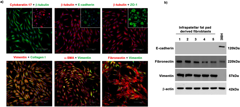
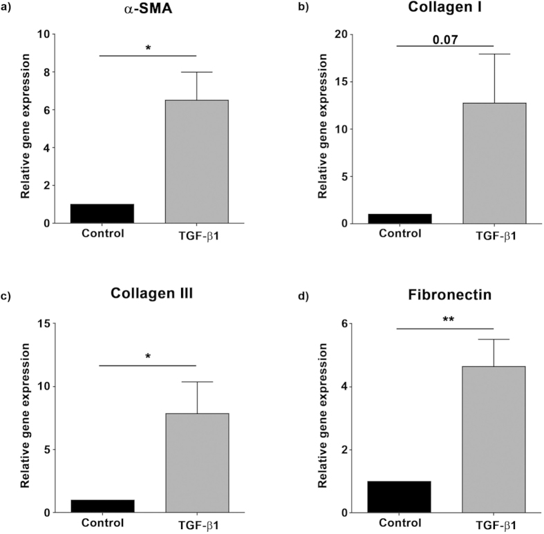
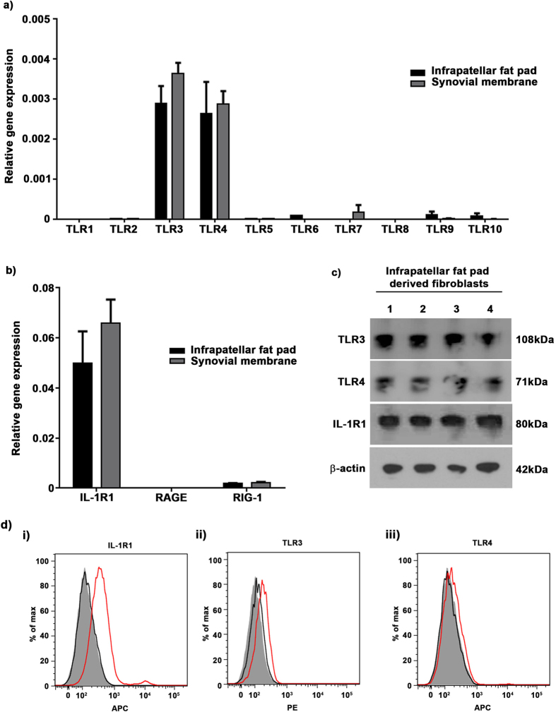
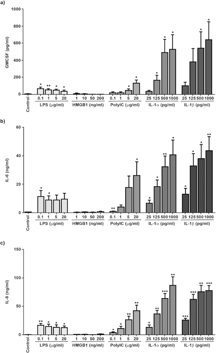
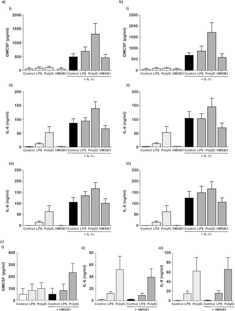
Arthrofibrosis is a fibroproliferative disease characterised by excessive deposition of extracellular matrix components intra-articularly leading to pain and restricted range of movement. Although frequently observed following total knee arthroplasty (TKA) no therapeutic options exist. A pilot study demonstrated that intra-articular injection of Anakinra, an IL-1R antagonist, improved range of movement and pain in patients with arthrofibrosis however the mechanism of action is unknown. We hypothesise that IL-1α/β will drive an inflammatory phenotype in fibroblasts isolated from the knee, therefore identifying a potential mechanism of action for Anakinra in arthrofibrosis following TKA. Fibroblasts isolated from synovial membranes and infra-patellar fat pad of patients undergoing TKA express high levels of IL-1R1. Stimulation with IL-1α/β induced a pro-inflammatory phenotype characterised by increased secretion of GMCSF, IL-6 and IL-8. No significant difference in the inflammatory response was observed between fibroblasts isolated from synovial membrane or infra-patellar fat pad. IL-1α/β treatments induced a pro-inflammatory phenotype in fibroblasts from both synovial membrane and infra-patellar fat pad and therefore Anakinra can likely have an inhibitory effect on fibroblasts present in both tissues in vivo. It is also likely that fibroblast responses in the tissues are controlled by IL-1α/β availability and not their ability to respond to it.


Conflict of interest statement

NTB has a knee consultancy contract with stryker. All other authors declare there are no competing financial interests.

Figures

Figure 1
Figure 1. Cells isolated from the infra-patellar fat pad demonstrate a mesenchymal phenotype.
(a) Cells isolated from the infra-patellar fat pad were cultured in vitro and the expression of epithelial and mesenchymal markers investigated by immunocytochemistry. Cells had little to no expression of the epithelial markers cytokeratin 17, E-cadherin and ZO-1. Insets show positive staining on human lung epithelial cells. β-tubulin was used to demonstrate cell morphology. In contrast cells expressed very high levels of the mesenchymal markers vimentin, collagen 1, α-SMA and fibronectin. DAPI was used as a nuclear counter stain. Images were acquired on a Leica TCS SP2 UV confocal microscope at x20 magnification. (b) Whole cell lysates of infra-patellar fat pad derived fibroblasts (n = 5) were investigated for the expression of epithelial and mesenchymal markers by Western blotting. Cells express high levels of fibronectin and vimentin but no E-cadherin. β-actin was used as a loading control. Human bronchial epithelial cells (16HBE14o- (HBE)) were used as a positive control for epithelial marker expression.
Figure 2
Figure 2. TGF-β1 treatment of infra-patellar fat pad derived fibroblasts upregulates fibrotic gene expression.
Fibroblasts isolated from the infra-patellar fat pad (n = 6) were cultured in vitro in the presence or absence of TGF-β1 (3 ng/ml) for 48 hours and the relative gene expression of (a) α-smooth muscle actin (α-SMA), (b) collagen I, (c) collagen III and (d) fibronectin quantified by qRT-PCR. The expression of all fibrotic genes was increased by TGF-β1. Data is presented as mean ± standard error of the mean with statistical significance indicated by; *p < 0.05 and **p < 0.01.
Figure 3
Figure 3. Interleukin-1 receptor 1 is highly expressed on infra-patellar pad derived fibroblasts.
Fibroblasts isolated from the infra-patellar fat pad and synovial membrane (both n = 5) were cultured in vitro and the relative gene expression of TLR1-10, RIG-1, RAGE and IL-1R1 quantified by qRT-PCR. (a) TLR expression was restricted mainly to TLR3 and TLR4 with little or no expression of other TLRs. (b) There was no detectable expression of RAGE but RIG-1 was expressed at levels comparable to TLR3 and TLR4. In contrast IL-1R1 expression was 100–150 fold greater than other receptors. Results are normalised to GAPDH as a loading control. Data is presented as mean ± standard error of the mean. Fibroblasts isolated from the infra-patellar fat pad were investigated for the expression of TLR3, TLR4 and IL-1R1 protein by Western Blotting (n = 4) (c) and flow cytometry (n = 6) (d). Cells expressed TLR3 and TLR4 at comparable levels at the cell surface. In contrast, expression of IL-1R1 was significantly higher than other receptors (red traces). β-actin was used as a loading control (c). Unstained cells (filled traces) and IgG controls (black traces) were used as flow cytometry controls (d).
Figure 4
Figure 4. Stimulation of infra-patellar fat pad derived fibroblasts with IL-1α and IL-1β increases secretion of inflammatory mediators.
Fibroblasts isolated from infra-patellar fat pad (n = 5) were stimulated with increasing doses of LPS (0.1–20 μg/ml), HMGB1 (1–200 ng/ml), PolyIC (0.1–20 μg/ml), IL-1α and IL-1β (both 25–1000 pg/ml) for 24 hours and the secreted levels of GM-CSF (a), IL-6 (b) and IL-8 (c) determined by ELISA. All three molecules demonstrated a dose dependant increase in secretion in response to PolyIC, IL-1α and IL-1β. LPS induced maximum release of all three molecules at the lowest concentration used (0.1 μg/ml) with higher concentrations having no additional effect. The cells showed no significant response to HMGB-1. Data is presented as mean ± standard error of the mean with statistical significance compared to control indicated by; *p < 0.05, **p < 0.01 and ***p < 0.001.
Figure 5
Figure 5. PolyIC and LPS increase IL-1α/IL-1β induced secretion of inflammatory mediators in infra-patellar fat pad derived fibroblasts.
Fibroblasts isolated from infra-patellar fat pads (n = 5) were stimulated with IL-1α, IL-1β (both 500 pg/ml), LPS, PolyIC (both 5 μg/ml) or HMGB-1 (50 ng/ml) alone or in combination for 24 hours and the secretion of IL-8, IL-6 and GM-CSF quantified by ELISA. (a,b) IL-1α and IL-1β induced secretion of GMCSF was accentuated by PolyIC. In contrast, Poly IC induced only an additive effect on IL-1α and IL-1β induced secretion of IL-6 and IL-8. LPS induced an additive effect on IL-1α and IL-1β induced secretion of GMCSF, IL-6 and IL-8 while HMGB1 had no effect. (c) Co-treatment with HMGB-1 and LPS or PolyIC was not significantly different to treatment with LPS or PolyIC alone. Data presented as mean ± standard error of the mean.

References

    1. Gallo J., Goodman S. B., Konttinen Y. T., Wimmer M. A. & Holinka M. Osteolysis around total knee arthroplasty: a review of pathogenetic mechanisms. Acta biomater. 9, 8046–8058 (2013). - PMC - PubMed
    1. Carr A. J. et al. Knee replacement. Lancet. 379, 1331–1340 (2012). - PubMed
    1. Schiavone Panni A., Cerciello S., Vasso M. & Tartarone M. Stiffness in total knee arthroplasty. J Orthop Traumatol. 10, 111–118 (2009). - PMC - PubMed
    1. Gandhi R. et al. Predictive risk factors for stiff knees in total knee arthroplasty. J Arthroplasty. 21, 46–52 (2006). - PubMed
    1. Gollwitzer H., Burgkart R., Diehl P., Gradinger R. & Buhren V. [Therapy of arthrofibrosis after total knee arthroplasty]. Orthopade. 35, 143–152 (2006). - PubMed

Publication types

MeSH terms