. 2015 Nov 10:5:16469.
doi: 10.1038/srep16469.

Fibrosis is a common outcome following total knee arthroplasty

Affiliations

Fibrosis is a common outcome following total knee arthroplasty

Nicole Abdul et al. Sci Rep. .

Abstract

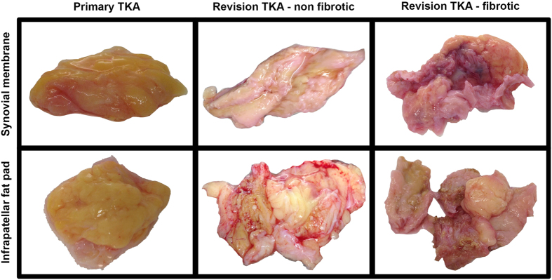
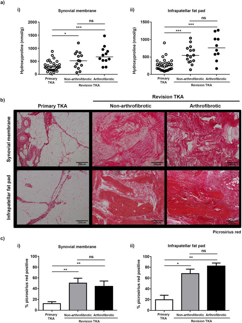
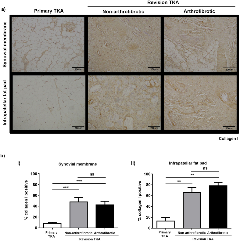
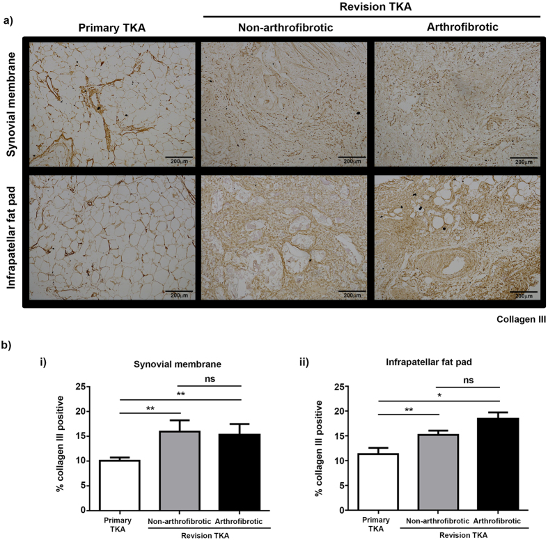
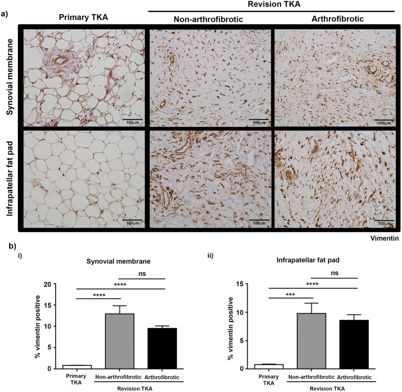
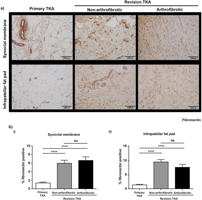
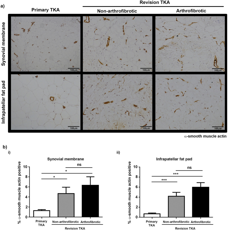
Total knee arthroplasty (TKA) is one of the most successful orthopaedic procedures that alleviates pain and restores function in patients with degenerative knee joint diseases. Arthrofibrosis, abnormal scarring in which dense fibrous tissue prevents normal range of motion, develops in ~3-10% of TKA patients. No prophylactic intervention is available and treatment is restricted to aggressive physiotherapy or revision surgery. Tissue was collected from patients undergoing primary (n = 30) or revision (n = 27) TKA. Revision patients were stratified as non-arthrofibrotic and arthrofibrotic. Tissue was macroscopically and histologically compared to improve our understanding of the pathophysiology of arthrofibrosis. Macroscopically, tissue from primary TKA presents as homogenous, fatty tissue whereas tissue from revision TKA presents as dense, pigmented tissue. Histologically, there was dramatic tissue remodelling, increased collagen deposition and increased (myo)fibroblast staining in tissue from revision TKA. Significantly, tissue architecture was similar between revision patients regardless of clinically diagnosis. There are significant differences in architecture and composition of tissue from revision TKA over primary TKA. Surprisingly, whether revision TKA were clinically diagnosed as arthrofibrotic or non-arthrofibrotic there were still significant differences in fibrotic markers compared to primary TKA suggesting an ongoing fibrotic process in all revision knees.


Conflict of interest statement

NB has a knee consultancy contract with Stryker.

Figures

Figure 1
Figure 1. Arthroscopic evidence of arthrofibrosis in the knee.
(a) Magnetic resonance image (MRI) of a knee joint. Red arrows indicate the location of the suprapatellar synovium and infrapatellar fat pad. (b) Arthroscopic view of an aberrant fibrous band bridging the space between the suprapatellar pouch roof (R) with evidence of neovascularisation (nV) and the superior articular margin of the trochlea (T) in a patient with severe arthrofibrosis (i). Surgical cautery with a diathermy probe (P) of dense layered adhesions (A) lying within the lateral synovial gutter of the knee (ii).
Figure 2
Figure 2. Macroscopic evaluation of tissue from primary and revision TKA.
Synovial membrane (upper panel) and infrapatellar fat pad (lower panel) were isolated from patients undergoing primary TKA (left panel), patients undergoing non-arthrofibrotic revision TKA (middle panel) and patients undergoing arthrofibrotic revision TKA (right panel). Both tissues from primary TKA patients appeared to be homogenous, fatty, non-fibrotic tissue. In contrast, tissue from revision TKA patients appeared to be dense, pigmented, fibrotic tissue regardless of clinical diagnosis. Images acquired by Dr Lee Borthwick.
Figure 3
Figure 3. Significant tissue remodelling and collagen deposition following TKA.
(a) In patients where sufficient tissue was available, hydroxyproline was measured as a surrogate for collagen content in tissue. There was a significant increase in the hydroxyproline content of synovial membrane (i) and infrapatellar fat pad (ii) from revision patients compared to primary TKA. There was no significant difference in the hydroxyproline content of either tissue between patients undergoing non-arthrofibrotic revision TKA and patients undergoing arthrofibrotic revision TKA. Synovial membrane; primary TKA n = 29, non-arthrofibrotic revision TKA n = 14, arthrofibrotic revision TKA n = 12. Infrapatellar fat pad; primary TKA n = 28, non-arthrofibrotic revision TKA n = 15, arthrofibrotic revision TKA n = 10. *p < 0.05. ***p < 0.001. (b) Representative images of picrosirius red stained synovial membrane (upper panel) and infrapatellar fat pad (lower panel) from patients undergoing primary TKA (left panel), patients undergoing non-arthrofibrotic revision TKA (middle panel) and patients undergoing arthrofibrotic revision TKA (right panel). There is evidence of significant tissue remodelling characterised by the loss of fat cells and the deposition of large quantities of densely packed collagen fibres in both tissues isolated from patients undergoing revision TKA regardless of clinical diagnosis. Images acquired on a Nikon inverted microscope at 10× magnification. (c) Picrosirius red positive staining was quantified using image analysis software. There was a significant increase in picrosirius red positive staining of synovial membrane (i) and infrapatellar fat pad (ii) from revision patients compared to primary TKA regardless of clinical diagnosis. There was no significant difference in picrosirius red positive staining of either tissue between patients undergoing non-arthrofibrotic revision TKA and patients undergoing arthrofibrotic revision TKA. Synovial membrane and Infrapatellar fat pad; primary TKA n = 9, non-arthrofibrotic revision TKA n = 10, arthrofibrotic revision TKA n = 10. *p < 0.05, **p < 0.01.
Figure 4
Figure 4. Collagen I is significantly increased in tissue from revision TKA.
Representative images of collagen I stained synovial membrane (upper panel) and infrapatellar fat pad (lower panel) from patients undergoing primary TKA (left panel), patients undergoing non-arthrofibrotic revision TKA (middle panel) and patients undergoing arthrofibrotic revision TKA (right panel). There is a significant increase in collagen I expression in revision TKA tissue compared to primary TKA tissue. Images acquired on a Nikon inverted microscope at 10× magnification. (b) Collagen I positive staining was quantified using image analysis software. There was a significant increase in collagen I positive staining of synovial membrane (i) and infrapatellar fat pad (ii) from revision patients compared to primary TKA regardless of clinical diagnosis. There was no significant difference in collagen I positive staining of either tissue between patients undergoing non-arthrofibrotic revision TKA and patients undergoing arthrofibrotic revision TKA. Synovial membrane and Infrapatellar fat pad; primary TKA n = 9, non-arthrofibrotic revision TKA n = 10, arthrofibrotic revision TKA n = 10. **p < 0.01, ***p < 0.001.
Figure 5
Figure 5. Increased collagen III expression in revision TKA tissue.
Representative images of collagen III stained synovial membrane (upper panel) and infrapatellar fat pad (lower panel) from patients undergoing primary TKA (left panel), patients undergoing non-arthrofibrotic revision TKA (middle panel) and patients undergoing arthrofibrotic revision TKA (right panel). Collagen III expression is significant increase in revision TKA tissue compared to primary TKA tissue. Images acquired on a Nikon inverted microscope at 10× magnification. (b) Collagen III positive staining was quantified using image analysis software. There was a significant increase in collagen III positive staining of synovial membrane (i) and infrapatellar fat pad (ii) from revision patients compared to primary TKA regardless of clinical diagnosis. There was no significant difference in collagen III positive staining of either tissue between patients undergoing non-arthrofibrotic revision TKA and patients undergoing arthrofibrotic revision TKA. Synovial membrane and Infrapatellar fat pad; primary TKA n = 9, non-arthrofibrotic revision TKA n = 10, arthrofibrotic revision TKA n = 10. *p < 0.05, **p < 0.01.
Figure 6
Figure 6. Increased expression of vimentin positive cells in tissue from revision TKA.
(a) Representative images of vimentin stained synovial membrane (upper panel) and infrapatellar fat pad (lower panel) from patients undergoing primary TKA (left panel), patients undergoing non-arthrofibrotic revision TKA (middle panel) and patients undergoing arthrofibrotic revision TKA (right panel). Vimentin staining is increase in revision TKA tissue compared to primary TKA tissue. Images acquired on a Nikon inverted microscope at 20× magnification. (b) Vimentin positive staining was quantified using image analysis software. There was a significant increase in vimentin positive staining of synovial membrane (i) and infrapatellar fat pad (ii) from revision patients compared to primary TKA regardless of clinical diagnosis. There was no significant difference in vimentin positive staining of either tissue between patients undergoing non-arthrofibrotic revision TKA and patients undergoing arthrofibrotic revision TKA. Synovial membrane and Infrapatellar fat pad; primary TKA n = 9, non-arthrofibrotic revision TKA n = 10, arthrofibrotic revision TKA n = 10. ***p < 0.001, ****p < 0.0001.
Figure 7
Figure 7. Increased expression of fibronectin positive cells in tissue from revision TKA.
(a) Representative images of fibronectin stained synovial membrane (upper panel) and infrapatellar fat pad (lower panel) from patients undergoing primary TKA (left panel), patients undergoing non-arthrofibrotic revision TKA (middle panel) and patients undergoing arthrofibrotic revision TKA (right panel). Fibronectin staining is increase in revision TKA tissue compared to primary TKA tissue. Images acquired on a Nikon inverted microscope at 20× magnification. (b) Fibronectin positive staining was quantified using image analysis software. There was a significant increase in fibronectin positive staining of synovial membrane (i) and infrapatellar fat pad (ii) from revision patients compared to primary TKA regardless of clinical diagnosis. There was no significant difference in fibronectin positive staining of either tissue between patients undergoing non-arthrofibrotic revision TKA and patients undergoing arthrofibrotic revision TKA. Synovial membrane and Infrapatellar fat pad; primary TKA n = 9, non-arthrofibrotic revision TKA n = 10, arthrofibrotic revision TKA n = 10. ****p < 0.0001.
Figure 8
Figure 8. Increased expression of α-SMA positive cells in tissue from revision TKA.
(a) Representative images of α-SMA stained synovial membrane (upper panel) and infrapatellar fat pad (lower panel) from patients undergoing primary TKA (left panel), patients undergoing non-arthrofibrotic revision TKA (middle panel) and patients undergoing arthrofibrotic revision TKA (right panel). α-SMA staining is increase in revision TKA tissue compared to primary TKA tissue. Images acquired on a Nikon inverted microscope at 20x magnification. (b) α-SMA positive staining was quantified using image analysis software. There was a significant increase in α-SMA positive staining of synovial membrane (i) and infrapatellar fat pad (ii) from revision patients compared to primary TKA regardless of clinical diagnosis. There was no significant difference in α-SMA positive staining of either tissue between patients undergoing non-arthrofibrotic revision TKA and patients undergoing arthrofibrotic revision TKA. Synovial membrane and Infrapatellar fat pad; primary TKA n = 10, non-arthrofibrotic revision TKA n = 5, arthrofibrotic revision TKA n = 5. *p < 0.05. ***p < 0.001.

Similar articles

Cited by

References

    1. Baker P. N. et al. Patient satisfaction with total knee replacement cannot be predicted from pre-operative variables alone: A cohort study from the National Joint Registry for England and Wales. Bone Joint J 95-B, 1359–1365 (2013). - PubMed
    1. Gallo J., Goodman S. B., Konttinen Y. T., Wimmer M. A. & Holinka M. Osteolysis around total knee arthroplasty: a review of pathogenetic mechanisms. Acta Biomater 9, 8046–8058 (2013). - PMC - PubMed
    1. Carr A. J. et al. Knee replacement. Lancet 379, 1331–1340 (2012). - PubMed
    1. Schiavone Panni A., Cerciello S., Vasso M. & Tartarone M. Stiffness in total knee arthroplasty. J Orthop Traumatol 10, 111–118 (2009). - PMC - PubMed
    1. Gandhi R. et al. Predictive risk factors for stiff knees in total knee arthroplasty. J arthroplasty 21, 46–52 (2006). - PubMed

Publication types

MeSH terms