. 2015 Nov 24;112(47):14581-6.
doi: 10.1073/pnas.1516925112. Epub 2015 Nov 9.

Guanine-vacancy-bearing G-quadruplexes responsive to guanine derivatives

Affiliations

Guanine-vacancy-bearing G-quadruplexes responsive to guanine derivatives

Xin-min Li et al. Proc Natl Acad Sci U S A. .

Abstract

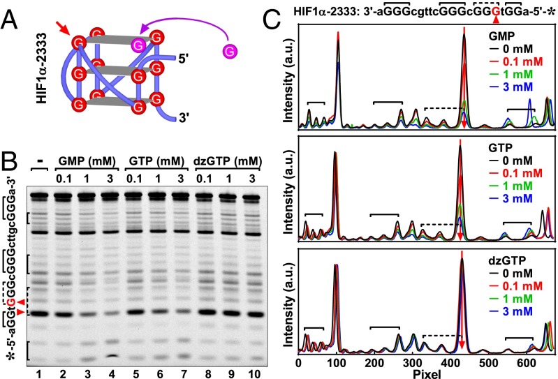
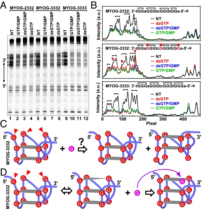
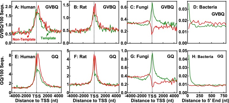
G-quadruplex structures formed by guanine-rich nucleic acids are implicated in essential physiological and pathological processes and nanodevices. G-quadruplexes are normally composed of four Gn (n ≥ 3) tracts assembled into a core of multiple stacked G-quartet layers. By dimethyl sulfate footprinting, circular dichroism spectroscopy, thermal melting, and photo-cross-linking, here we describe a unique type of intramolecular G-quadruplex that forms with one G2 and three G3 tracts and bears a guanine vacancy (G-vacancy) in one of the G-quartet layers. The G-vacancy can be filled up by a guanine base from GTP or GMP to complete an intact G-quartet by Hoogsteen hydrogen bonding, resulting in significant G-quadruplex stabilization that can effectively alter DNA replication in vitro at physiological concentration of GTP and Mg(2+). A bioinformatic survey shows motifs of such G-quadruplexes are evolutionally selected in genes with unique distribution pattern in both eukaryotic and prokaryotic organisms, implying such G-vacancy-bearing G-quadruplexes are present and play a role in gene regulation. Because guanine derivatives are natural metabolites in cells, the formation of such G-quadruplexes and guanine fill-in (G-fill-in) may grant an environment-responsive regulation in cellular processes. Our findings thus not only expand the sequence definition of G-quadruplex formation, but more importantly, reveal a structural and functional property not seen in the standard canonical G-quadruplexes.

Keywords: G-quadruplex; G-vacancy; guanine-responsive; nucleic acids.


Conflict of interest statement

The authors declare no conflict of interest.

Figures

Fig. 1.
Fig. 1.
Scheme of a stacked core unit (A) of a parallel G-quadruplex with three G-quartet layers. Each G-quartet (B) consists of four guanine bases (dashed polygon) connected by eight Hoogsteen hydrogen bonds (hashed bonds). The N7 in each guanine base is indicated in green. Removing one guanine from a G-quartet exposes the corresponding N7 (C, red circle).
Fig. 2.
Fig. 2.
G-quadruplex formation in MYOG G-core ssDNAs detected by (A) CD spectroscopy and (B and C) DMS footprinting. (A) CD spectra of MYOG G-quadruplexes. (B) DNA cleavage fragments resolved by denaturing gel electrophoresis. (C) Digitization of the gel in B. (D) Scheme of G-quartet completion by G fill-in in the GVBQ of MYOG-3332. (E) Structure of MYOG-3333 G-quadruplex. Red arrowhead in BD indicates hyper-cleaved guanine residue that was protected by a G fill-in with GMP in MYOG-3332.
Fig. S1.
Fig. S1.
G-quadruplexes of DNAs resolved by native gel electrophoresis. The intramolecular nature of the G-quadruplexes is indicated by their faster migration in comparison with that of the M-21 random oligomer. Sequence and size of each DNA are listed below the gel. DNA (0.5 μM) was prepared in the same way as for the CD analysis and electrophoresed at pH 7.4 in the presence of 50 mM K+ and 40% (wt/vol) PEG 200.
Fig. 3.
Fig. 3.
G-quadruplex formation in HIF1α G-core ssDNAs detected by (A) CD spectroscopy and (B and C) DMS footprinting. (A) CD spectra of HIF1α-2333 G-quadruplexes. (B) DNA cleavage fragments resolved by denaturing gel electrophoresis. (C) Digitization of the gel in B. (D) Scheme of G-quartet completion by G fill-in in the GVBQ of HIF1α-2333. (E) Structure of HIF1α-3333 G-quadruplex. Red arrowhead in BD indicates hyper-cleaved guanine residue that was protected by a G fill-in with GMP in HIF1α-2333. Black arrowhead indicates the orphan guanine that was not assembled in the G-quadruplex and hence not protected by GMP from cleavage in the HIF1α-2333 (blue vs. green peak).
Fig. 4.
Fig. 4.
Protection of the hyper-cleave prone guanine in the GVBQ of MYOG-3332 by G fill-in with different guanine derivatives in DMS footprinting. (A) Structure of the GVBQ of MYOG-3332 with a G fill-in (B) DNA cleavage fragments resolved by denaturing gel electrophoresis. (C) Digitization of the gel in B. Red arrowhead indicates hyper-cleaved guanine residue that was protected by G fill-in with a guanine derivative.
Fig. 5.
Fig. 5.
Protection of the hyper-cleave-prone guanine in the GVBQ of HIF1α-2333 by G fill-in with different guanine derivatives in DMS footprinting. (A) Structure of the GVBQ of HIF1α-2333 with a G fill-in. (B) DNA cleavage fragments resolved by denaturing gel electrophoresis. (C) Digitization of the gel in B. Red arrowhead indicates hyper-cleaved guanine residue that was protected by G-fill-in with a guanine derivative.
Fig. S2.
Fig. S2.
Quantitation of protection of hyper-cleavages in (A and B) MYOG-3332 and (C and D) HIF1α-2333 by GTP and GMP. Guanine hyper-cleavage in DMS footprinting was assayed as in Figs. 4 and 5. Intensity ratio of the hyper-cleavage (HC) band over full-length (FL) band (top band on gel) with SD was obtained from three independent experiments.
Fig. 6.
Fig. 6.
G fill-in stabilized the GVBQ of MYOG-3332 in FRET melting. (A) Melting of MYOG GVBQ in the absence of guanine derivatives. Curves were obtained in a solution containing 50 mM K+ (solid) or in 1 mM K+ plus 49 mM Li+ (dashed). T1/2 gives the temperature for the fluorescence to reach the midvalue between the minimum and maximum values. (B) Effect of guanine derivatives on the T1/2 of G-quadruplex from MYOG-2332, MYOG-2333, and MYOG-3333. Assay was carried out in a solution containing 1 mM K+ plus 49 mM Li+ for MYOG-3333 and 50 mM K+ for the others at various concentrations of guanine derivatives.
Fig. S3.
Fig. S3.
G fill-in enhanced the stability of the GVBQ of (A) HIF1α-2333, (B) LRRC42, (C) ABTB2, and (D) TSC22D3 in FRET melting. Assay was carried out in a solution containing 50 mM K+ at various concentrations of guanine derivatives as in Fig. 6B. T1/2 indicates the temperature for the fluorescence to reach midvalue between the minimum and maximum values.
Fig. 7.
Fig. 7.
Confirmation of G fill-in in the GVBQ of MYOG-3332 by photo-cross-linking and subsequent electrophoretic mobility shift. (A) Trifunctional SBED-GMP used for cross-linking. (B) Cross-linking between SBED-GMP and MYOG-3332 DNA detected by denaturing gel electrophoresis. (C) Mobility shift of cross-linked MYOG-3332 DNA by streptavidin (SA) detected by native gel electrophoresis. Schemes at the left side of gel indicate the structure of the corresponding DNA bands. Open triangle indicates cross-linked DNA.
Fig. S4.
Fig. S4.
(A) Synthesis of SBED-GMP from Sulfo-SBED and 5′-amino-GMP and full MS scan of the synthesized SBED-GMP. m/z 1,125.4 represents the precursor ion of SBED-GMP. Sulfo-SBED reacts with the amine (-NH2) residue on 5′-amino-GMP, resulting in a trifunctional SBED-GMP. (B) Mass spectrum of the fragment ions of SBED-GMP.
Fig. S5.
Fig. S5.
Confirmation of G fill-in the GVBQ of HIF1α-2333 by photo-cross-linking and subsequent electrophoretic mobility shift. (A) Cross-linking between SBED-GMP and HIF1α-2333 DNA detected by denaturing gel electrophoresis. (B) Mobility shift of cross-linked HIF1α-2333 DNA by streptavidin (SA) detected by native gel electrophoresis. Schemes at the left side of gel indicate the structure of the corresponding DNA bands. Open triangle indicates cross-linked DNA.
Fig. S6.
Fig. S6.
Formation of GVBQ in LRRC42, ABTB2, and TSC22D3 ssDNA detected by (A) CD spectroscopy and (B–G) DMS footprinting. (B–D) DNA was incubated in a solution containing 50 mM of the indicated cation in the absence or presence of GMP before DMS treatment. (E–G) Digitization of the gels in B–D). Schemes at the right side of E–G indicate the structure of the G-quadruplexes based on the CD and footprinting results. Red arrowhead indicates hyper-cleaved guanine residue that was protected by a G fill-in with GMP. Black arrowhead indicates the orphan guanine that was not assembled in the G-quadruplex and hence not protected by GMP from cleavage (blue vs. green peak).
Fig. S7.
Fig. S7.
Confirmation of G fill-in in the GVBQ of LRRC42, ABTB2, and TSC22D3 by photo-cross-linking and subsequent electrophoretic mobility shift. (A) Cross-linking between SBED-GMP and DNAs detected by denaturing gel electrophoresis. (B) Mobility shift of cross-linked DNA by streptavidin (SA) detected by native gel electrophoresis. Schemes at the left side of gel indicate the structure of the corresponding DNA bands. Open triangle indicates cross-linked DNA.
Fig. S8.
Fig. S8.
(A) Formation of GVBQ in MYOG-3332, LRRC42, ABTB2, and TSC22D3 ssDNA detected by (A) CD spectroscopy, (B–F) DMS footprinting, and (G) photo-cross-linking and electrophoretic mobility shift with streptavidin (SA). (A) CD spectra were obtained in a solution containing 150 mM K+ without PEG. (B–F) DNA was incubated in a solution containing 150 mM of the indicated cation in the absence or presence of GMP without PEG before DMS treatment. Red arrowhead indicates hyper-cleaved guanine residue that was protected by a G fill-in with GMP. (G) DNA was incubated in a solution containing 150 mM K+ without PEG before cross-linking. Open triangle indicates cross-linked DNA.
Fig. 8.
Fig. 8.
G-quadruplex formation in transcribed MYOG dsDNAs detected by DMS footprinting. (A) DNA cleavage fragments resolved by denaturing gel electrophoresis. (B) Digitization of the gel in A. (C) Possible alternative G2 tract alignment that could lead to G3 tracts protection. (D) G fill-in at the G-vacancy by GMP/GTP that could lead to G3 tracts protection. DNA was not transcribed (NT) or transcribed in the presence of the indicated guanine derivative(s). Red arrowhead in BD indicates the guanine residue attacked in the absence and protected in the presence of GMP/GTP. Blue curves in B largely overlap with the green ones such that they are barely visible.
Fig. 9.
Fig. 9.
Effect of G fill-in in GVBQ on in vitro DNA replication and stability in the presence of 2 mM Mg2+. (A) Inhibition of DNA primer extension by stabilization of GVBQ of MYOG-3332 and TSC22D3 with GMP/GTP, assayed by DNA-templated primer extension. Marker was made by a same extension reaction with a template without the G-core and its 3′ flanking sequence. (B) Ratio of premature termination (PT) over full-length (FL) replicon (±SD) in three independent experiments in K+ solution demonstrated in A. (C) Stabilization of the GVBQ of MYOG-3332 and TSC22D3 as a function of GMP/GTP concentration assayed by FRET melting.
Fig. 10.
Fig. 10.
Distribution of potential GVBQ-forming motifs in comparison with the canonical G-quadruplex–forming sequences (GQ) in prokaryotic and eukaryotic genes. Motifs bearing one G2 and three G3–4 tracts, with loop sizes from one to seven nucleotides, are counted on both the nontemplate (red) and template (green) DNA strands. Survey on fungi and bacteria covered genes from 53 strains and 4,222 chromosomes, respectively, whose sequences were available in the Ensembl and NCBI database. Frequency was normalized to the number of sequences and expressed as the number of occurrences in 100 sequences in a 100- (human, rat, and fungi) or 25-nt (bacteria) window.

Similar articles

Cited by

References

    1. Burge S, Parkinson GN, Hazel P, Todd AK, Neidle S. Quadruplex DNA: Sequence, topology and structure. Nucleic Acids Res. 2006;34(19):5402–5415. - PMC - PubMed
    1. Balasubramanian S, Hurley LH, Neidle S. Targeting G-quadruplexes in gene promoters: A novel anticancer strategy? Nat Rev Drug Discov. 2011;10(4):261–275. - PMC - PubMed
    1. Patel DJ, Phan AT, Kuryavyi V. Human telomere, oncogenic promoter and 5′-UTR G-quadruplexes: Diverse higher order DNA and RNA targets for cancer therapeutics. Nucleic Acids Res. 2007;35(22):7429–7455. - PMC - PubMed
    1. Tarsounas M, Tijsterman M. Genomes and G-quadruplexes: For better or for worse. J Mol Biol. 2013;425(23):4782–4789. - PubMed
    1. Cahoon LA, Seifert HS. An alternative DNA structure is necessary for pilin antigenic variation in Neisseria gonorrhoeae. Science. 2009;325(5941):764–767. - PMC - PubMed

Publication types