. 2015 Nov 24;112(47):14711-6.
doi: 10.1073/pnas.1520029112. Epub 2015 Nov 9.

Conformational signaling required for synaptic plasticity by the NMDA receptor complex

Affiliations

Conformational signaling required for synaptic plasticity by the NMDA receptor complex

Jonathan Aow et al. Proc Natl Acad Sci U S A. .

Abstract

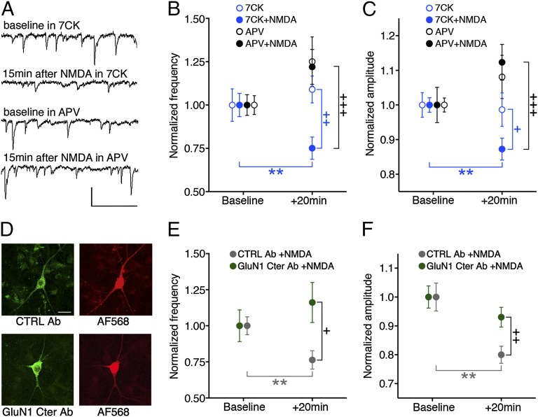
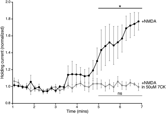
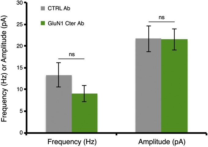
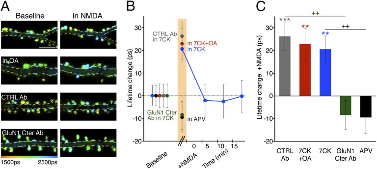
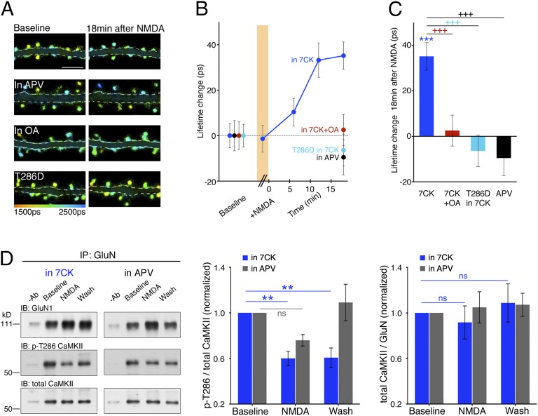
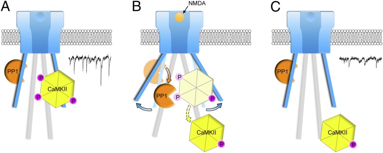
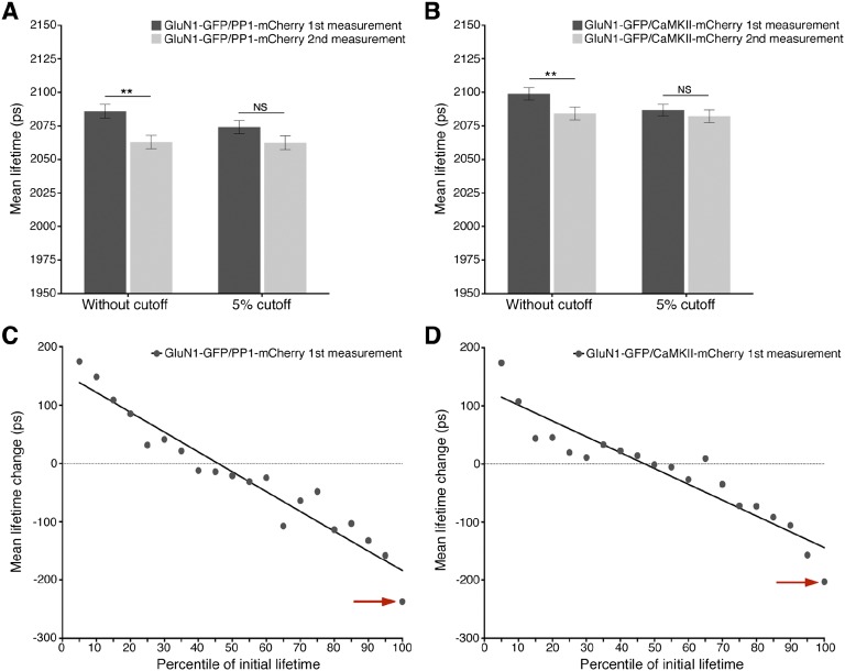
The NMDA receptor (NMDAR) is known to transmit important information by conducting calcium ions. However, some recent studies suggest that activation of NMDARs can trigger synaptic plasticity in the absence of ion flow. Does ligand binding transmit information to signaling molecules that mediate synaptic plasticity? Using Förster resonance energy transfer (FRET) imaging of fluorescently tagged proteins expressed in neurons, conformational signaling is identified within the NMDAR complex that is essential for downstream actions. Ligand binding transiently reduces FRET between the NMDAR cytoplasmic domain (cd) and the associated protein phosphatase 1 (PP1), requiring NMDARcd movement, and persistently reduces FRET between the NMDARcd and calcium/calmodulin-dependent protein kinase II (CaMKII), a process requiring PP1 activity. These studies directly monitor agonist-driven conformational signaling at the NMDAR complex required for synaptic plasticity.

Keywords: LTD; NMDAR-CaMKII interaction; NMDAR-PP1 FRET; ion-flow independent; long-term depression.


Conflict of interest statement

The authors declare no conflict of interest.

Figures

Fig. 1.
Fig. 1.
Transient NMDA application produces reduced transmission in absence of ion flow through NMDARs blocked by an antibody to GluN1 C-terminal domain (GluN1 Cter Ab). (A) Representative traces of spontaneous EPSCs from primary hippocampal neurons at indicated times and conditions. (Scale bar, 50 pA, 100 ms.) (B and C) Plot of spontaneous EPSC frequency (B) or amplitude (C), normalized (to baseline period) at indicated times and conditions; N (neurons) for 7CK 16, +NMDA 23; APV 15, +NMDA 15. (D) Representative images of neurons infused with fluorescent control (CTRL) Ab (Top) or with GluN1 Cter Ab, immunostained with fluorescent secondary Ab (Bottom), along with red dye (Right). (Scale bar, 20 μm.) (E and F) Plot of spontaneous EPSC frequency (E) or amplitude (F) (normalized to baseline period) 20 min after NMDA application in 7CK for neurons infused with CTRL Ab (n = 14) or GluN1 Cter Ab (n = 15). +P < 0.05, ++P < 0.01, +++P < 0.001 compared between conditions (Mann–Whitney); **P < 0.01 compared with baseline value (Wilcoxon).
Fig. S1.
Fig. S1.
NMDA-induced NMDAR conductance is blocked by 50 μM 7CK. Graph of normalized holding current against time for primary hippocampal neurons held at +40 mV after 25 μM NMDA application (added to the perfusion at time 0) in the absence (black circles, n = 7) or presence (light gray circles, n = 6) of 50 μM 7CK. *P < 0.05; ns, not significant; Wilcoxon test (comparison between 1–2 min and 5–7 min).
Fig. S2.
Fig. S2.
Synaptic transmission is unaffected by GluN1 C-terminus antibody infusion. Spontaneous EPSCs from primary hippocampal neurons were recorded for 5 min in 7CK after the infusion of a CTRL antibody (n = 14) or GluN1 Cter Ab (n = 15) for 7 min. ns, not significant; unpaired t-test.
Fig. 2.
Fig. 2.
NMDA drives FRET changes between PP1 and the NMDARcd. (A) Representative FLIM images of neurons expressing GluN1-GFP/PP1-mCherry at indicated times and conditions. OA (1 µM), okadaic acid. (B) Plot of GluN1-GFP lifetime change, relative to baseline, before, during (orange bar), and after NMDA application. N >15 neurons; > 400 spines per condition. (C) Bar graph of GluN1-GFP lifetime change, relative to baseline, during NMDA application for the indicated conditions. N > 15 neurons; > 400 spines per condition. ++P < 0.01 compared between conditions (Mann–Whitney); **P < 0.01, ***P < 0.001 compared with baseline value (Wilcoxon).
Fig. 3.
Fig. 3.
NMDA drives dephosphorylation and movement of CaMKII bound to NMDARcd. (A) Representative FLIM images of neurons expressing GluN1-GFP/CaMKII-mCherry at indicated times and conditions. T286D indicates Thr286Asp mutant of CaMKII. (B) Plot of GluN1-GFP lifetime change, relative to baseline, before, during (orange bar), and after NMDA application. N > 16 neurons; > 400 spines per condition. (C) Bar graph of GluN1-GFP lifetime change, relative to baseline, during NMDA application for the indicated conditions. N > 16 neurons; > 400 spines per condition. (D, left) Representative immunoblots (IB) of GluN1, phospho-CaMKII-Thr286 (p-T286), and total CaMKII from cortical neuronal lysates coimmunoprecipitated with a GluN1 or GluN2B antibody under the indicated conditions. −Ab, no primary antibody during immunoprecipitation. (Middle) Average p-T286/total CaMKII for the indicated conditions; n ≥ 6 experiments, including GluN1 or GluN2B for IP. (Right) Average total CaMKII levels relative to GluN1 or GluN2B. ns, not significant. +++P < 0.001 compared between conditions (Mann–Whitney); **P < 0.01, ***P < 0.001 compared with baseline value (Wilcoxon).
Fig. 4.
Fig. 4.
Model consistent with imaging, electrophysiological, and biochemical data. (A) CaMKII phosphorylated (P) at Thr286 and PP1 bound to NMDAR-complex; PP1 lacks catalytic access to phospho-CaMKII-T286; in this conformation, NMDAR-complex signaling maintains basal transmission (trace). (B) NMDARcd moves permitting PP1 catalytic access to phospho-CaMKII-T286. (C) CaMKII lacking phosphate on Thr286 is relocated on the NMDARcd, which returns to baseline conformation; CaMKII, along with other signaling molecules activated by metabotropic NMDAR signaling, contributes to synaptic depression (trace). Trace examples taken from Fig. 1A.
Fig. S3.
Fig. S3.
GluN1-GFP, coexpressed with PP1-mCherry or CaMKII-mCherry, lifetimes in spines decrease during the second measurement because of a disproportionate drop in spines with the highest initial lifetimes. (A) Average initial GluN1-GFP lifetimes in spines coexpressing PP1-mCherry before and after exclusion of the highest 5% lifetime measurements (dark gray bars); average final GluN1-GFP lifetimes before and after exclusion of those spines with the highest 5% initial lifetimes (light gray bars). Average spine lifetimes are significantly different between the initial and final measurements (2,086 ± 5 ps vs. 2,063 ± 5 ps; n = 544; P < 0.01) before exclusion, but not after exclusion (2,074 ± 4 ps vs. 2,063 ± 4 ps; n = 517; P > 0.05). (B) As with A, but with spines coexpressing GluN1-GFP and CaMKII-mCherry. Average spine lifetimes are significantly different between the initial and final measurements (2,098 ± 5 ps vs. 2,084 ± 5 ps; n = 623; P < 0.01) before exclusion, but not significant after exclusion (2,086 ± 5 ps vs. 2,082 ± 6 ps; n = 592; P > 0.05). (C) Plot of mean lifetime change (ps) per GluN1-GFP expressing spine versus initial lifetime measurement percentile. Spines were sorted into 5% bins according to the percentile of the initial lifetime measurement, and the mean lifetime change of spines in each bin was plotted against the percentile. Note the presence of the outlier at the 96th–100th percentile lifetimes (indicated with red arrow). (D) As in C, but with spines coexpressing GluN1-GFP and CaMKII-mCherry.

References

    1. Malenka RC, Bear MF. LTP and LTD: An embarrassment of riches. Neuron. 2004;44(1):5–21. - PubMed
    1. Malenka RC. Synaptic plasticity in the hippocampus: LTP and LTD. Cell. 1994;78(4):535–538. - PubMed
    1. Lisman J. A mechanism for the Hebb and the anti-Hebb processes underlying learning and memory. Proc Natl Acad Sci USA. 1989;86(23):9574–9578. - PMC - PubMed
    1. Nabavi S, et al. Metabotropic NMDA receptor function is required for NMDA receptor-dependent long-term depression. Proc Natl Acad Sci USA. 2013;110(10):4027–4032. - PMC - PubMed
    1. Nabavi S, Fox R, Alfonso S, Aow J, Malinow R. GluA1 trafficking and metabotropic NMDA: Addressing results from other laboratories inconsistent with ours. Philos Trans R Soc Lond B Biol Sci. 2014;369(1633):20130145. - PMC - PubMed

Publication types

MeSH terms

Substances