. 2015 Dec 22;112(51):E7138-47.
doi: 10.1073/pnas.1511670112. Epub 2015 Nov 9.

Amphetamine activates Rho GTPase signaling to mediate dopamine transporter internalization and acute behavioral effects of amphetamine

Affiliations

Amphetamine activates Rho GTPase signaling to mediate dopamine transporter internalization and acute behavioral effects of amphetamine

David S Wheeler et al. Proc Natl Acad Sci U S A. .

Abstract

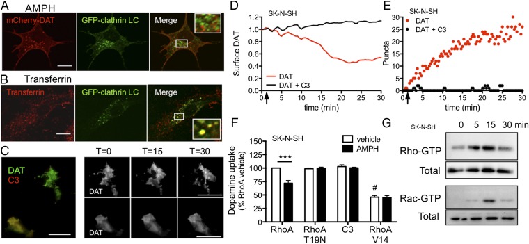
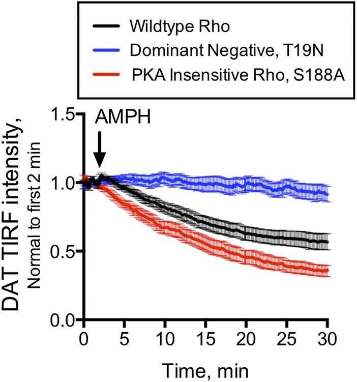
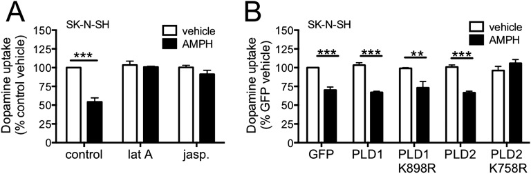
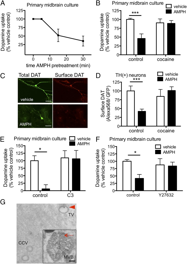
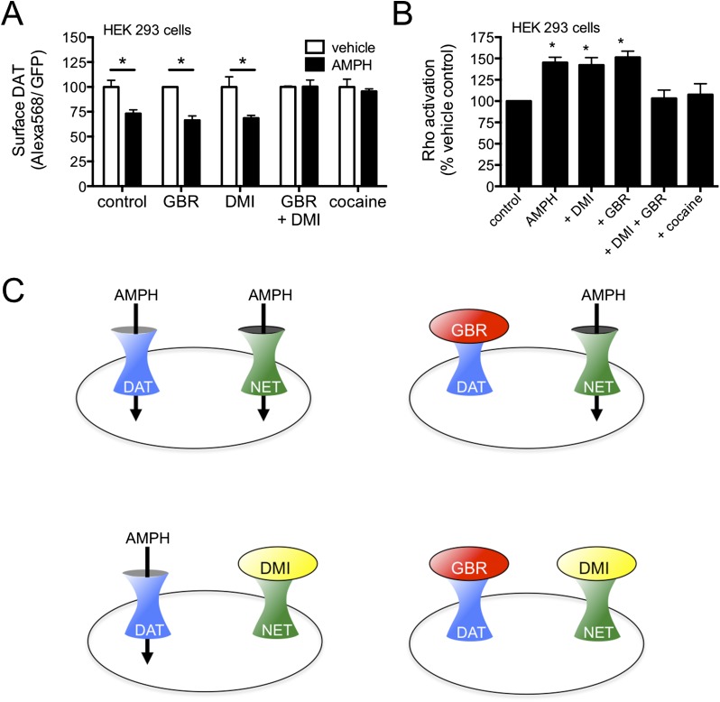
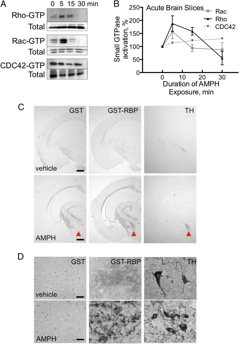
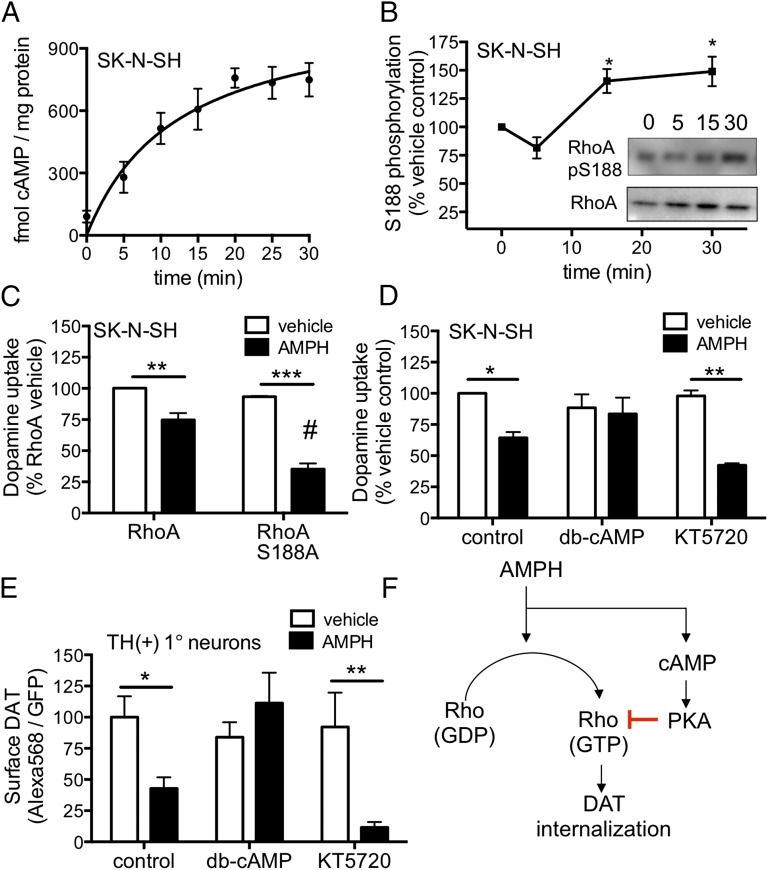
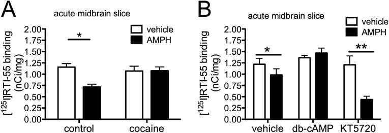
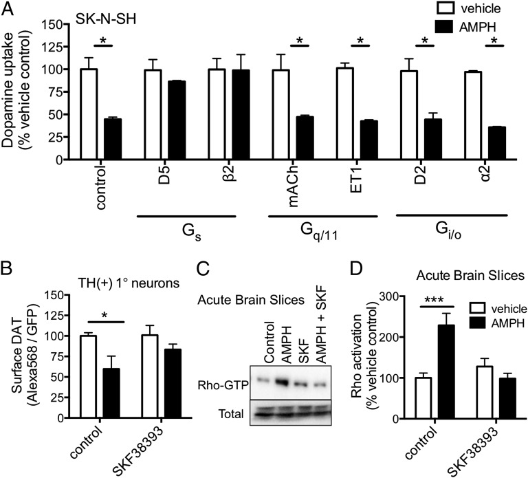
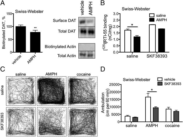
Acute amphetamine (AMPH) exposure elevates extracellular dopamine through a variety of mechanisms that include inhibition of dopamine reuptake, depletion of vesicular stores, and facilitation of dopamine efflux across the plasma membrane. Recent work has shown that the DAT substrate AMPH, unlike cocaine and other nontransported blockers, can also stimulate endocytosis of the plasma membrane dopamine transporter (DAT). Here, we show that when AMPH enters the cytoplasm it rapidly stimulates DAT internalization through a dynamin-dependent, clathrin-independent process. This effect, which can be observed in transfected cells, cultured dopamine neurons, and midbrain slices, is mediated by activation of the small GTPase RhoA. Inhibition of RhoA activity with C3 exotoxin or a dominant-negative RhoA blocks AMPH-induced DAT internalization. These actions depend on AMPH entry into the cell and are blocked by the DAT inhibitor cocaine. AMPH also stimulates cAMP accumulation and PKA-dependent inactivation of RhoA, thus providing a mechanism whereby PKA- and RhoA-dependent signaling pathways can interact to regulate the timing and robustness of AMPH's effects on DAT internalization. Consistent with this model, the activation of D1/D5 receptors that couple to PKA in dopamine neurons antagonizes RhoA activation, DAT internalization, and hyperlocomotion observed in mice after AMPH treatment. These observations support the existence of an unanticipated intracellular target that mediates the effects of AMPH on RhoA and cAMP signaling and suggest new pathways to target to disrupt AMPH action.

Keywords: Rho GTPase; amphetamine; dopamine transporter; endocytosis; protein kinase A.


Conflict of interest statement

The authors declare no conflict of interest.

Figures

Fig. 1.
Fig. 1.
AMPH-induced DAT internalization is clathrin-independent but Rho-dependent. (A) SK-N-SH cells were cotransfected with mCherry-DAT and a clathrin light chain-GFP fusion protein and stimulated with AMPH for 5 min. AMPH causes DAT (red) to internalize but, once endocytosed, DAT fails to colocalize with clathrin (green). (B) SK-N-SH cells were transfected with a GFP-clathrin light chain and treated with transferrin conjugated to Alexa-Fluor-568 for 5 min. These cells exhibit colocalization of clathrin with the transferrin-bound receptors. (C) SK-N-SH cells were cotransfected with mCherry-DAT (green) and GFP-C3 (red; note that pseudocoloring has been reversed for consistency throughout the figures). The field shown here is of a cell expressing DAT and C3 (yellow) and a cell expressing DAT alone (green) at T = 0 (Movie S2). In the absence of C3 coexpression, AMPH causes robust clustering and internalization of DAT at T = 15 and loss of surface DAT by T = 30 (Right, Top). However, AMPH fails to alter DAT at either T = 15 or T = 30 in a cell coexpressing C3 with DAT (Right, Bottom). The time course of AMPH-induced internalization in control DAT- and DAT-C3–expressing SKNSH cells assessed by (D) surface DAT fluorescence and (E) the number of internalizing puncta. The arrow indicates the start of AMPH treatment. (F) AMPH pretreatment decreases [3H]-dopamine uptake by DAT. Coexpression of a dominant-negative RhoA (T19N) or the Rho inhibitor C3 blocks this effect. The constitutively active form of RhoA (V14) decreases dopamine uptake and does not exhibit any further decrease in response to AMPH pretreatment. (G) Isolation of activated Rho and Rac by GST-RBD and GST-p21, respectively, from DAT-transfected SK-N-SH cells shows AMPH-mediated activation of Rho and Rac1 GTPases over time. (Scale bars: 25 μm in A and B, and the insets have been enlarged 3-fold; 30 μm in C.) ***P < 0.001, two-way ANOVA; #P < 0.001, one-way ANOVA compared with vehicle RhoA control.
Fig. S1.
Fig. S1.
Rho mutants modulate AMPH-induced trafficking of DAT. TIRF microscopy of SK-N-SH cells transiently transfected with mCherry-DAT and GFP-tagged wild-type Rho (black) had a loss of cell-surface DAT localization in response to AMPH (arrow). The dominant-negative Rho mutant, T19N (blue), blocked AMPH-induced DAT internalization. The DAT cotransfected with the PKA-insensitive Rho mutant, S188A (red), had an enhanced response to AMPH.
Fig. S2.
Fig. S2.
AMPH-induced DAT internalization has properties characteristic of other Rho-dependent internalization processes. Rho-dependent internalization requires actin skeleton reorganization and activation of phospholipase D. (A) Disruption of actin remodeling with either 200 ng/mL latrunculin A (lat A, inhibitor of actin filament polymerization) or 500 nM jasplakinolide (jasp, actin filament stabilizer) blocks AMPH-induced DAT internalization as measured by dopamine uptake. (B) Expression of a dominant-negative isoform of phospholipase D2 (PLD2 K758R) inhibits AMPH-induced DAT internalization whereas the dominant-negative phospholipase D1 (PLD1 K898R) construct does not (**P < 0.01 and ***P < 0.001, two-way ANOVA).
Fig. 2.
Fig. 2.
AMPH causes internalization of DAT in dopamine neurons. (A) In midbrain cultures, AMPH pretreatment causes a loss of DAT-mediated dopamine transport in a time-dependent process. (B) Pretreatment with 10 μM AMPH for 30 min leads to a substantial loss of dopamine transport in primary cultures. Coapplication of the DAT inhibitor cocaine (100 μM) blocks this effect of AMPH pretreatment. (C) Primary murine midbrain neurons transfected with the GFP-AP-DAT construct (green) demonstrate DAT internalization by AMPH through decreased Alexa Fluor 568-streptavidin (red) surface labeling. (D) The ratio of surface GFP-AP-DAT (Alexa Fluor 568) to total GFP-AP-DAT (GFP) in TH(+) neurons is normalized to vehicle control. AMPH (10 μM) treatment reduces surface DAT by 58 ± 6%, and this effect is blocked by 100 μM cocaine. (E) Bar graph showing dopamine uptake in control primary midbrain cultures with either vehicle control or the Rho inhibitor exotoxin C3 introduced into the cells by the scrape method (12). Approximately 18 h after the neurons were scrape-loaded in the presence or absence of the C3 exotoxin, neuronal cultures were treated with AMPH for 30 min. AMPH significantly decreases dopamine transport in the sham-treated control cultures but has no effect on the cells treated with C3. (F) The ROCK inhibitor Y27632 also attenuates the AMPH-induced decrease in [3H]-dopamine uptake in primary cultures. (G) Primary midbrain neurons expressing GFP-AP-DAT labeled with streptavidin-conjugated gold particles. Transmission electron micrographs of AMPH-stimulated neurons reveal gold particles (arrowhead) in tubulovesicular bodies (TV) and multivesicular bodies (MVB, Inset). Gold particles are absent from clathrin-coated pits or vesicles (CCV). (Scale bars: 50 μm in C, 100 nm in G, and 75 nm in the Inset of G). *P < 0.05, ***P < 0.001, two-way ANOVA.
Fig. S3.
Fig. S3.
AMPH requires a monoamine transporter to gain entry into the cytoplasm and trigger DAT internalization. (A) AMPH acts intracellularly in the absence of monoamine vesicles to stimulate DAT internalization. HEK293 cells expressing both GFP-AP-DAT and NET were exposed to AMPH in the presence of a DAT inhibitor (GBR12909, 100 nM) or the NET inhibitor desipramine (DMI, 10 nM) at concentrations that selectively block only one of the transporters. Blockade of either DAT or NET alone has no effect on AMPH-induced DAT internalization as measured by the ratio of cell-surface streptavidin–Alexa Fluor 568 labeling to the total GFP fluorescence. Blocking both DAT and NET with either cocaine or the combination of GBR and DMI completely blocks AMPH-mediated internalization of the DAT (*P < 0.05, two-way ANOVA). (B) AMPH entering the cell through either DAT or NET can trigger Rho activation in HEK293 cells. Cells were cotransfected with GFP-AP-DAT and NET and treated with transport inhibitors as above. Rho activation was assessed by GST-RBD–binding assay (*P < 0.05, one-way ANOVA). (C) Schematic illustrating the experimental design for A and B. AMPH transport by DAT (blue) or NET (green) can be blocked by GBR12909 (red) or DMI (yellow), respectively.
Fig. 3.
Fig. 3.
AMPH activates Rho and Rac in dopamine neurons in midbrain slices. (A) Acute midbrain slices from adult Swiss-Webster mice were treated with 10 μM AMPH for the indicated time. Activated GTP-bound Rho, Rac, and CDC42 were affinity-purified with the GST fusion proteins and analyzed by Western blot. (B) Blots of activated GTPases were quantified by densitometry. AMPH treatment causes maximal activation of Rho and Rac at 5 min. (C) Frozen sections of vehicle- and AMPH-treated acute midbrain slices were stained with biotinylated GST-RBD (Rho-binding domain) to anatomically localize activated Rho. In vehicle-treated sections, staining with GST and GST-RBD were almost identical, indicating that Rho activity is minimal at baseline. In AMPH-treated sections, GST-RBD selectively stained a cluster of cells (arrowheads) in the midbrain. This staining overlaps with TH staining of dopamine neurons in adjacent sections. (D) Higher magnification of the region containing TH(+) cell bodies. Rho activation appears predominantly within neuronal processes. (Scale bar: 1 mm in C and 75 μm in D.)
Fig. 4.
Fig. 4.
AMPH stimulates cAMP, which terminates Rho signaling and DAT internalization. (A) Cumulative cAMP generation was measured in DAT-transfected SK-N-SH cell lysates in the presence of 3-isobutyl-1-methylxanthine (IBMX). AMPH (10 μM) treatment of the cells causes a time-dependent increase in cAMP levels with a τ1/2 of 12.5 min. (B) AMPH treatment increases the amount of phospho-Ser188 RhoA. (Inset) Immunoblot of SK-N-SH protein lysates probed with an antibody that recognizes P-Ser188 RhoA (Top) and total RhoA (Bottom). (C) Expression of RhoA S188A, a mutant that cannot be inactivated by PKA, enhances AMPH-induced DAT internalization as assessed by uptake of [3H]-dopamine. (D) Activation of PKA by 10 μM dibutyryl-cAMP blocks AMPH-mediated internalization of DAT. In contrast, inhibition of PKA with 600 nM KT5720 enhances DAT internalization. (E) In primary neurons, the actions of db-cAMP and KT5720 parallel the results seen in SK-N-SH cells as assessed by membrane labeling of the GFP-AP-DAT protein (F) A schematic illustrating the crosstalk between AMPH-induced Rho activation and cAMP signaling. AMPH activates Rho but also stimulates cAMP production, which in turn activates PKA to terminate Rho activation and DAT internalization. *P < 0.05, **P < 0.01, ***P < 0.001, two-way ANOVA; #P < 0.01 compared with RhoA + AMPH in C.
Fig. S4.
Fig. S4.
[125I]RTI-55 binding to murine acute brain slices confirms AMPH-induced internalization of endogenous DAT. (A) Acute midbrain slices were incubated in oxygenated aCSF containing vehicle, 10 μM AMPH, 100 μM cocaine, or both AMPH and cocaine. After 30 min, [125I]RTI-55 labeling decreases by 38.2%. Cocaine pretreatment of midbrain slices has no effect on subsequent RTI binding, but does block the AMPH-mediated decrease binding. (B) Acute slices pretreated with vehicle, 10 μM db-cAMP, or 600 nM KT5720 for 15 min before addition of vehicle or 10 μM AMPH indicate that AMPH-induced DAT internalization is blocked by activation of PKA with db-cAMP and is enhanced by inhibition of PKA with KT5720 (*P < 0.05 and **P < 0.01, two-way ANOVA).
Fig. 5.
Fig. 5.
Activation of endogenous Gs-coupled GPCRs prevents AMPH effects. (A) Stimulation of Gs-coupled receptors blocks the AMPH-mediated decrease in dopamine uptake in SK-N-SH cells. D1/D5 dopamine receptors were stimulated with 100 nM SKF38393, and β2 adrenergic receptors were stimulated with 10 μM isoproterenol. Activation of muscarinic (mAch; 10 μM carbachol), endothelin (ET1; 1 μM endothelin), D2 dopamine (D2; 10 nM bromocriptine), or α2 adrenergic (α2; 100 nM norepinephrine) receptors does not alter the effect of AMPH treatment. Cells were treated with AMPH (10 μM) with or without GPCR agonist for 30 min, washed, and assayed for uptake of [3H]-dopamine. (B) D1/D5 receptor activation with 100 nM SKF38393 is able to suppress AMPH-induced GFP-AP-DAT internalization in primary neurons. (C and D) AMPH-induced Rho activation is also inhibited by D1/D5 receptor stimulation. A 30-min exposure of midbrain slices to AMPH leads to increased GTP-bound activated Rho that is blocked by SKF38393. *P < 0.05 and ***P < 0.001, two-way ANOVA.
Fig. 6.
Fig. 6.
Activation of endogenous Gs-coupled GPCRs prevents AMPH effects in vivo. (A) Western blot analysis of biotinylated tissue from adult male Swiss-Webster mice that were IP-injected with saline or AMPH (2 mg/kg). After 30 min, midbrain tissue was isolated, biotinylated, and assessed for plasma membrane localization of the DAT. (B) Adult Swiss-Webster mice were IP-injected with saline or the D1/D5 agonist SKF38393 (3 mg/kg) followed 5 min later by a second injection of saline or AMPH (2 mg/kg). Animals were killed after 45 min, and acute midbrain sections were incubated with radiolabeled [125I]RTI-55. AMPH decreased [125I]RTI-55 binding. This effect was blocked by SKF38393 pretreatment. (C) Mice received IP injections of AMPH (2 mg/kg) or cocaine (20 mg/kg) with or without the D5 agonist, SKF38393 (3 mg/kg). Representative traces of animals’ movements in the test arena after injection show comparable spatial distributions of activity. Variations in trace density reflect differences in total ambulation. (D) Quantification of ambulatory activity for all groups. Pretreatment with SKF38393 significantly decreases AMPH-induced ambulation, but it does not affect cocaine-induced activity or have a significant effect on its own **P < 0.0001 by paired t test; *P < 0.05, two-way ANOVA.

Comment in

Similar articles

Cited by

References

    1. Gonzalez Castro F, Barrington EH, Walton MA, Rawson RA. Cocaine and methamphetamine: Differential addiction rates. Psychol Addict Behav. 2000;14(4):390–396. - PubMed
    1. Kalechstein AD, et al. Psychiatric comorbidity of methamphetamine dependence in a forensic sample. J Neuropsychiatry Clin Neurosci. 2000;12(4):480–484. - PubMed
    1. Rawson R, et al. Methamphetamine and cocaine users: Differences in characteristics and treatment retention. J Psychoactive Drugs. 2000;32(2):233–238. - PubMed
    1. Mahoney JJ, III, Kalechstein AD, De La Garza R, II, Newton TF. Presence and persistence of psychotic symptoms in cocaine- versus methamphetamine-dependent participants. Am J Addict. 2008;17(2):83–98. - PMC - PubMed
    1. Yamamoto BK, Moszczynska A, Gudelsky GA. Amphetamine toxicities: Classical and emerging mechanisms. Ann N Y Acad Sci. 2010;1187:101–121. - PMC - PubMed

Publication types

MeSH terms

Substances

LinkOut - more resources