. 2015 Nov 24;112(47):14599-604.
doi: 10.1073/pnas.1516401112. Epub 2015 Nov 9.

Directed evolution of the tryptophan synthase β-subunit for stand-alone function recapitulates allosteric activation

Affiliations

Directed evolution of the tryptophan synthase β-subunit for stand-alone function recapitulates allosteric activation

Andrew R Buller et al. Proc Natl Acad Sci U S A. .

Abstract

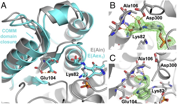
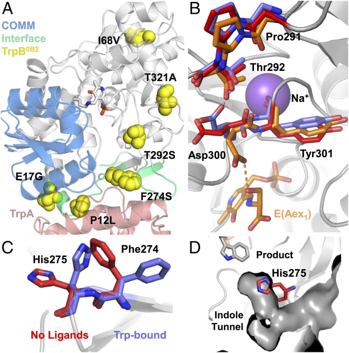
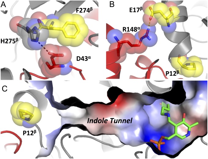
Enzymes in heteromeric, allosterically regulated complexes catalyze a rich array of chemical reactions. Separating the subunits of such complexes, however, often severely attenuates their catalytic activities, because they can no longer be activated by their protein partners. We used directed evolution to explore allosteric regulation as a source of latent catalytic potential using the β-subunit of tryptophan synthase from Pyrococcus furiosus (PfTrpB). As part of its native αββα complex, TrpB efficiently produces tryptophan and tryptophan analogs; activity drops considerably when it is used as a stand-alone catalyst without the α-subunit. Kinetic, spectroscopic, and X-ray crystallographic data show that this lost activity can be recovered by mutations that reproduce the effects of complexation with the α-subunit. The engineered PfTrpB is a powerful platform for production of Trp analogs and for further directed evolution to expand substrate and reaction scope.

Keywords: PLP; allostery; noncanonical amino acid; protein engineering.


Conflict of interest statement

The authors declare no conflict of interest.

Figures

Fig. 1.
Fig. 1.
Catalytic cycle of the native TrpS complex. (A) Indole (4) is released through a retro-aldol reaction in TrpA (red) and then diffuses through a 25-Å tunnel into TrpB (black), where a PLP-mediated β-substitution reaction occurs with l-serine (2), yielding l-tryptophan (1). The COMM domain is indicated in blue. Scheme is superimposed over PfTrpS with Lys-PLP internal aldimine, E(Ain), shown in green sticks. The native complex is an αββα heterotetramer; a single αβ pair is shown for clarity. (B) Mechanism of l-tryptophan formation. Transimination of l-serine to form an external aldimine, E(Aex1), followed by dehydration across Cα–Cβ through a quinonoid intermediate, E(Q1), is designated stage I of the overall reaction. A mechanistically similar process occurs in reverse for the addition of indole into E(A-A) and subsequent release of l-tryptophan, designated stage II of the reaction.
Fig. S1.
Fig. S1.
Distribution of activating mutations identified through random mutagenesis and screening. (A) Positions on PfTrpB where activating mutations were discovered are displayed as spheres. The mutations are W2R, G4C, V11A, E17G, K20E, E23V, F35S, N35S, Y41C, I68V, M123V, I127S, M144T, N150T, N166D, Y178C, H180R, Y181C, L182P, M233V, M233I, F274S, F274L, D284G, T292S, T321A, and T323A. Mutations located in the COMM domain are colored blue, those within 5 Å of PfTrpA (red) are cyan, those present in PfTrpB4D11 are yellow, and the remainder are gray. (B) Positions on PfTrpB where activating mutations were discovered using PfTrp4D11 as the parent for random mutagenesis. The mutations are G4D, E5K, Y10H, P12L, E13G, E21V, L59Q, K67I, L146V, D220E, N267S, G272D, and D284E. Color scheme is the same as A; yellow spheres are mutations present in the final variant PfTrpB0B2.
Fig. 2.
Fig. 2.
UV-vis absorption spectra of native and engineered PfTrpB. (A) The PLP absorption spectrum of PfTrpB (black) has a λmax = 412 nm, characteristic of E(Ain). Addition of 20 mM l-serine causes a red-shift to 428 nm (gray), consistent with E(Aex1) formation. (B) Addition of l-serine to PfTrpS causes a shift in λmax to 350 nm, which is attributed to the E(A-A). A residual peak at 428 nm indicates population of E(Aex1). (C) Like PfTrpS, addition of 20 mM l-serine to PfTrpB0B2 shows λmax = 350 nm as well as contributions from E(Aex1). All spectra were collected with 20 µM of enzyme. See Materials and Methods for details.
Fig. S2.
Fig. S2.
UV-vis absorption spectra for native and engineered TrpB. A, D, and E are the same as in Fig. 2. PLP absorption spectra for each enzyme are shown in black and share λmax = 412 nm, characteristic of E(Ain). Addition of 20 mM l-Ser causes a red-shift to 420 nm (red), consistent with E(Aex1) formation for PfTrpB and PfTrpB2G9 (B), which also has an absorbance band at ∼320 nm that we attribute to formation of pyruvate through serine deaminase activity. l-Ser addition to PfTrpS, PfTrpB4D11 (C), and PfTrpB0B2 causes a shift in λmax to 350 nm and a broad shoulder extending to 550 nm. The 350-nm absorption is attributed to the E(A-A), and the shoulder indicates a mixed population of E(Aex1) and E(A-A). All spectra collected with 20 µM enzyme. To limit the amount of pyruvate production, spectra were taken as quickly as possible (<10 s) after addition of l-Ser.
Fig. 3.
Fig. 3.
Structural transitions upon ligand binding in PfTrpB. (A) Superimposition of PfTrpB-E(Ain) and PfTrpB-E(Aex1) in gray and cyan, respectively. Overlay shows the 2.1-Å displacement of the COMM domain upon E(Aex1) formation. This closure moves the side chain of Glu104 by 3.7 Å, toward its catalytic orientation (orange dashes). (B) Structure of Ser-bound PfTrpB with a Fo–Fc map of E(Aex1) contoured at 3.0 σ (green). (C) Structure of l-tryptophan–bound PfTrpB with Fo–Fc map of Trp ligand contoured at 3.0 σ.
Fig. 4.
Fig. 4.
Distribution of PfTrpB0B2 mutations and interaction networks altered by mutational reactivation. (A) PfTrpB residues within 5 Å of PfTrpA (red) are colored cyan and the COMM domain is colored blue. (B) A hydrogen bond between D300 and E(Aex1) in the Ser-bound structure (orange) is formed transiently during the catalytic cycle. When this H-bond is severed, D300 may interact with T292 (no ligands, red, or Trp-bound, blue). This complex network is centered on a monovalent cation cofactor, shown here as Na+, which is known to mediate allosteric interactions between the α- and β-subunits (6). (C) Residues F274 and H275 undergo a rotameric shift upon substrate or product binding into an open state (blue). (D) In the closed state (red, no ligands bound), H275 blocks access to the active site.
Fig. S3.
Fig. S3.
Structural comparison between StTrpS and PfTrpB in E(Ain) and E(Aex1) forms. (A) Alignment of StTrpB (green; PDB ID code 1BKS) and PfTrpB (light blue) in their substrate-free [E(Ain)] forms yields an rmsd of 0.5 Å. Small differences exist in the structures near the α-subunit (red) binding site, and the COMM domain of PfTrpB (dark blue) is in a slightly more open conformation than for StTrpB. (B) Alignment of StTrpB (green; PDB ID code 2CLL) and PfTrpB (light blue) with l-serine covalently bound as the external aldimine, E(Aex1), yields an rmsd of 0.5 Å.
Fig. S4.
Fig. S4.
Interactions between PfTrpA and PfTrpB that are disrupted in PfTrpB0B2. (A) Hydrogen bond between H275β (gray) and D43α (red) in the gate-open conformation is adjacent F274β (yellow), which is mutated to Ser in the PfTrpB4D11 and PfTrpB0B2 enzymes. (B) A salt bridge (dashes) between R148α (red) and E17β (yellow) is removed with the E17G mutation present in PfTrpB4D11 and PfTrpB0B2. P12β is located along the indole tunnel (C) between the subunits. Steric constraints indicate that the P12L mutation of PfTrpB0B2 would disrupt this tunnel, which might contribute to the large drop in Trp synthase activity of PfTrpS0B2.
Fig. 5.
Fig. 5.
Substrate profile of native and engineered TrpB enzymes. (A) Indole analogs that have been reported to react with StTrpS were tested for reactivity with the PfTrpB, PfTrpS, and PfTrpB0B2 enzymes. The nucleophilic atom is indicated with a gray circle. (B) Relative activities of enzyme complex PfTrpS (black) and PfTrpB0B2 (gray) compared with PfTrpB. Reactions performed in duplicate with 20 mM of each substrate and varying enzyme concentrations to ensure incomplete conversion after 1 h. Products were later confirmed in scaled-up reactions using PfTrpB0B2. See Materials and Methods for details.
Fig. S5.
Fig. S5.
Hypothetical reaction coordinate diagram to illustrate inversion of TrpA effector activation to inhibition. Native enzyme, such as TrpB (blue), with multiple reaction transition states. Effector addition or introduction of mutations (green) increases the rate of the reaction by lowering the free energy of the rate-limiting step (RLS), while simultaneously raising the energy of a different transition state thereby generating a new RLS. The same changes applied a second time (red), as might occur with PfTrpA addition to the engineered TrpB, can lead to a free-energy barrier higher than for the native enzyme and a reduction in overall reaction rate.
Fig. S6.
Fig. S6.
SDS/PAGE of PfTrpA, PfTrpB, and PfTrpS. 1, PfTrpA; 2, PfTrpB; 3–5, aliquots from 1.5-mL fractions from PfTrpA pull-down using His-tagged PfTrpB; 4–20% gradient, SDS/PAGE. Molecular weights from ColorPlus Prestained Protein Ladder (New England Biosystems) listed on the right.

Similar articles

Cited by

References

    1. Du L, Lou L. PKS and NRPS release mechanisms. Nat Prod Rep. 2010;27(2):255–278. - PubMed
    1. Dunn MF. Allosteric regulation of substrate channeling and catalysis in the tryptophan synthase bienzyme complex. Arch Biochem Biophys. 2012;519(2):154–166. - PMC - PubMed
    1. Davidsen JM, Bartley DM, Townsend CA. Non-ribosomal propeptide precursor in nocardicin A biosynthesis predicted from adenylation domain specificity dependent on the MbtH family protein NocI. J Am Chem Soc. 2013;135(5):1749–1759. - PMC - PubMed
    1. Niks D, et al. Allostery and substrate channeling in the tryptophan synthase bienzyme complex: Evidence for two subunit conformations and four quaternary states. Biochemistry. 2013;52(37):6396–6411. - PMC - PubMed
    1. Hyde CC, Ahmed SA, Padlan EA, Miles EW, Davies DR. Three-dimensional structure of the tryptophan synthase alpha 2 beta 2 multienzyme complex from Salmonella typhimurium. J Biol Chem. 1988;263(33):17857–17871. - PubMed

Publication types