. 2015 Nov 24;112(47):14723-7.
doi: 10.1073/pnas.1515228112. Epub 2015 Nov 9.

Unmyelinated type II afferent neurons report cochlear damage

Affiliations

Unmyelinated type II afferent neurons report cochlear damage

Chang Liu et al. Proc Natl Acad Sci U S A. .

Abstract

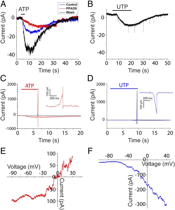
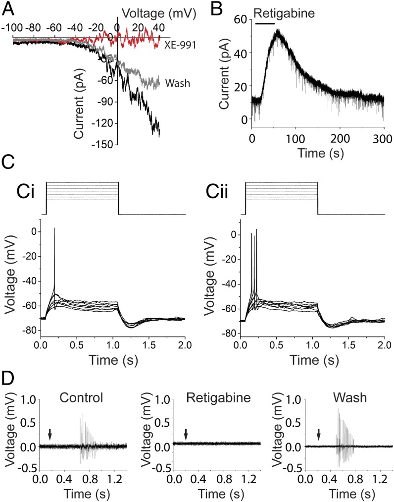
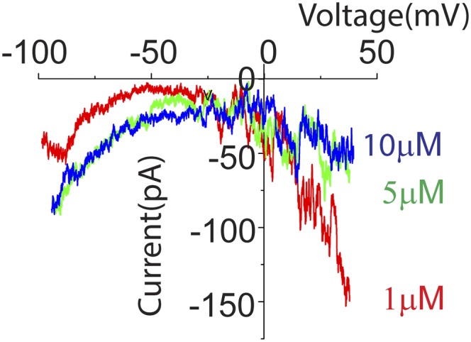
In the mammalian cochlea, acoustic information is carried to the brain by the predominant (95%) large-diameter, myelinated type I afferents, each of which is postsynaptic to a single inner hair cell. The remaining thin, unmyelinated type II afferents extend hundreds of microns along the cochlear duct to contact many outer hair cells. Despite this extensive arbor, type II afferents are weakly activated by outer hair cell transmitter release and are insensitive to sound. Intriguingly, type II afferents remain intact in damaged regions of the cochlea. Here, we show that type II afferents are activated when outer hair cells are damaged. This response depends on both ionotropic (P2X) and metabotropic (P2Y) purinergic receptors, binding ATP released from nearby supporting cells in response to hair cell damage. Selective activation of P2Y receptors increased type II afferent excitability by the closure of KCNQ-type potassium channels, a potential mechanism for the painful hypersensitivity (that we term "noxacusis" to distinguish from hyperacusis without pain) that can accompany hearing loss. Exposure to the KCNQ channel activator retigabine suppressed the type II fiber's response to hair cell damage. Type II afferents may be the cochlea's nociceptors, prompting avoidance of further damage to the irreparable inner ear.

Keywords: ATP; acoustic trauma; hyperacusis; noxacusis; type II cochlear afferents.


Conflict of interest statement

The authors declare no conflict of interest.

Figures

Fig. 1.
Fig. 1.
The experimental preparation. (A) Whole mount of the apical turn of a P8 rat cochlea (apex to the Left), type II afferent filled with biocytin. Typical recording site (arrow) and OHC damage site were within the branched synaptic input zone. (Scale bar: 250 µm.) (B) Electrode (Right) recording from a type II afferent (out of focus) under OHC rows (stars). The glass needle (arrow) poised to ablate one to three OHCs per trial. (C) Two FM1-43–loaded OHCs [arrowheads (Ci)] were ruptured and lost fluorescence (Cii).
Fig. 2.
Fig. 2.
ATP contributes to cell damage-induced response. (A) OHC ablation depolarized type II afferents and triggered action potentials on the rising phase (Inset). (B) Representative traces of damage-induced currents (at −70 mV) from five different type II afferents (gray) and the average (black). Identical needle movement that failed to ablate OHCs induced no current (magenta). (C) Average ablation-induced current in PPADS (red, three trials in three afferents) compared with average control current from B. (D) Average ablation-induced current in CBX (blue, five trials in three afferents) compared with control average.
Fig. S1.
Fig. S1.
Damage-induced responses of type II afferents were insensitive to block of glutamate receptors. (A) Loose-patch extracellular recording of action potentials from a type II afferent following OHC ablation (at arrow). (B) A mixture of glutamate receptor blockers, including 50 µM APV, 50 µM CNQX, and 1 mM MCPG to block NMDA, AMPA/kainate, and metabotropic glutamate receptors had no effect (n = 4 afferents). (C) Type II response to OHC ablation after removal of glutamate receptor blockers. This burst of action potentials occurred <1 s after OHC ablation, during the peak depolarization of the type II fiber (Fig. 2A). Similar to the early inward current seen in voltage-clamp recording, this damaged-induced burst of action potentials also was unaffected by the purinergic antagonist PPADS (n = 2 afferents).
Fig. S2.
Fig. S2.
Potassium ions contribute to the peak damage-induced current. (A) External potassium was elevated from 5.8 to 40 mM by substitution with sodium, shifting the potassium equilibrium (EK) potential from −80 to −31 mV. Thus, the effect of any additional potassium released by hair cell damage will be reduced in this condition. In normal saline, hair cell rupture might expose nearby tissue to ∼150 mM K+, producing an inward current due to the change in EK from −80 to 0 mV. In the presence of 40 mM potassium, the same bolus of high potassium will change the driving force from −31 to 0 mV. Assuming the induced inward current is purely due to K+, the evoked current should be 2.6-fold smaller. The peak amplitude of the fast component was reversibly decreased 1.7-fold (from 94.1 ± 14.1 to 54.5 ± 5.9 pA; three experiments in three afferents) by prior exposure to 40 mM K+. Given that the actual changes in potassium concentration are unknown, this result supports the suggestion that the early inward current is carried at least in part by potassium ions. (B) Superfusion with “internal solution” (150 mM K-methanesulfonate, buffered calcium, no ATP) induced large inward currents in type II afferents. In 10 trials in three afferents, the maximum response induced by puffing internal solution averaged 97.4 ± 6.7 pA, not significantly different from the response induced by OHC ablation (P = 0.103), suggesting potassium release is able to depolarize type II afferents.
Fig. 3.
Fig. 3.
Purinergic receptors in type II afferents. (A) ATP-evoked inward current could be blocked by the P2X antagonist PPADS (red), with partial recovery (blue). (B) UTP (agonist for P2Y2 and P2Y4 receptor) induced inward current in type II afferents. Excitatory postsynaptic currents were recorded, but their frequency was not significantly increased during UTP application. (C) Overlay of responses from a type II afferent when the holding potential was ramped from −90 to +30 mV in normal external solution (black), or after ATP was applied (red). (Inset) Difference current during voltage ramp with and without ATP application. (D) Overlay of responses from a type II afferent when the holding potential was ramped from −110 to +30 mV in normal external solution (black), or after UTP was applied (blue). (Inset) Difference current during voltage ramp with and without UTP application. (E and F) I–V relation of ATP and UTP response revealed by voltage-ramp recordings. The current evoked by ATP reversed at 0 mV (red; n = 3 afferents) and reversal of UTP-evoked current extrapolated to near −70 mV (blue; n = 6 afferents).
Fig. 4.
Fig. 4.
KCNQ channels regulate type II afferent excitability. (A) XE-991 (KCNQ blocker) reversibly eliminated the UTP-evoked ramp current. I–V relation of UTP before (black), during (red), and after (gray) XE-991 application. (B) Retigabine (KCNQ opener) induced outward current at −60 mV in type II cochlear afferents. (C) Current step protocol (1pA steps) to evoke action potentials (Ci). In UTP, from the same resting membrane potential, action potentials were evoked by smaller current steps (Cii). (D) The KCNQ channel activator retigabine reversibly prevented the response of type II afferents to OHC ablation (n = 5 afferents).
Fig. S3.
Fig. S3.
Low concentration of ATP preferentially activates “P2Y-like” ramp currents. A voltage-ramp protocol (as in Fig. 3) was used to examine membrane currents evoked in type II fibers by lower concentrations of ATP (1, 5, and 10 µM) that might differentially activate lower (P2X)- and higher-affinity (P2Y) receptors. As ATP concentration was decreased, the evoked ramp current changed from “P2X-like” to P2Y-like. With the lowest concentration tested (1 µM), the ATP-dependent current was inward at positive voltages (i.e., reduced outward current), suggesting that P2Y receptors may be preferentially activated. With increasing concentration, the ATP-evoked current became more inward at negative voltages, and less inward at positive voltages, suggesting the P2X response (increased cation conductance) overwhelms the P2Y response in higher concentrations of ATP (two experiments in two cells).

Similar articles

Cited by

References

    1. Wong AC, Ryan AF. Mechanisms of sensorineural cell damage, death and survival in the cochlea. Front Aging Neurosci. 2015;7:58. - PMC - PubMed
    1. Brask T. The noise protection effect of the stapedius reflex. Acta Otolaryngol Suppl. 1979;360:116–117. - PubMed
    1. Guinan JJ., Jr Olivocochlear efferents: Anatomy, physiology, function, and the measurement of efferent effects in humans. Ear Hear. 2006;27(6):589–607. - PubMed
    1. Dauman R, Bouscau-Faure F. Assessment and amelioration of hyperacusis in tinnitus patients. Acta Otolaryngol. 2005;125(5):503–509. - PubMed
    1. Hébert S, Fournier P, Noreña A. The auditory sensitivity is increased in tinnitus ears. J Neurosci. 2013;33(6):2356–2364. - PMC - PubMed

Publication types

MeSH terms