. 2015 Nov 24;112(47):E6466-75.
doi: 10.1073/pnas.1508760112. Epub 2015 Nov 9.

HAX-1 regulates cyclophilin-D levels and mitochondria permeability transition pore in the heart

Affiliations

HAX-1 regulates cyclophilin-D levels and mitochondria permeability transition pore in the heart

Chi Keung Lam et al. Proc Natl Acad Sci U S A. .

Abstract

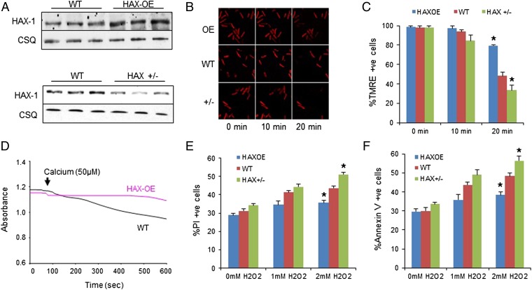
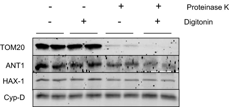
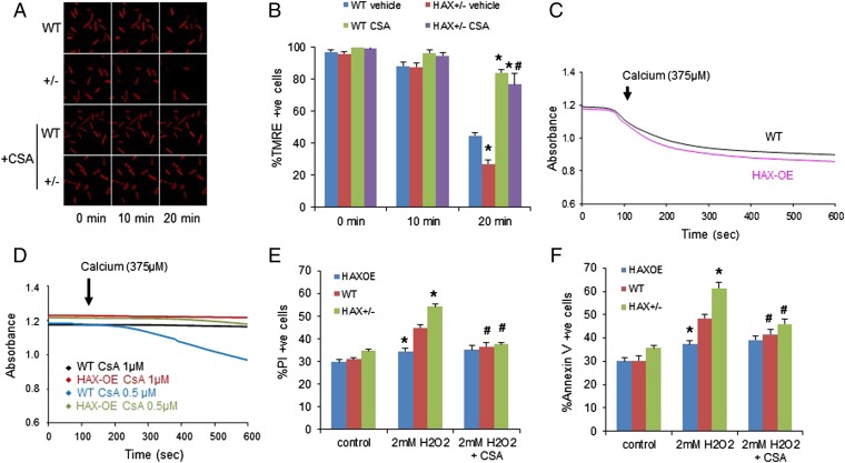
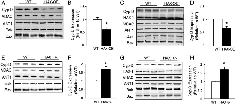
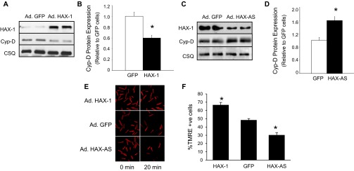
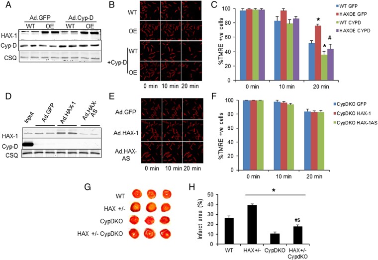
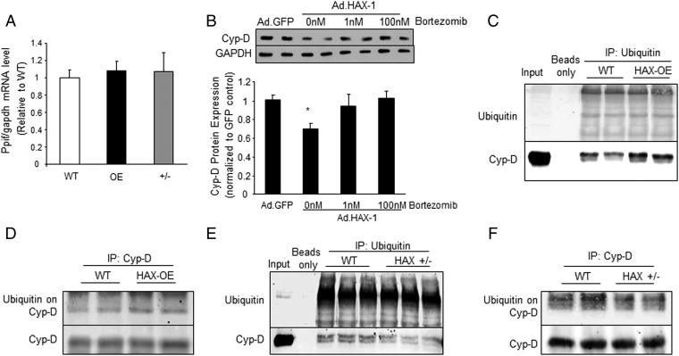
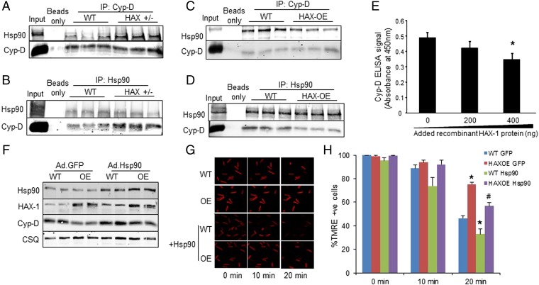
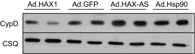
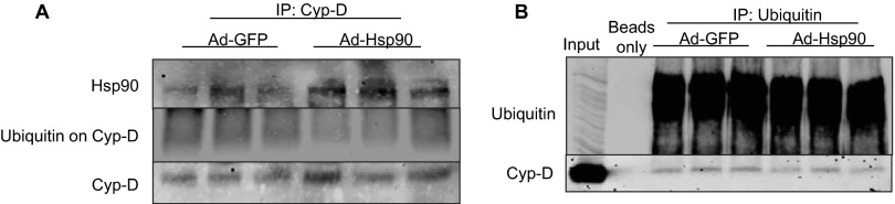
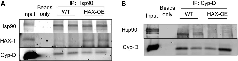
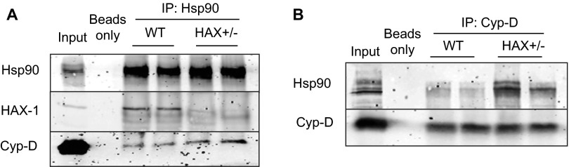
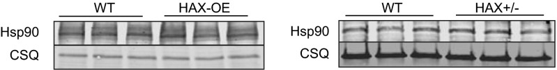
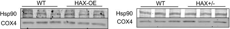
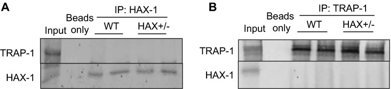
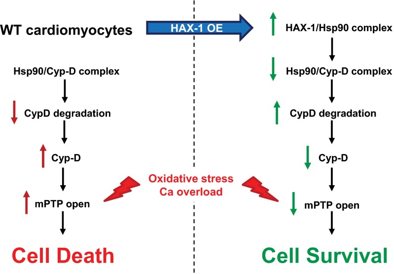
The major underpinning of massive cell death associated with myocardial infarction involves opening of the mitochondrial permeability transition pore (mPTP), resulting in disruption of mitochondria membrane integrity and programmed necrosis. Studies in human lymphocytes suggested that the hematopoietic-substrate-1 associated protein X-1 (HAX-1) is linked to regulation of mitochondrial membrane function, but its role in controlling mPTP activity remains obscure. Herein we used models with altered HAX-1 expression levels in the heart and uncovered an unexpected role of HAX-1 in regulation of mPTP and cardiomyocyte survival. Cardiac-specific HAX-1 overexpression was associated with resistance against loss of mitochondrial membrane potential, induced by oxidative stress, whereas HAX-1 heterozygous deficiency exacerbated vulnerability. The protective effects of HAX-1 were attributed to specific down-regulation of cyclophilin-D levels leading to reduction in mPTP activation. Accordingly, cyclophilin-D and mPTP were increased in heterozygous hearts, but genetic ablation of cyclophilin-D in these hearts significantly alleviated their susceptibility to ischemia/reperfusion injury. Mechanistically, alterations in cyclophilin-D levels by HAX-1 were contributed by the ubiquitin-proteosomal degradation pathway. HAX-1 overexpression enhanced cyclophilin-D ubiquitination, whereas proteosomal inhibition restored cyclophilin-D levels. The regulatory effects of HAX-1 were mediated through interference of cyclophilin-D binding to heat shock protein-90 (Hsp90) in mitochondria, rendering it susceptible to degradation. Accordingly, enhanced Hsp90 expression in HAX-1 overexpressing cardiomyocytes increased cyclophilin-D levels, as well as mPTP activation upon oxidative stress. Taken together, our findings reveal the role of HAX-1 in regulating cyclophilin-D levels via an Hsp90-dependent mechanism, resulting in protection against activation of mPTP and subsequent cell death responses.

Keywords: HAX-1; cyclophilin-D; heat shock protein-90; mitochondrial permeability transition; necrosis.


Conflict of interest statement

The authors declare no conflict of interest.

Figures

Fig. 1.
Fig. 1.
Overexpression of HAX-1 protects mitochondrial membrane integrity against oxidative stress and calcium overload, whereas heterozygous ablation of HAX-1 has opposite effects. (A) HAX-1 protein expression in HAX-OE and HAX+/− hearts. CSQ was used as loading control. (B and C) Isolated WT, HAX-OE, and HAX+/− cardiomyocytes were loaded with mitochondrial membrane potential fluorescent dye, TMRE. Upon 2 mM H2O2 administration for 20 min, membrane potential was diminished in all groups, but this effect was most pronounced in the heterozygous cells. HAX-OE exhibited more than 80% preserved TMRE signals. n = 7 hearts for HAX-OE, 16 hearts for WT, and 9 hearts for HAX+/− (>8 cells per heart); *P < 0.05 vs. WT at 20 min. (D) HAX-1 overexpressing mitochondria demonstrated resistance to swelling induced by 50 μM calcium. n = 3 hearts for each group. (E and F) HAX-1 overexpression in cardiomyocytes prevented cell death due to loss of plasma membrane integrity (E) and apoptosis (F) after 20 min of 2 mM H2O2 treatment, whereas HAX-1 heterozygous ablation increased cell death. n = 8 hearts for HAX-OE, 12 hearts for WT, and 13 hearts for HAX+/− (>10,000 cells per heart); *P < 0.05 vs. WT at 2 mM H2O2. Data are expressed as mean ± SEM.
Fig. S1.
Fig. S1.
The purity of the mitochondrial fraction. Based on the use of HDAC5, GAPDH, and COX4 as nuclear, cytosolic and mitochondrial markers, respectively, the purity of mitochondrial fraction appeared high.
Fig. S2.
Fig. S2.
HAX-1 is localized to mitochondria. Isolated WT mouse mitochondria were subjected to protein digestion by proteinase K in the presence or absence of the outer membrane disruptor, digitonin. Translocase of the outer membrane 20 Homolog Type (TOM20), adenine nucleotide translocase 1 (ANT1), and cyclophilin-D (Cyp-D) were used as markers for mitochondrial outer membrane, inner membrane, and matrix, respectively. Proteinase K can only digest proteins that are accessible and not protected by the mitochondrial membrane. HAX-1 was not digested by proteinase K when the outer membrane was disrupted by digitonin, suggesting that it is localized to the inner membrane of mitochondria.
Fig. 2.
Fig. 2.
HAX-1 regulates the mitochondrial permeability transition pore. (A and B) Cyclosporine A abrogated the detrimental effect of HAX-1 heterozygous ablation on mitochondrial membrane potential after H2O2 treatment for 20 min. n = 4–8 hearts (>12 cells per heart); *P < 0.05 vs. WT with vehicle (DMSO) at 20 min; #P < 0.05 vs. HAX+/− with vehicle at 20 min. (C) Swelling induced by high calcium concentration in HAX-OE and WT mitochondria. n = 3 hearts for each group. (D) Inhibition of mitochondrial swelling required a lower cyclosporine A dose in HAX-1 overexpressing mitochondria. n = 3 hearts for each group. (E and F) Cyclosporine A abrogated the effect of HAX-1 on necrotic (E) and apoptotic (F) cell death. n = 8–15 hearts (>10,000 cells per heart); *P < 0.05 vs. WT at 2 mM H2O2; #P < 0.05 vs. the respective untreated group at 2 mM H2O2. Data are expressed as mean ± SEM.
Fig. 3.
Fig. 3.
HAX-1 specifically alters cyclophilin-D levels in the heart. (A and B) Cyp-D levels were reduced in HAX-OE cardiac homogenates. n = 4 hearts for each group. (C and D) Cyp-D levels were reduced in HAX-OE cardiac mitochondrial fraction. n = 5 hearts for HAX-OE, 6 hearts for WT. (E and F) Cyp-D levels were increased in HAX +/− cardiac homogenates. n = 6 hearts for each group. (G and H) Cyp-D levels were increased in the HAX+/− mitochondrial fraction. n = 9 hearts for each group. *P < 0.05 vs. WT. Data are expressed as mean ± SEM.
Fig. S3.
Fig. S3.
HAX-1 alters cyclophilin-D levels in infected cardiomyocytes. (A and B) Cyclophilin-D levels were reduced when HAX-1 was acutely overexpressed in rat cardiomyocytes. n = 4; *P < 0.05 vs. GFP. (C and D) Cyclophilin-D levels were increased when HAX-1 was acutely down-regulated in rat cardiomyocytes. n = 4; *P < 0.05 vs. GFP. (E and F) Mitochondrial membrane potential was protected with increased HAX-1 expression levels. n = 6; *P < 0.05 vs. GFP.
Fig. 4.
Fig. 4.
The effects of HAX-1 are mainly mediated by cyclophilin-D. (A) Cyp-D expression was increased to similar levels in WT and HAX-OE cells after adenoviral infection of adult rat cardiomyocytes. (B and C) Adenoviral delivery of Cyp-D removed the protection mediated by HAX-1 overexpression upon 2 mM hydrogen peroxide administration. CSQ was used as a loading control. n = 4–6 hearts (>12 cells per heart); *P < 0.05 vs. WT GFP at 2 min; #P < 0.05 vs. HAX-OE GFP at 20 min. (D) HAX-1 levels after adenoviral delivery of Ad.GFP, Ad.HAX-1, and Ad.HAX-AS in CypD-KO cells. WT cardiac homogenate was loaded to serve as input. (E and F) Cyp-D ablation abolished the effect of different HAX-1 levels on the maintenance of mitochondrial membrane potential upon 2 mM hydrogen peroxide challenge. CSQ was used as a loading control. n = 4–5 hearts (>16 cells per heart). (G and H) Cyp-D ablation significantly reduced infarct size in HAX-1 heterozygous knockout hearts. n = 4 for each group; *P < 0.05 vs. WT; #P < 0.05 vs. HAX+/−; $P < 0.05 vs. CypD-KO. Data are expressed as mean ± SEM.
Fig. 5.
Fig. 5.
HAX-1 enhances the degradation of cyclophilin-D. (A) Cyp-D mRNA transcript, or ppif levels were similar in WT, HAX-OE, and HAX+/− hearts. n = 4 hearts for WT, and 5 hearts for each of the HAX-OE and HAX+/−. (B) Proteasomal inhibitor, Bortezomib, restored Cyp-D levels in HAX-1 overexpressing cells. Glyceraldehyde 3-phosphate dehydrogenase (GAPDH) was used as a loading control. n = 4 hearts for each group; *P < 0.05 vs. Ad.GFP. (C and D) Ubiquitination of Cyp-D was enhanced in HAX-OE hearts. Three independent experiments were performed. n = 4 hearts for each group. (E and F) Ubiquitination of Cyp-D was reduced in HAX+/− hearts. Three independent experiments were performed. n = 4 hearts for each group. Data are expressed as mean ± SEM.
Fig. 6.
Fig. 6.
HAX-1 displaces Hsp90 from cyclophilin-D, rendering it susceptible to degradation. (A and B) Immunoprecipitations using Cyp-D (A) or Hsp90 (B) as a bait, demonstrated that the Cyp-D/Hsp90 association was enhanced in HAX+/− hearts. WT cardiac homogenate served as input. (C and D) Immunoprecipitations, using Cyp-D (C) or Hsp90 (D) as a bait, demonstrated that the Cyp-D/Hsp90 association was reduced in HAX-OE hearts. WT cardiac homogenate served as input. (E) The Cyp-D/Hsp90 interaction was examined in the presence of increasing levels of HAX-1, using recombinant proteins. ELISA signals were calculated after subtraction of the mean value of controls, represented by GST-coated wells (n = 25 wells). (F) Increases in Hsp90 levels by adenoviral infection of cultured adult WT and HAX-OE cardiomyocytes enhanced Cyp-D protein expression. CSQ was used as a loading control. (G and H) Hsp90 overexpression exacerbated the loss of mitochondrial membrane potential in both WT and HAX-OE cells. n = 4–7 hearts (>10 cells per heart); *P < 0.05 vs. WT GFP at 20 min; #P < 0.05 vs. HAX-OE GFP at 20 min. Data are expressed as mean ± SEM.
Fig. S4.
Fig. S4.
Adenoviral delivery of Hsp90 results in increased Cyp-D levels in rat cardiomyocytes. Rat cardiomyocytes were isolated, cultured and infected with Ad.HAX-1, Ad.GFP, Ad.HAX-AS (HAX-1 antisense), or Ad.Hsp90 for 24 h. CypD levels were increased in Ad.Hsp90 cells to a comparable level as in Ad.HAX-AS cells.
Fig. S5.
Fig. S5.
Adenoviral delivery of Hsp90 results in reduced Cyp-D ubiquitination in rat cardiomyocytes. Rat cardiomyocytes were isolated, cultured, and infected with Ad.GFP or Ad.Hsp90 for 24 h. Cells were then collected and lysed for immunoprecipitation experiments. Using either Cyp-D (A) or Ubiquitin (B) as bait, ubiquitination of Cyp-D was reduced when Hsp90 levels were enhanced.
Fig. S6.
Fig. S6.
HAX-1 overexpression reduced mitochondrial Hsp90/Cyp-D association. Using either Hsp90 (A) or Cyp-D (B) as bait, a reduction of Hsp90/Cyp-D binding was observed in HAX-OE mitochondrial fraction.
Fig. S7.
Fig. S7.
Heterozygous ablation of HAX-1 enhanced mitochondrial Hsp90/Cyp-D association. Using either Hsp90 (A) or Cyp-D (B) as bait, an increase of Hsp90/Cyp-D binding was observed in HAX+/− mitochondrial fraction.
Fig. S8.
Fig. S8.
Expression of Hsp90 in cardiac homogenates. Hsp90 expression was not altered by overexpression or heterozygous deficiency of HAX-1 in cardiac homogenates.
Fig. S9.
Fig. S9.
Expression of Hsp90 in cardiac mitochondrial fraction. Hsp90 expression was not altered by HAX-1 overexpression or heterozygous deficiency in cardiac mitochondrial fraction.
Fig. S10.
Fig. S10.
HAX-1 does not appear to associate with TRAP-1 in mouse cardiac homogenates. Using either HAX-1 (A) or TRAP-1 (B) as bait, we did not detect HAX-1/TRAP-1 association in cardiac homogenate.
Fig. S11.
Fig. S11.
Proposed HAX-1 protective mechanism against the opening of mitochondrial permeability transition pore in cardiomyocytes.

Similar articles

Cited by

References

    1. Carley AN, Taegtmeyer H, Lewandowski ED. Matrix revisited: Mechanisms linking energy substrate metabolism to the function of the heart. Circ Res. 2014;114(4):717–729. - PMC - PubMed
    1. Chen L, Knowlton AA. Mitochondria and heart failure: New insights into an energetic problem. Minerva Cardioangiol. 2010;58(2):213–229. - PMC - PubMed
    1. Galluzzi L, Kepp O, Trojel-Hansen C, Kroemer G. Mitochondrial control of cellular life, stress, and death. Circ Res. 2012;111(9):1198–1207. - PubMed
    1. Chiong M, et al. Cardiomyocyte death: Mechanisms and translational implications. Cell Death Dis. 2011;2:e244. - PMC - PubMed
    1. Murphy E, Steenbergen C. Mechanisms underlying acute protection from cardiac ischemia-reperfusion injury. Physiol Rev. 2008;88(2):581–609. - PMC - PubMed

MeSH terms

LinkOut - more resources