. 2015 Nov 24;112(47):E6446-55.
doi: 10.1073/pnas.1512088112. Epub 2015 Nov 9.

Global shape mimicry of tRNA within a viral internal ribosome entry site mediates translational reading frame selection

Affiliations

Global shape mimicry of tRNA within a viral internal ribosome entry site mediates translational reading frame selection

Hilda H Au et al. Proc Natl Acad Sci U S A. .

Abstract

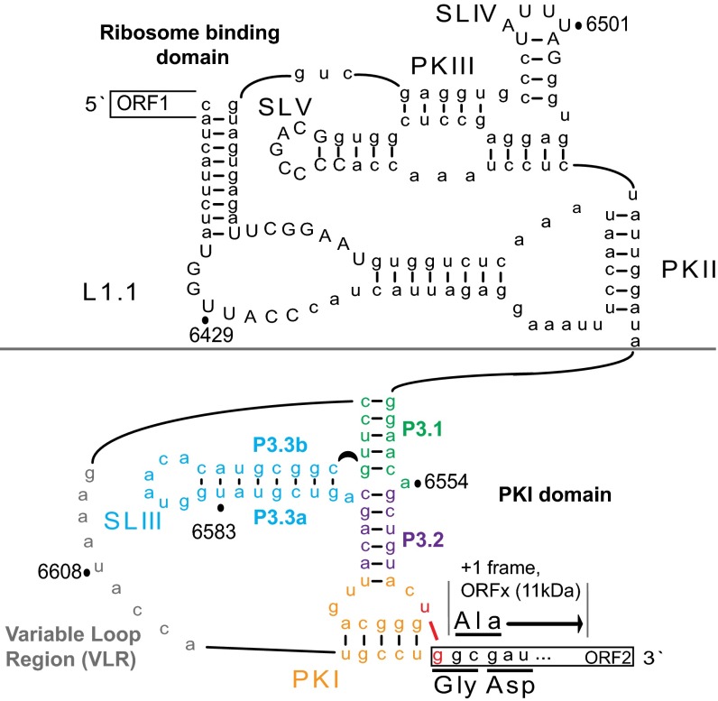
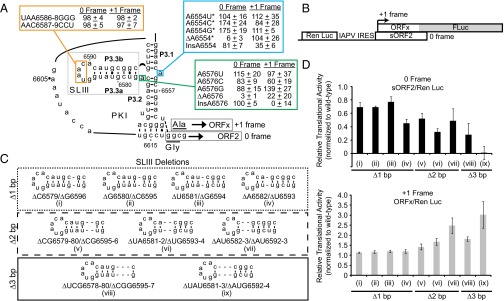
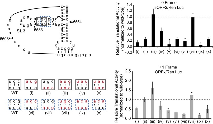
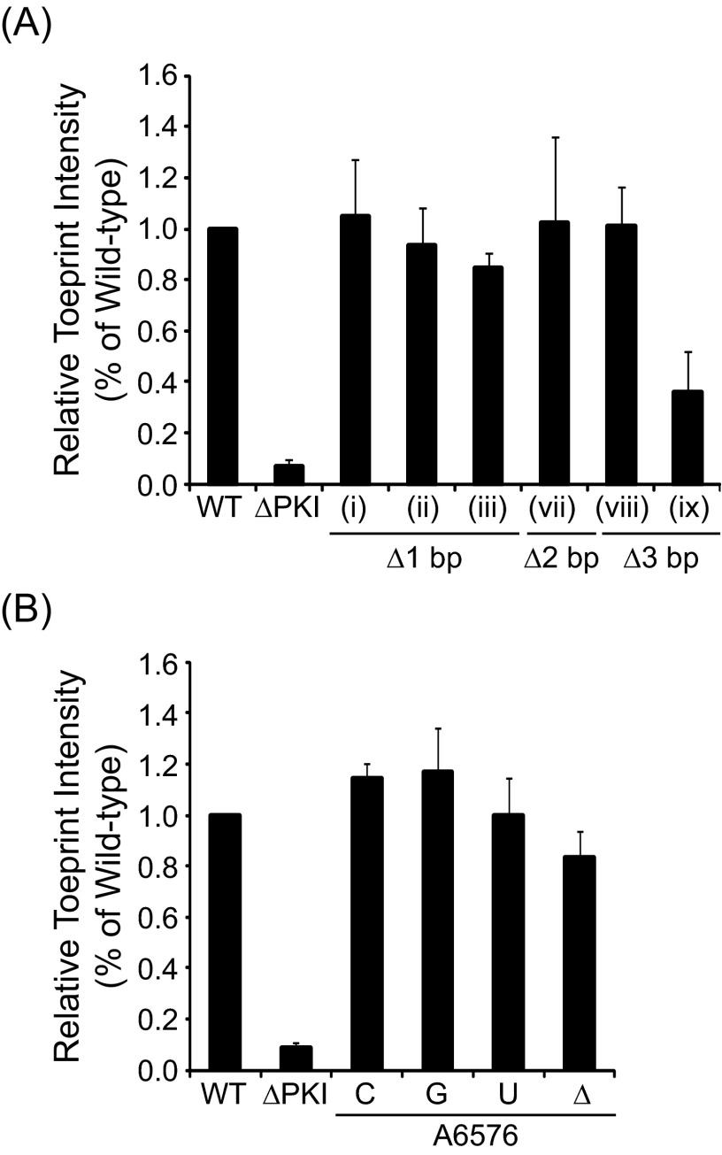
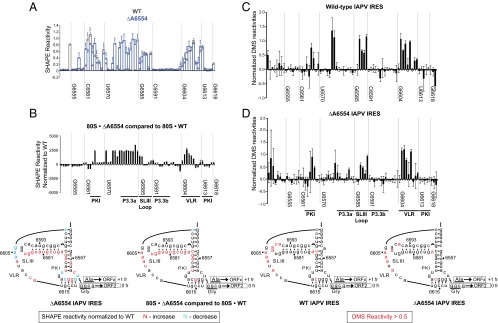
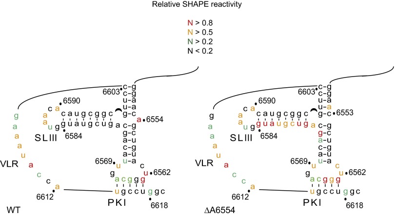
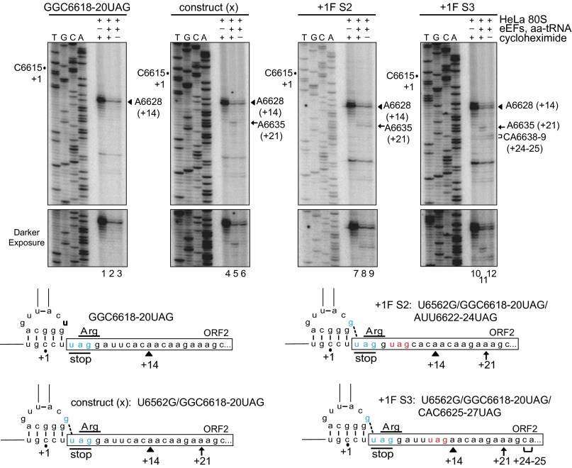
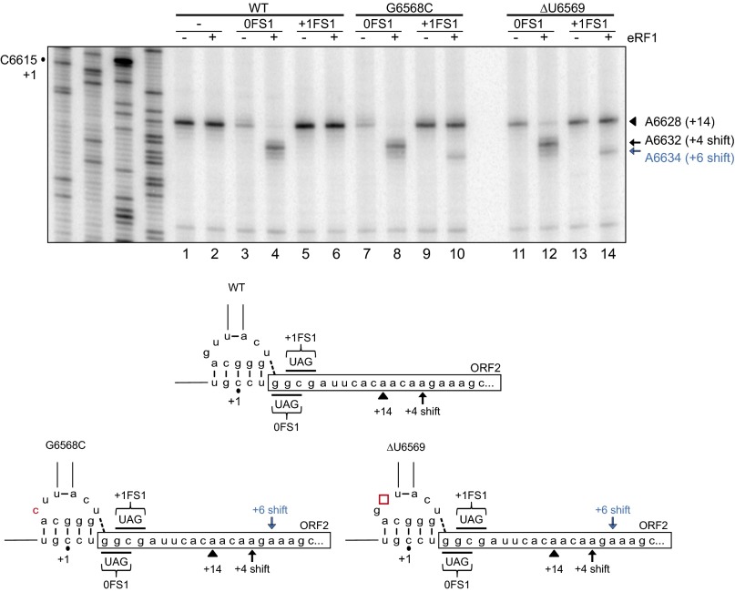
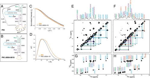
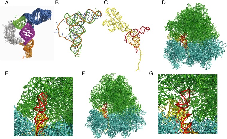
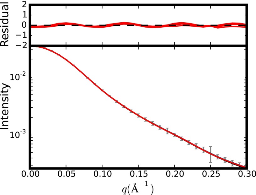
The dicistrovirus intergenic region internal ribosome entry site (IRES) adopts a triple-pseudoknotted RNA structure and occupies the core ribosomal E, P, and A sites to directly recruit the ribosome and initiate translation at a non-AUG codon. A subset of dicistrovirus IRESs directs translation in the 0 and +1 frames to produce the viral structural proteins and a +1 overlapping open reading frame called ORFx, respectively. Here we show that specific mutations of two unpaired adenosines located at the core of the three-helical junction of the honey bee dicistrovirus Israeli acute paralysis virus (IAPV) IRES PKI domain can uncouple 0 and +1 frame translation, suggesting that the structure adopts distinct conformations that contribute to 0 or +1 frame translation. Using a reconstituted translation system, we show that ribosomes assembled on mutant IRESs that direct exclusive 0 or +1 frame translation lack reading frame fidelity. Finally, a nuclear magnetic resonance/small-angle X-ray scattering hybrid approach reveals that the PKI domain of the IAPV IRES adopts an RNA structure that resembles a complete tRNA. The tRNA shape-mimicry enables the viral IRES to gain access to the ribosome tRNA-binding sites and form intermolecular contacts with the ribosome that are necessary for initiating IRES translation in a specific reading frame.

Keywords: RNA; internal ribosome entry site; ribosome; translation; virus.


Conflict of interest statement

The authors declare no conflict of interest.

Figures

Fig. 1.
Fig. 1.
Secondary structure of the IAPV IGR IRES. Pseudoknots PKI, PKII, and PKIII; stem loops SLIII, SLIV, and SLV and loop L1.1; and the variable loop region (VLR) are indicated. The PKI domain comprises a three-way junction involving helices P3.1 (green), P3.2 (purple), and P3.3 (blue). The IAPV IRES can mediate translation of ORFx in the +1 reading frame, which overlaps the viral structural protein coding region. IRES-mediated translation in the 0 and +1 frames starts from the GGC glycine and GCG alanine codons, respectively. Translation of the +1 frame ORFx is directed by a U6562/G6618 base pair adjacent to PKI (red nucleotides). Conserved nucleotides within the type II IGR IRESs are shown in capital letters.
Fig. 2.
Fig. 2.
Translational activities of IAPV IRES PKI mutants. (A) Summary of translational activities of WT and mutant IRESs. Translational activities are normalized to the WT IRES, which is set to 100% for both the 0 and +1 frames. For the WT IRES, the +1 frame translation is ∼20% of the 0 frame translation in vitro. Shown are the average ± 1 SD values from at least three independent experiments. *Data from Ren et al. (26). (B) Bicistronic reporter construct. The bicistronic reporter contains an upstream Renilla luciferase reporter (Ren Luc) and a downstream firefly luciferase reporter (FLuc), which are expressed by cap-dependent and IRES-dependent translation, respectively. FLuc is fused in the +1 reading frame, downstream of the ORFx coding sequence to generate a full-length protein of 76 kDa. The 0 frame translation results in a truncated protein (sORF2) of 14 kDa. (C) Schematics of IRES mutants harboring systematic one-bp (i–iv), two-bp (v–vii), or three-bp (viii–ix) deletions at various positions along helix P3.3 of SLIII. (D) Relative translational activities of helix P3.3 deletion mutants, expressed as a ratio of 0 frame translation to Ren Luc expression (Top) or as a ratio of +1 frame translation to Ren Luc expression (Bottom).
Fig. S1.
Fig. S1.
Translational activities of helix P3.3 mutants. Mutations are introduced at two positions along helix P3.3 of SLIII, as indicated on the PKI secondary structure. Specific mutations at each position are denoted by boxes in the corresponding color, with mutated bases shown in red. Relative translational activities of the helix P3.3 mutants are expressed as a ratio of 0 frame translation (sORF2) to Ren Luc expression, or as a ratio of +1 frame translation (ORFx) to Ren Luc expression, normalized to the WT IRES, set as 100% for both reading frames.
Fig. 3.
Fig. 3.
Toeprinting/primer extension analysis. Toeprinting analysis of IAPV IRES/ribosome complexes for helix P3.3 deletion mutations as depicted in Fig. 2C (A) and A6576 mutants (B). Bicistronic RNAs harboring the WT or mutant IRESs were incubated alone (−) or with salt-washed HeLa ribosomes (+) and analyzed by primer extension. The sequencing reactions of the WT IRES are shown on the left, with the position of the +1 nucleotide indicated for reference. The position of the observed toeprint is as denoted.
Fig. S2.
Fig. S2.
Quantitation of the relative toeprint intensities in Fig. 3 for helix P3.3 (A) and A6576 mutants (B), normalized to the WT IRES. Shown are the average ± SD values from at least three independent experiments.
Fig. 4.
Fig. 4.
Structural probing analyses of WT and ΔA6554 IRESs. (A and B) SHAPE modification profiles of WT and ΔA6554 IRESs in solution (A) and bound to the ribosome (B). (C and D) DMS modification profiles of the WT (C) and ΔA6554 (D) IRESs in solution. Normalized reactivities are shown as a function of the nucleotide position. The difference in normalized SHAPE reactivities between the mutant and WT IRESs or the normalized DMS reactivities are summarized on the secondary structure according to the legend indicated (Bottom). Specific nucleotide positions are indicated for reference, and major IRES structural elements are denoted.
Fig. S3.
Fig. S3.
SHAPE analysis of the WT and ΔA6554 IRESs in solution. Normalized SHAPE reactivies for each IRES RNA are summarized on the corresponding secondary structure according to the legend indicated.
Fig. 5.
Fig. 5.
Reconstitution of IRES-mediated translation. (Top) Bicistronic IRES RNAs were incubated with purified, salt-washed human ribosomes in the presence or absence of yeast elongation factors, bulk aminoacyl-tRNAs, and the translation inhibitor cycloheximide, as indicated. Ribosome positioning was monitored by primer extension analysis, and the resultant cDNA products were resolved by denaturing PAGE. Sequencing reactions are shown on the left, with the position of the +1 nucleotide as denoted. The locations of major toeprints including A6628 (+14), A6635 (+21), and CA6638-9 (+24–25), are indicated on the right. (Bottom) Schematics of IRES mutants with the locations of the major toeprints shown.
Fig. S4.
Fig. S4.
Reconstitution of translocation using eRF1. (Top) Bicistronic IRES RNAs were incubated with purified human ribosomes, GTP, and eukaryotic elongation factor 2, in the presence or absence of eRF1, as indicated. Ribosome positioning was monitored by primer extension analysis, and the resultant cDNA products were analyzed by denaturing polyacrylamide gel electrophoresis. Sequencing reactions are shown on the left, with the position of the +1 nucleotide as denoted. The locations of the major toeprints, including A6628 (+14), A6632 (+4 shift), and A6634 (+6 shift), are indicated. (Bottom) Schematics of IRES mutants and locations of the major toeprints.
Fig. 6.
Fig. 6.
SAXS and NMR analyses of the IAPV IGR IRES PKI domain. (A and B) Secondary structures of the WT IAPV IRES PKI domain (A) and PKIΔ6604–6618 (B). (C and D) SAXS profile (C) and pair distance distribution function plot (D) of the WT and Δ6604–6618 IAPV IRES PKI domains. (E and F) One-dimensional 1H spectrum and 2D 1H-1H NOESY of the Δ6604–6618 (E) and WT (F) IAPV IRES PKI domains in 20 mM potassium phosphate (pH 6.3), 200 mM KCl, and 0.5 μM EDTA. (G and H) 1H and 15N imino chemical shift assignments for Δ6604–6618 (G) and WT (H) IAPV IRES PKI domains. Assignments and connecting lines are color-coded according to secondary structure, as in A and B. Base pairs confirmed by 2D 1H-1H NOESY are indicated in A and B by black lines or circles, and base pairs inferred by chemical shift agreement are indicated with gray lines.
Fig. 7.
Fig. 7.
Structural model of the IAPV PKI domain. (A) Structural ensemble of the IAPV IRES, as determined by NMR/SAXS. The structural elements are colored as in Fig. 1. (B) Averaged structure of the IAPV PKI domain (orange) overlaid onto the structure of a Phe-tRNA (green). SLIII mimics the tRNA acceptor stem. (C) The IAPV PKI domain (red) superimposed onto the structure of the CrPV IGR IRES in the posttranslocated state (yellow) [Protein Data Bank (PDB) ID code 4D5Y)] (23). (D) The IAPV PKI domain (red) superimposed onto the structure of the CrPV IGR IRES bound in the A site of the yeast ribosome (PDB ID code 4V91) (20). The CrPV IRES (yellow), large ribosomal subunit RNA (green), and small ribosomal subunit RNA (cyan) are shown. (E) Zoom-in view of D, showing that SLIII (red) can be accommodated by occupying the space within the large ribosomal subunit normally occupied by the acceptor stem of a ribosomal A site tRNA (23). (F) The IAPV PKI domain (red) superimposed onto the structure of the CrPV IGR IRES bound in the P site of the O. cuniculus ribosome (PDB ID code 4V91) (20). (G) Zoom-in view of F.
Fig. S5.
Fig. S5.
Plot of the SAXS data. Shown are the experimental (in gray) and back-calculated data (in red) by X-PLOR-NIH from the 10 lowest-energy NMR structures refined with SAXS and RDCs. (Top) Residuals are given by [I_calc(i) − I_exp(i)]/error(i).

Similar articles

Cited by

References

    1. Dinman JD. Mechanisms and implications of programmed translational frameshifting. Wiley Interdiscip Rev RNA. 2012;3(5):661–673. - PMC - PubMed
    1. Firth AE, Brierley I. Non-canonical translation in RNA viruses. J Gen Virol. 2012;93(Pt 7):1385–1409. - PMC - PubMed
    1. Ren Q, et al. Alternative reading frame selection mediated by a tRNA-like domain of an internal ribosome entry site. Proc Natl Acad Sci USA. 2012;109(11):E630–E639. - PMC - PubMed
    1. Hellen CU, Sarnow P. Internal ribosome entry sites in eukaryotic mRNA molecules. Genes Dev. 2001;15(13):1593–1612. - PubMed
    1. Kieft JS. Viral IRES RNA structures and ribosome interactions. Trends Biochem Sci. 2008;33(6):274–283. - PMC - PubMed

Publication types

Substances

Associated data

LinkOut - more resources