

Unlocking the early fossil record of the arthropod central nervous system
- PMID: 26554038
- PMCID: PMC4650122
- DOI: 10.1098/rstb.2015.0038
Unlocking the early fossil record of the arthropod central nervous system
Abstract
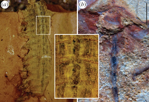
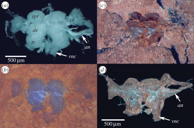
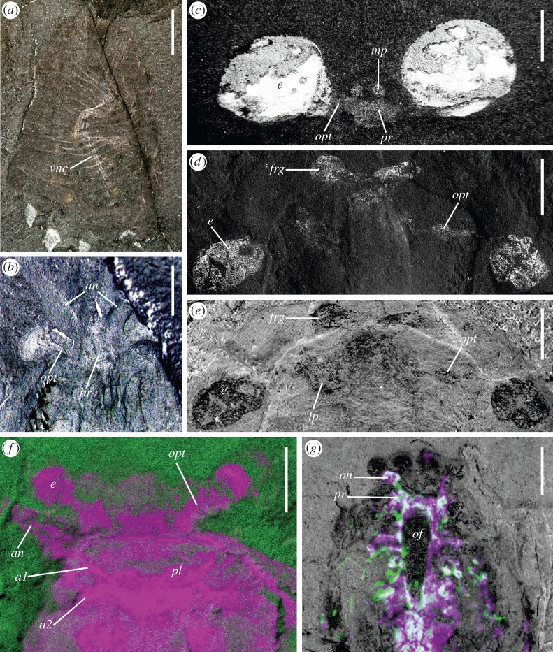
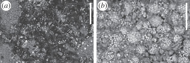
Extant panarthropods (euarthropods, onychophorans and tardigrades) are hallmarked by stunning morphological and taxonomic diversity, but their central nervous systems (CNS) are relatively conserved. The timing of divergences of the ground pattern CNS organization of the major panarthropod clades has been poorly constrained because of a scarcity of data from their early fossil record. Although the CNS has been documented in three-dimensional detail in insects from Cenozoic ambers, it is widely assumed that these tissues are too prone to decay to withstand other styles of fossilization or geologically older preservation. However, Cambrian Burgess Shale-type compressions have emerged as sources of fossilized brains and nerve cords. CNS in these Cambrian fossils are preserved as carbon films or as iron oxides/hydroxides after pyrite in association with carbon. Experiments with carcasses compacted in fine-grained sediment depict preservation of neural tissue for a more prolonged temporal window than anticipated by decay experiments in other media. CNS and compound eye characters in exceptionally preserved Cambrian fossils predict divergences of the mandibulate and chelicerate ground patterns by Cambrian Stage 3 (ca 518 Ma), a dating that is compatible with molecular estimates for these splits.
Keywords: Arthropoda; Burgess Shale; Cambrian; Chengjiang; brains.
© 2015 The Authors.
Figures
Similar articles
-
Preservational Pathways of Corresponding Brains of a Cambrian Euarthropod.Curr Biol. 2015 Nov 16;25(22):2969-75. doi: 10.1016/j.cub.2015.09.063. Epub 2015 Oct 29. Curr Biol. 2015. PMID: 26526373
-
Proclivity of nervous system preservation in Cambrian Burgess Shale-type deposits.Proc Biol Sci. 2019 Dec 18;286(1917):20192370. doi: 10.1098/rspb.2019.2370. Epub 2019 Dec 11. Proc Biol Sci. 2019. PMID: 31822253 Free PMC article.
-
Fossils and the Evolution of the Arthropod Brain.Curr Biol. 2016 Oct 24;26(20):R989-R1000. doi: 10.1016/j.cub.2016.09.012. Curr Biol. 2016. PMID: 27780074 Review.
-
Mandibulate convergence in an armoured Cambrian stem chelicerate.BMC Evol Biol. 2017 Dec 21;17(1):261. doi: 10.1186/s12862-017-1088-7. BMC Evol Biol. 2017. PMID: 29262772 Free PMC article.
-
Origin and evolution of the panarthropod head - A palaeobiological and developmental perspective.Arthropod Struct Dev. 2017 May;46(3):354-379. doi: 10.1016/j.asd.2016.10.011. Epub 2016 Dec 18. Arthropod Struct Dev. 2017. PMID: 27989966 Review.
Cited by
-
Internal anatomy of a fossilized embryonic stage of the Cambrian-Ordovician scalidophoran Markuelia.R Soc Open Sci. 2022 Oct 5;9(10):220115. doi: 10.1098/rsos.220115. eCollection 2022 Oct. R Soc Open Sci. 2022. PMID: 36249341 Free PMC article.
-
Human brains preserve in diverse environments for at least 12 000 years.Proc Biol Sci. 2024 Mar 27;291(2019):20232606. doi: 10.1098/rspb.2023.2606. Epub 2024 Mar 20. Proc Biol Sci. 2024. PMID: 38503334 Free PMC article.
-
Assessing segmental versus non-segmental features in the ventral nervous system of onychophorans (velvet worms).BMC Evol Biol. 2017 Jan 3;17(1):3. doi: 10.1186/s12862-016-0853-3. BMC Evol Biol. 2017. PMID: 28049417 Free PMC article.
-
Preservation of three-dimensional anatomy in phosphatized fossil arthropods enriches evolutionary inference.Elife. 2016 Feb 5;5:e12129. doi: 10.7554/eLife.12129. Elife. 2016. PMID: 26854367 Free PMC article.
-
Early animal evolution and the origins of nervous systems.Philos Trans R Soc Lond B Biol Sci. 2015 Dec 19;370(1684):20150037. doi: 10.1098/rstb.2015.0037. Philos Trans R Soc Lond B Biol Sci. 2015. PMID: 26554037 Free PMC article. Review.
References
Publication types
MeSH terms
LinkOut - more resources
Full Text Sources
Other Literature Sources
