Review
. 2015 Nov;11(11):1859-98.
doi: 10.1166/jbn.2015.2145.

Recent Developments in Active Tumor Targeted Multifunctional Nanoparticles for Combination Chemotherapy in Cancer Treatment and Imaging

Review

Recent Developments in Active Tumor Targeted Multifunctional Nanoparticles for Combination Chemotherapy in Cancer Treatment and Imaging

Micah D K Glasgow et al. J Biomed Nanotechnol. 2015 Nov.

Abstract

Nanotechnology and combination therapy are two major fields that show great promise in the treatment of cancer. The delivery of drugs via nanoparticles helps to improve drug's therapeutic effectiveness while reducing adverse side effects associated wifh high dosage by improving their pharmacokinetics. Taking advantage of molecular markers over-expressing on tumor tissues compared to normal cells, an "active" molecular marker targeted approach would be-beneficial for cancer therapy. These actively targeted nanoparticles would increase drug concentration at the tumor site, improving efficacy while further reducing chemo-resistance. The multidisciplinary approach may help to improve the overall efficacy in cancer therapy. This review article summarizes recent developments of targeted multifunctional nanoparticles in the delivery, of various drugs for a combinational chemotherapy approach to cancer treatment and imaging.


Figures

Figure 1
Figure 1
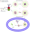
Schematic illustration of nanoscale drug carriers used for combinatorial drug delivery: (a) liposome, (b) polymeric micelle, (c) polymer-drug conjugate, (d) dendrimer, (e) oil nanoemulsion, (f) mesoporous silica nanoparticle, and (g) iron oxide nanoparticle. Reprinted with permission from [98], C. M. Hu and L. Zhang, Nanoparticle-based combination therapy toward overcoming drug resistance in cancer. Biochemical pharmacology 83, 1104 (2012). © 2012, Elsevier.
Figure 2
Figure 2
Schematic representation of SPIO/PTX-loaded PLGA-based nanoparticles. Reprinted with permission from [277], N. Schleich, et al., Dual anticancer drug/superparamagnetic iron oxide-loaded PLGA-based nanoparticles for cancer therapy and magnetic resonance imaging. Int. J. Pharm. 447, 94 (2013). © 2013, Elsevier.
Figure 3
Figure 3
Schematic graph of TPGS functions as a pore former and promotes anti-tumor activity of DTX-loaded Nps. Reprinted with permission from [110], H. Zhu, et al., Co-delivery of chemotherapeutic drugs with vitamin E TPGS by porous PLGA nanoparticles for enhanced chemotherapy against multi-drug resistance. Biomaterials 35, 2391 (2014). © 2014, Elsevier.
Figure 4
Figure 4
Illustration of biodegradable amphiphilic copolymer NPs loaded with both DOX and TAX using improved double emulsion method. Emulsification procedure used to generate double emulsions. Step (I), generating water-in-oil for encapsulations of DOX; Step (II), generating water-in-oil-in-water for encapsulations of TAX. Green represents the oil phase containing amphiphilic copolymer and red the aqueous phase containing DOX. Reprinted with permission from [111], H. Wang, et al., Enhanced anti-tumor efficacy by co-delivery of doxorubicin and paclitaxel with amphiphilic methoxy PEG-PLGA copolymer nanoparticles. Biomaterials 32, 8281 (2011). © 2011, Elsevier.
Figure 5
Figure 5
(A) Examples of bonds utilized in the synthesis of biodegradable polymer-drug conjugates. Biodegradation typically occurs via hydrolysis (via reduction for disulfides). (B) Overall strategy for the synthesis of multiblock polyHPMA copolymers. HPMA copolymer blocks are linked together via lysosomally degradable Gly-Phe-Leu-Gly (GFLG) linkages introduced via a combination of RAFT polymerization and click chemistry. Reprinted with permission from [137], J. Yang, et al., Synthesis of biodegradable multiblock copolymers by click coupling of RAFT-generated heterotelechelicpolyhpma conjugates. Reactive and Functional Polymers 71, 294 (2011). © 2011, Elsevier.
Figure 6
Figure 6
(A) Dendrimers are hyperbranched, star-link polymers. Drugs can be either conjugated to the dendrimer surface or encapsulated within “void” spaces between adjacent branches. (B) Dendrimers grow linearly in size and exponentially in surface area with each successive “generation.” They can be utilized as multifunctional nanocarriers, bearing drugs, imaging agents, and/or targeting moieties. (C) Synthesis of poly(amido amine) (PAMAM) dendrimers occurs from a ethylenediamine core with alternating reactions with methyl acrylate and ethylenediamine to produce each generation. Reprinted with permission from [278], N. Larson and H. Ghandehari, Polymeric conjugates for drug delivery. Chem. Mater. 24, 840 (2012). © 2012, American Chemical Society.
Figure 7
Figure 7
(A) Schematic diagram showing EGFRvIII-IONPs. (B)–(F) Survival studies of nude mice implanted with the U87ΔEGFRvIII glioma model. (B) T2-weighted MRI showing a tumor region with a bright signal 7 days after tumor implantation (arrow). (C) A tumor is shown (arrow) after injection of a gadolinium contrast agent (Gd-DTPA). (D) The MRI signal decreased (arrow) after CED of EGFRvIIIAb-IONPs. (E) EGFRvIIIAb-IONP dispersion and T2 signal decrease (arrow) 4 days after CED. (F) Survival curve of the nude mice bearing U87ΔEGFRvIII cells after a treatment regimen of MRI-guided CED: the untreated control, IONPs, EGFRvIIIAb, or EGFRvIIIAb-IONPs. Reprinted with permission from [194], C. G. Hadjipanayis, et al., EGFRvIII antibody-conjugated iron oxide nanoparticles for magnetic resonance imaging-guided convection-enhanced delivery and targeted therapy of glioblastoma. Cancer Res. 70, 6303 (2010). © 2010, Cancer Research.
Figure 8
Figure 8
(A) Schematic diagram showing the synthesis of MN-EPPT-siBIRC5. (B) Representative pre-contrast images and 24 h post-contrast T2-weighted images (top), and color-coded T2 maps (bottom) of the tumor-bearing mice i.v. injected with MN-EPPT-siBIRC5 (10 mg/kg Fe). (C) Relative tumor volume measurements of MN-EPPT-siBIRC5- and MN-EPPT-siSCR-injected animals over the course of treatment. Reprinted with permission from [212], M. Kumar, et al., Image-guided breast tumor therapy using a small interfering RNA nanodrug. Cancer Res. 70, 7553 (2010). © 2010, Cancer Research.
Figure 9
Figure 9
Schematic illustration of the silica nanoporous particle-supported lipid bilayer, depicting the disparate types of therapeutic and diagnostic agents that can be loaded within the nanoporous silica core, as well as the ligands that can be displayed on the surface of the nanoparticle. Reprinted with permission from [279], C. E. Ashley, et al., The targeted delivery of multicomponent cargos to cancer cells by nanoporous particle-supported lipid bilayers. Nature materials 10, 389 (2011). © 2011, Nature Publishing Group.
Figure 10
Figure 10
(A) Schematic representation of nanoparticle communication to achieve amplified tumor targeting. Tumor-targeted signaling nanoparticles (blue) broadcast the tumor location to the receiving nanoparticles (red) present in circulation. (B) Shown are the harnessing of the biological cascade to transmit and amplify nanoparticle communication and the molecular signaling pathway between the signaling and receiving components. (C) Thermographic images of the photothermal NRs with heating. Seventy-two hours after NR or saline injection, mice were co-injected with FXIII-NWs and untargeted control-NWs, and their right flanks were broadly irradiated (top). Twenty-four hours post-irradiation, whole-animal fluorescence imaging revealed the distribution of the receiving nanoparticles (bottom). (D) Amplified tumor therapy with communicating nanoparticles. Tumor volumes following a single treatment with the communicating nanoparticle systems and controls. Reprinted with permission from [280], G. von Maltzahn, et al., Nanoparticles that communicate in vivo to amplify tumour targeting. Nature Materials 10, 545 (2011). © 2011, Nature Publishing Group.
Figure 11
Figure 11
Schematic illustration of (A) the intercalation of a hydrophilic anthracycline drug, such as DOX within the A10 PSMA aptamer; (B) the encapsulation of a hydrophobic drug, such as Dtxl, within the PLGA-b-PEG nanoparticles using the nano-precipitation method; and (C) nanoparticle–aptamer (NP–Apt) bioconjugates comprised of PLGA-b-PEG nanoparticles surface functionalized with the A10 PSMA aptamer for co-delivery of Dtxl and DOX. Both drugs can be released from the bioconjugates over time. Reprinted with permission from [281], J. Zhou, et al., Current progress of RNA aptamer-based therapeutics. Frontiers in Genetics 3, 234 (2012). © 2012, Cancer Research.
Figure 12
Figure 12
Schematic illustration of multifunctional core–shell hybrid nanogels. The Ag–Au bimetallic NP (10 ± 3 nm) core is NIR resonant and highly fluorescent. The thermo-responsive nonlinear PEG-based gel shell cannot only manipulate the fluorescence intensity of Ag–Au NP core, but also trigger the release of drug molecules encapsulated in the gel shell under the local temperature increase of targeted pathological zones or the heat generated upon NIR irradiation. HA, a known targeting ligand, can be readily semi-interpenetrated into the surface networks of gel shell at a light penetration depth. Reprinted with permission from [254], W. Wu, et al., Core–shell hybrid nanogels for integration of optical temperature-sensing, targeted tumor cell imaging, and combined chemo-photothermal treatment. Biomaterials 31, 7555 (2010). © 2010, Elsevier.
Figure 13
Figure 13
(A) Conceptual description of a theragnostic nanoscale particle designed for cancer imaging and treatment. The theragnostic chitosan-based nanoparticles (CNPs) can preferentially accumulate at the tumor tissue by the enhanced permeation and retention (EPR) effect due to their unique properties, such as stability in blood, deformability, and fast cellular uptake. (B) Chemical structure of the glycol chitosan conjugates labeled with Cy5.5, a near-infrared fluorescent (NIRF) dye, and modified with hydrophobic 5β-cholanic acid. (C) A TEM image of Cy5.5-labeled CNPs (1 mg/ml) in distilled water. (D) Bright field and NIRF images of the Cy5.5-labeled CNPs in PBS. The NIRF image was obtained using a Cy5.5 filter set (ex = 674 nm, em = 695 nm). (E) Time-dependant size distribution of Cy5.5-labeled CNPs in PBS at 37 °C was confirmed using dynamic light scattering. (F) Filtration of water-soluble glycol chitosan (GC), CNPs, and polystyrene (PS) beads through filters of different pore sizes (0.8 μm, 0.45 μm, and 0.2 μm). The amount of each particle passed through the filters was quantified through NIRF intensity of the filtrate. (G) In vitro stability of the Cy5.5-labeled CNPs was determined using an SDS-PAGE test. Cy5.5-labeled GC polymers and CNPs were incubated in 10% serum for 6 h at 37 °C and their migratory positions were monitored using a Cy5.5 filter set. Reprinted with permission from [265], K. Kim, et al., Tumor-homing multifunctional nanoparticles for cancer theragnosis: Simultaneous diagnosis, drug delivery, and therapeutic monitoring. J. Cntrl. Release 146, 219 (2010). © 2010, Elsevier.
Figure 14
Figure 14
(A) In vivo biodistribution of Cy5.5-labeled CNPs in SCC7 tumor-bearing mice. After tumor diameters reached 7–8 mm, 3.3 μmol of Cy5.5-labeled CNPs (5 mg/kg) were i.v. injected into the tumor-bearing C3H/HeN nude mice. (NIRF signal scale: 45–2680). (B) Time-dependent tumor contrast after administration of Cy5.5-labeled CNPs into tumor-bearing mice (n = 3). (C) NIRF images of the dissected major organs harvested from Cy5.5-labeled CNP-treated mice. The first row represents the bright field images of individual organs. A strong NIRF signal was observed in tumor tissues at all experimental times. (D) Ex vivo imaging of SCC7 xenograft tumor showed higher NIRF signal than other organs at all time points. A quantification of in vivo targeting characteristics of Cy5.5-labeled CNPs was recorded as total photons per centimeter squared per steradian (p/s/cm2/sr) per milligram of each organ at all time points (n = 3 mice per group). All data represent mean ± s.e. Reprinted with permission from [265], K. Kim, et al., Tumor-homing multifunctional nanoparticles for cancer theragnosis: Simultaneous diagnosis, drug delivery, and therapeutic monitoring. J. Cntrl. Release 146, 219 (2010). © 2010, Elsevier.
Figure 15
Figure 15
Schematic illustrations of the concept of the study. pH-responsive DOX (DOX)-loaded nanoparticles (NPs), made of N-palmitoyl chitosan bearing a Cy5 moiety (Cy5–NPCS), were prepared as an anticancer delivery device. Using the technique of Förster resonance energy transfer (FRET), the drug release behavior of DOX-loaded Cy5–NPCS NPs can be monitored/imaged intracellularly. Reprinted with permission from [269], K. J. Chen, et al., Intracellularly monitoring/imaging the release of doxorubicin from pH-responsive nanoparticles using Forster resonance energy transfer. Biomaterials 32, 2586 (2011). © 2011, Elsevier.
Figure 16
Figure 16
Dual-emission fluorescence images of HT1080 cells; cells were incubated with DOX-loaded Cy5–NPCS nanoparticles (NPs) for distinct durations and fluorescence images were then taken by CLSM in optical windows between 560–600 nm (DOX imaging channel) and 660–700 nm (Cy5 imaging channel) when irradiating NP suspensions at 488 nm. Reprinted with permission from [269], K. J. Chen, et al., Intracellularly monitoring/imaging the release of doxorubicin from pH-responsive nanoparticles using Forster resonance energy transfer. Biomaterials 32, 2586 (2011). © 2011, Elsevier.
Figure 17
Figure 17
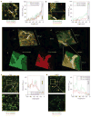
In vivo real-time microdistribution of DACHPt/m with different diameters in tumours. (a), (b) Microdistribution of fluorescently labelled 30 nm (green) and 70 nm (red) micelles 1 h after injection into C26 (a) and BxPC3 (b) tumours. Their colocalization is shown in yellow. Right panels in (a) and (b) show fluorescence intensity profile from the blood vessel (0–10 mm; grey area) to the tumour tissue (10–100 mm) in the selected region (indicated by a white rectangle) expressed as a percentage of the maximum fluorescence intensity attained in the vascular region (%Vmax). (c), (d) Z-stack volume reconstruction of C26 (c) and BxPC3 (d) tumours 1 h after co-injection of the fluorescent micelles. (e) Magnification of the perivascular region (indicated by a white trapezium) of the z-stack volume image of BxPC3 tumours. (f), (g) Distribution of 30 and 70 nm micelles 24 h after injection into C26 tumours (f) and BxPC3 tumours (g). White arrows in (g) indicate 70 nm micelles localizing at perivascular regions. Right panels show fluorescence intensity profile from the blood vessel (0–10 mm; grey area) to the tumour tissue (10–100 mm) in the selected region (indicated by white rectangle). Reprinted with permission from [275], H. Cabral, et al., Accumulation of sub-100 nm polymeric micelles in poorly permeable tumours depends on size. Nature nanotechnology 6, 815 (2011). © 2011, Nature Publishing Group.

Similar articles

Cited by

References

    1. Bangham A, Horne R. Negative staining of phospholipids and their structural modification by surface-active agents as observed in the electron microscope. J Mol Biol. 1964;8:660. - PubMed
    1. Zhang L, Radovic-Moreno AF, Alexis F, Gu FX, Basto PA, Bagalkot V, Jon S, Langer RS, Farokhzad OC. Co-delivery of hydrophobic and hydrophilic drugs from nanoparticle-aptamer bioconjugates. Chem Med Chem. 2007;2:1268. - PubMed
    1. Davis M, Chen Z, Shin D. Nanoparticle therapeutics: An emerging treatment modality for cancer. Nat Rev Drug Discov. 2008;7:771. - PubMed
    1. Wagner V, Dullaart A, Bock AK, Zweck A. The emerging nanomedicine landscape. Nat Biotechnol. 2006;24:1211. - PubMed
    1. Northfelt DW, Dezube BJ, Thommes JA, Miller BJ, Fischl MA, Friedman-Kien A, Kaplan LD, Du Mond C, Mamelok RD, Henry DH. Pegylated-liposomal doxorubicin versus doxorubicin, bleomycin, and vincristine in the treatment of AIDS-related Kaposi’s sarcoma: Results of a randomized phase III clinical trial. J Clin Oncol. 1998;16:2445. - PubMed

Publication types

LinkOut - more resources