. 2015 Dec 29;6(42):44728-44.
doi: 10.18632/oncotarget.6296.

Expression of androgen receptor splice variants in clinical breast cancers

Affiliations

Expression of androgen receptor splice variants in clinical breast cancers

Theresa E Hickey et al. Oncotarget. .

Abstract

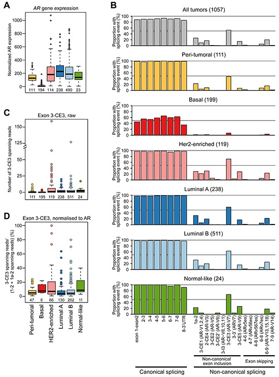
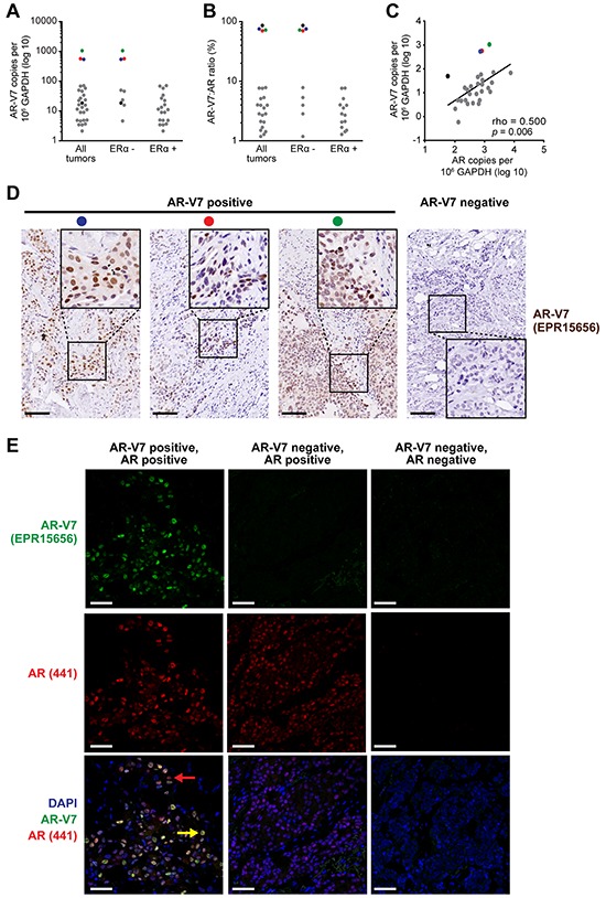
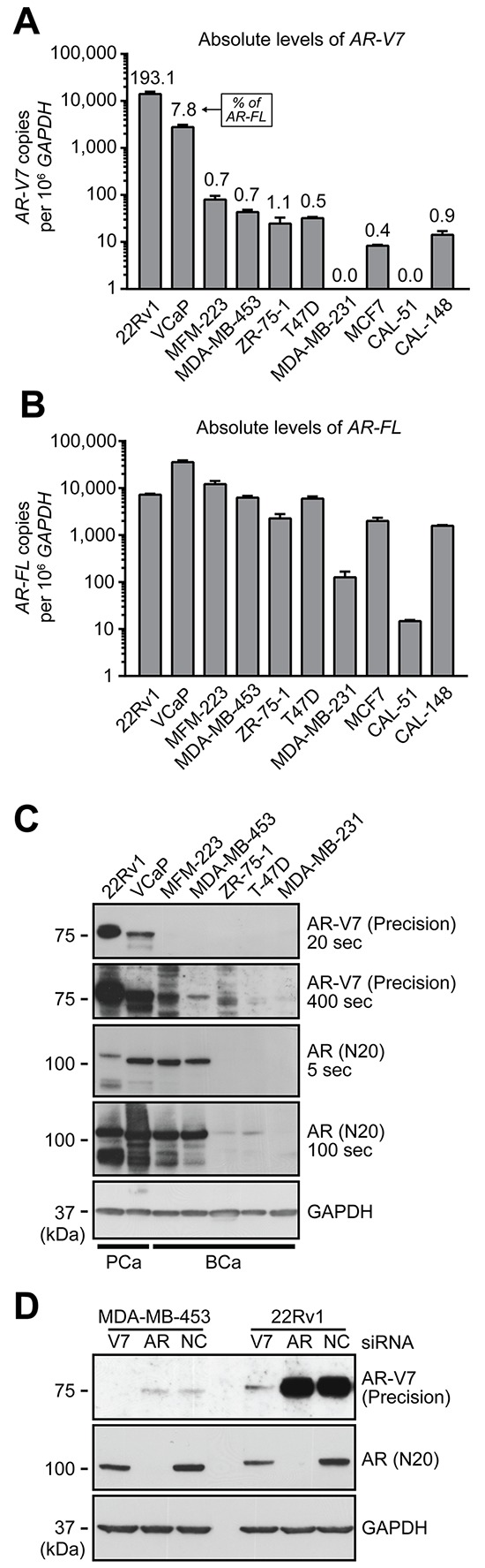
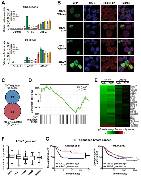
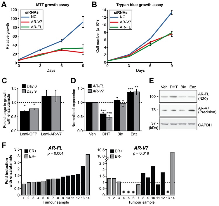
The importance of androgen receptor (AR) signaling is increasingly being recognized in breast cancer, which has elicited clinical trials aimed at assessing the efficacy of androgen deprivation therapy (ADT) for metastatic disease. In prostate cancer, resistance to ADT is frequently associated with the emergence of androgen-independent splice variants of the AR (AR variants, AR-Vs) that lack the LBD and are constitutively active. Women with breast cancer may be prone to a similar phenomenon. Herein, we show that in addition to the prototypical transcript, the AR gene produces a diverse range of AR-V transcripts in primary breast tumors. The most frequently and highly expressed variant was AR-V7 (exons 1/2/3/CE3), which was detectable at the mRNA level in > 50% of all breast cancers and at the protein level in a subset of ERα-negative tumors. Functionally, AR-V7 is a constitutively active and ADT-resistant transcription factor that promotes growth and regulates a transcriptional program distinct from AR in ERα-negative breast cancer cells. Importantly, we provide ex vivo evidence that AR-V7 is upregulated by the AR antagonist enzalutamide in primary breast tumors. These findings have implications for treatment response in the ongoing clinical trials of ADT in breast cancer.

Keywords: alternative splicing; androgen deprivation therapy; androgen receptor; biomarker; breast cancer.


Conflict of interest statement

CONFLICTS OF INTEREST

No potential conflicts of interest were disclosed.

Figures

Figure 1
Figure 1. The diversity and frequency of AR splicing in breast cancer
A. Relative expression of AR by PAM50 subtype in the TCGA cohort. B. Proportion of TCGA tumor or normal samples with the indicated splicing event. Tumors were subtyped using the PAM50 classification system. C. Quantitation of exon 3-CE3 spanning reads, predicted to encode the AR-V7 variant, in breast cancer and peri-tumoral breast tissues. D. Normalized quantitation of exon 3-CE3 spanning reads in breast cancer and peri-tumoral breast tissues. Exon 3-CE3 reads were scaled by the sum of exon 1–2 and exon 1a-2 reads (see Materials and Methods for more detail).
Figure 2
Figure 2. The AR variant AR-V7 is expressed in clinical breast cancer samples
A. Expression of AR-V7 in ERα-positive (n = 35) and ERα-negative (n = 19) breast cancers, as determined by qRT-PCR. A Cq < 35 was used as a cut-off for determining AR-V7 positivity. AR-V7 copy number was calculated as described in Materials and Methods. B. Ratio of AR-V7 as a proportion of AR-FL (in AR-V7-positive tumors). C. Correlation between AR-V7 and AR-FL copy number in AR-V7-positive tumors. Pearson's correlation rho and p values are shown. Note that three tumors with high AR-V7 mRNA levels (red, blue green) or a high AR-V7:AR-FL ratio (black) are identifiable in (A)-(C) by color. D. Detection of AR-V7 protein by IHC in 3 breast tumors with high levels of AR-V7 mRNA expression (left). Positive staining was not detected in specimens that do not express AR-V7 mRNA (a representative is shown on the right). Inset images of higher magnification demonstrate strong nuclear staining. Colored dots enable matching of samples to (A)-(C) Scale bars are 100 microns. E. Detection of AR-V7 protein by IF and co-localization with AR-FL. Shown are an AR-V7 positive tumor (left), an AR-V7-negative but AR-positive tumor (middle), and a dual-negative tumor (right). Nuclei were stained with DAPI. In the bottom left image, a yellow arrow indicates a representative cell with strong AR-FL and AR-V7 staining and a red arrow indicates a representative cell with strong AR-FL staining but weak or no AR-V7 staining. Scale bars are 50 microns.
Figure 3
Figure 3. Expression of AR-V7 in breast cancer cell line models
A–B. qRT-PCR was used to quantify AR-V7 (A) and AR-FL (B) in a panel of 8 breast cancer cell lines and 2 prostate cancer cell line controls (22Rv1 and VCaP). AR-V7 levels as a percentage of AR-FL transcript are shown above the columns in panel A. Values are the mean (± SEM) of triplicate samples. C. Protein lysates were collected from the indicated cell lines used above and subjected to Western blot analysis using AR-V7 (Precision Biosciences), AR (N20) and GAPDH (loading control) antibodies. Short and long exposures are shown. Note that AR-FL from 22Rv1 cells migrates more slowly because it contains two copies of the second zinc finger domain as a result of a genomic duplication. D. MDA-MB-453 and 22Rv1 cells were transfected with a non-specific control siRNA (NC) or siRNAs specific for AR-FL or AR-V7. After 72 h, protein lysates were analyzed by Western blotting as in (C).
Figure 4
Figure 4. AR-V7 is constitutively active, resistant to AR antagonists and regulates a transcriptome distinct from AR-FL
A. MDA-MB-453 and MFM-223 cells were transfected with plasmids expressing full-length AR-FL or AR-V7 and an AR-responsive reporter construct and subsequently treated with 1 nM DHT, 1 μM bicalutamide and/or 1 μM enzalutamide. Luciferase activity values represent the mean (±SEM) of 6 biological replicates; results are representative of three independent experiments. ANOVA followed by Tukey's multiple comparisons tests were used to assess changes in luciferase activity (**,p < 0.01; *,p < 0.05): for AR-FL, comparisons were with DHT; for AR-V7, comparisons were with vehicle (Veh). None of the treatments caused a statistically significant change in AR-V7 activity. B. MDA-MB-453 cells grown in androgen-depleted media were transfected with GFP-tagged forms of full-length AR-FL or AR-V7 and treated with 10 nM DHT or vehicle control (ethanol) for 24 h. Nuclei were stained with DAPI and cytoskeletons with phalloidin. Representative images are shown for four color channels (GFP, DAPI, phalloidin and merge). C. Venn diagram showing the overlap between genes differentially expressed in response to AR-V7 expression and DHT treatments. D. No association between DHT-regulated and AR-V7-regulated genes in the MDA-MB-453 model, as demonstrated by gene set enrichment analysis (GSEA). E. Heat map demonstrating that AR-FL over-expression does not alter expression of the core AR-V7 regulated gene set. F. Expression of the core AR-V7-regulated gene set according to PAM50 breast cancer subtype in the GOBO cohort. The gene set was highest in basal tumors compared to all other subtypes (ANOVA, followed by Tukey's multiple comparisons test). G. Kaplan-Meier survival plot showing metastasis-free survival in HER2-enriched patients from the Ringner et al cohort (left) and disease-specific survival in HER2-enriched patients from the METABRIC cohort (right). Patients were stratified by median expression of the core AR-V7 regulated gene set into low and high groups. Log rank test p values: ****,p < 0.0001; *,p < 0.05.
Figure 5
Figure 5. AR-V7 regulates the growth of MDA-MB-453 cells and response to enzalutamide
A–B. MDA-MB-453 cells were transfected with siRNAs specific for AR-FL and AR-V7 or a control siRNA and growth was assessed in androgen-replete media using MTT (A) and Trypan Blue (B) growth assays. Values are the mean (±SEM) of 3 biological replicates; results are representative of three independent experiments. C. MDA-MB-453 cells were transduced with lentivirus designed to overexpress AR-V7 or GFP (negative control) and cell growth assessed using Crystal Violet assays in response to the enzalutamide. The relative change in growth compared to vehicle control is shown at 6 and 9 days. Values are the mean (±SEM) of 6 biological replicates comprising two independent experiments. D. Treatment with AR antagonists induces AR gene transcription in MDA-MB-453 cells. Cells were grown in androgen depleted media and treated with 10 nM DHT, 10 μM bicalutamide, 10 μM enzalutamide or vehicle control for 24 h. AR-FL and AR-V7 mRNA was measured by qRT-PCR. Values are normalized to GAPDH and represent the mean (± SEM) of triplicate samples, with vehicle control treatment set to 1. Results are representative of three independent experiments. E. Matched samples from (D) were analyzed by Western blotting using AR-V7 (Precision Biosciences), AR (N20) and GAPDH (loading control) antibodies. F. Human breast tumor explants were cultured with vehicle (DMSO) or enzalutamide (50 μM) for 48 h. AR-FL and AR-V7 transcripts were measured by qRT-PCR and normalized to GAPDH. Values are expressed as fold-change relative to DMSO for each individual tumor. Statistically significant differences compared to the control treatment were assessed using a Wilcoxon signed rank test; p values are shown. Four of the tumors did not express AR-V7 (shown by a # symbol).

References

    1. Bauer KR, Brown M, Cress RD, Parise CA, Caggiano V. Descriptive analysis of estrogen receptor (ER)-negative, progesterone receptor (PR)-negative, and HER2-negative invasive breast cancer, the so-called triple-negative phenotype: a population-based study from the California cancer Registry. Cancer. 2007;109:1721–1728. - PubMed
    1. Hudis CA, Gianni L. Triple-negative breast cancer: an unmet medical need. The oncologist. 2011;16:1–11. - PubMed
    1. Green SM, Mostaghel EA, Nelson PS. Androgen action and metabolism in prostate cancer. Molecular and cellular endocrinology. 2012;360:3–13. - PMC - PubMed
    1. Hickey TE, Robinson JL, Carroll JS, Tilley WD. Minireview: The androgen receptor in breast tissues: growth inhibitor, tumor suppressor, oncogene? Molecular endocrinology. 2012;26:1252–1267. - PMC - PubMed
    1. Agoff SN, Swanson PE, Linden H, Hawes SE, Lawton TJ. Androgen receptor expression in estrogen receptor-negative breast cancer. Immunohistochemical, clinical, and prognostic associations. American journal of clinical pathology. 2003;120:725–731. - PubMed

Publication types

MeSH terms