. 2015 Nov;7(11):928-36.
doi: 10.18632/aging.100839.

OTX2 regulates the expression of TAp63 leading to macular and cochlear neuroepithelium development

Affiliations

OTX2 regulates the expression of TAp63 leading to macular and cochlear neuroepithelium development

Ramona Palombo et al. Aging (Albany NY). 2015 Nov.

Abstract

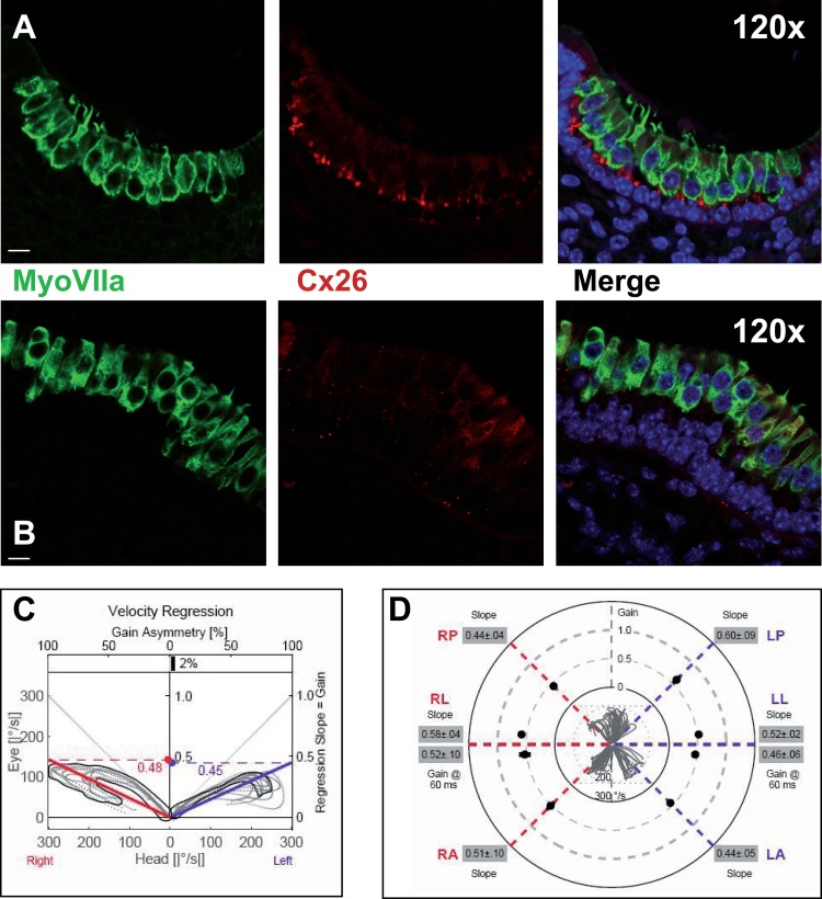
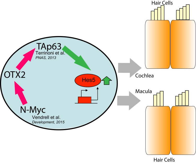
OTX proteins, homologs of the Drosophila orthodenticle (Otd), are important for the morphogenesis of the neuroectoderm, and for the central nervous system formation. OTX1 and OTX2 are important for the cochlea and macula development, indeed when OTX1 is knocked down, these organs undergo developmental failure. Moreover OTX2 transfection revert this effect in OTX1(-/-) mice. The TA isoform of TP63, involved in Notch regulation pathway, has a critical function in the cochlear neuroepithelium differentiation. TAp63 positively regulates Hes5 and Atoh1 transcription. This pathway has been also demonstrated in p63(-/-) mice, and in patients p63 mutated, affected by Ectodermal Dysplasia (ED, OMIM 129810). These patients are affected by mild sensorineural deafness, most likely related to the mutation in p63 gene impairing the Notch pathway. We demonstrated the role of OTX2 on TAp63 regulation necessary for the correct formation of macular neuroepithelium and we confirmed the impairment of vestibular function caused by p63 mutations. Although the abnormalities found in our patient were still at a subclinical extent, aging could exacerbate this impairment and cause a decrease in quality of life.

Keywords: Cochlea; Ectodermal Dysplasia; Macula; OTX2; TAp63.


Conflict of interest statement

Conflict of interest statement

The authors have no conflicts of interest to declare.

Figures

Figure 1
Figure 1. Promoter analysis of p63 gene
(A) Genomic location and structure of human p63 gene, showing the position and size of the different intron and exons. In lower panels are identified, the location position of OTX responsive elements, and their conservation in different species. (B) Detailed structure of p63 gene. (C) OTX2 binding element, matrix Jaspar PH0130.1.
Figure 2
Figure 2. Molecular study of OTX2 responsive elements in the p63 gene
(A) The H1299 cell line has been transfected using OTX2 cDNA, and the expression of p63, p53 and p73 analyzed at 24 and 48 hours by RTqPRC analysis. The graphs show that OTX2 nicely induces the transcription of TAp63 isoform, with very low effects on p53 and none on TAp73, *t-test on DDCt values, coupled, two tails, *p = 0,0005, **p = 0,00073. (B) Induction control respect to DeltaN isoforms, the graph show that 48 hours and none of these isoform are transactivated. (C) ChIP analysis, showing the binding of OTX2 at the responsive element, located at the 5′ (upper panel), respect to that located within intron 5. In the first case the PCR product is detectable at cycle 31 whereas the other is only detectable later after 43 cycles. (D) qPCR analysis using DNA pulled down using OTX antibody. t-test on DDCt values, coupled, two tails, *p = 0,00057.( E) Western blot of protein extracted from cells after the transfection. A band corresponding to TAp63 molecular weight is visible. The control has been obtained using protein extracts from a SaOS TET-On cell line.
Figure 3
Figure 3. Macular structure in p63 defective mice and vestibular function in EEC patient
(A) Representative picture of a macula of the vestibule from mice normal controls stained with MyoVIIa and Connexin 26 antibody. Dapi has been used for nuclei staining. Bars =20 μm. (B) Macular neuroepithelium from p63−/− mice. (C) Results of vHIT testing of the lateral semicircular canal (LSC) for the patient described in the article; the regression analysis of the vestibulo-ocular reflex (VOR) gain (eye angular velocity/head angular velocity) shows values of 0.48 and 0.45 for the right and left LSC, respectively. This shows VOR hyporeflectivity (average gain between 0.8 and 1.1). (D) The vestibular hyporeflectivity for both sides was confirmed by studying VOR gain for all the semicircular canals, with instant gain at 60 milliseconds for the lateral semicircular canals: gain values were all under average, without any significant asymmetry involving one side with respect to the other. Canals positioned on the same plane are connected by lines; RA= right anterior, RP= right posterior, RL=right lateral; LA= left anterior, LP= left posterior, LL=left lateral.
Figure 4
Figure 4. Schematic representation of OTX2 pathway
N-Myc has been demonstrated to drive the expression of OTX2, and the latter is able to transactivate TAp63, inducing Hes5 and leading to the differentiation of both cochlear and macular neuroepithelium.

References

    1. Boncinelli E, Simeone A, Acampora D, Gulisano M. Homeobox genes in the developing central nervous system. Annales de genetique. 1993;36:30–37. - PubMed
    1. Klein WH, Li X. Function and evolution of Otx proteins. Biochemical and biophysical research communications. 1999;258:229–233. - PubMed
    1. Simeone A, Acampora D. The role of Otx2 in organizing the anterior patterning in mouse. The International journal of developmental biology. 2001;45:337–345. - PubMed
    1. Larsen KB, Lutterodt MC, Mollgard K, Moller M. Expression of the homeobox genes OTX2 and OTX1 in the early developing human brain. The journal of histochemistry and cytochemistry : official journal of the Histochemistry Society. 2010;58:669–678. - PMC - PubMed
    1. Hartman BH, Durruthy-Durruthy R, Laske RD, Losorelli S, Heller S. Identification and characterization of mouse otic sensory lineage genes. Frontiers in cellular neuroscience. 2015;9:79. - PMC - PubMed

Publication types

MeSH terms

LinkOut - more resources