. 2015 Nov 10;10(11):e0142118.
doi: 10.1371/journal.pone.0142118. eCollection 2015.

A Naturally Occurring rev1-vpu Fusion Gene Does Not Confer a Fitness Advantage to HIV-1

Affiliations

A Naturally Occurring rev1-vpu Fusion Gene Does Not Confer a Fitness Advantage to HIV-1

Simon M Langer et al. PLoS One. .

Abstract

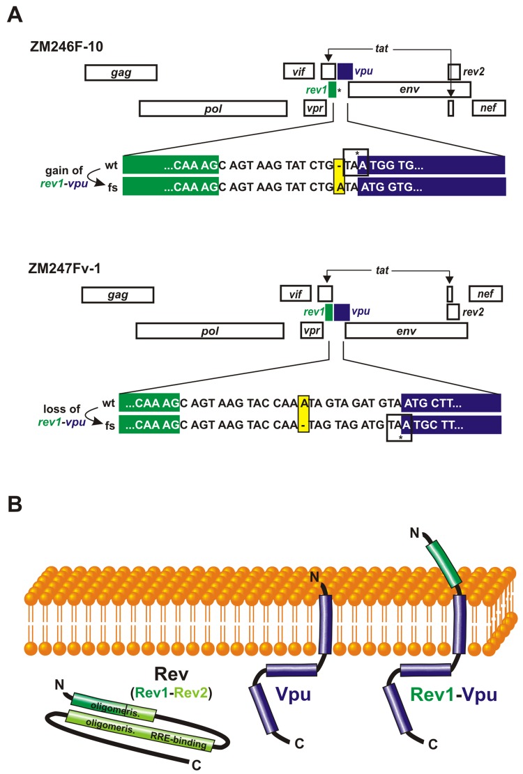
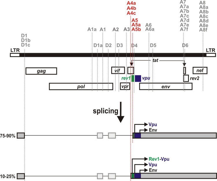
Background: Pandemic strains of HIV-1 (group M) encode a total of nine structural (gag, pol, env), regulatory (rev, tat) and accessory (vif, vpr, vpu, nef) genes. However, some subtype A and C viruses exhibit an unusual gene arrangement in which the first exon of rev (rev1) and the vpu gene are placed in the same open reading frame. Although this rev1-vpu gene fusion is present in a considerable fraction of HIV-1 strains, its functional significance is unknown.

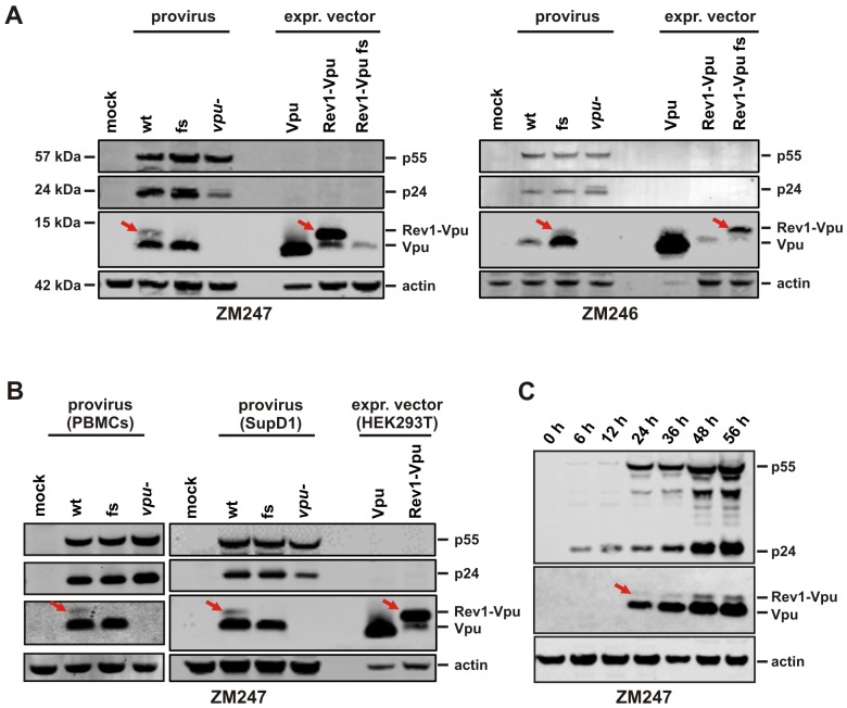
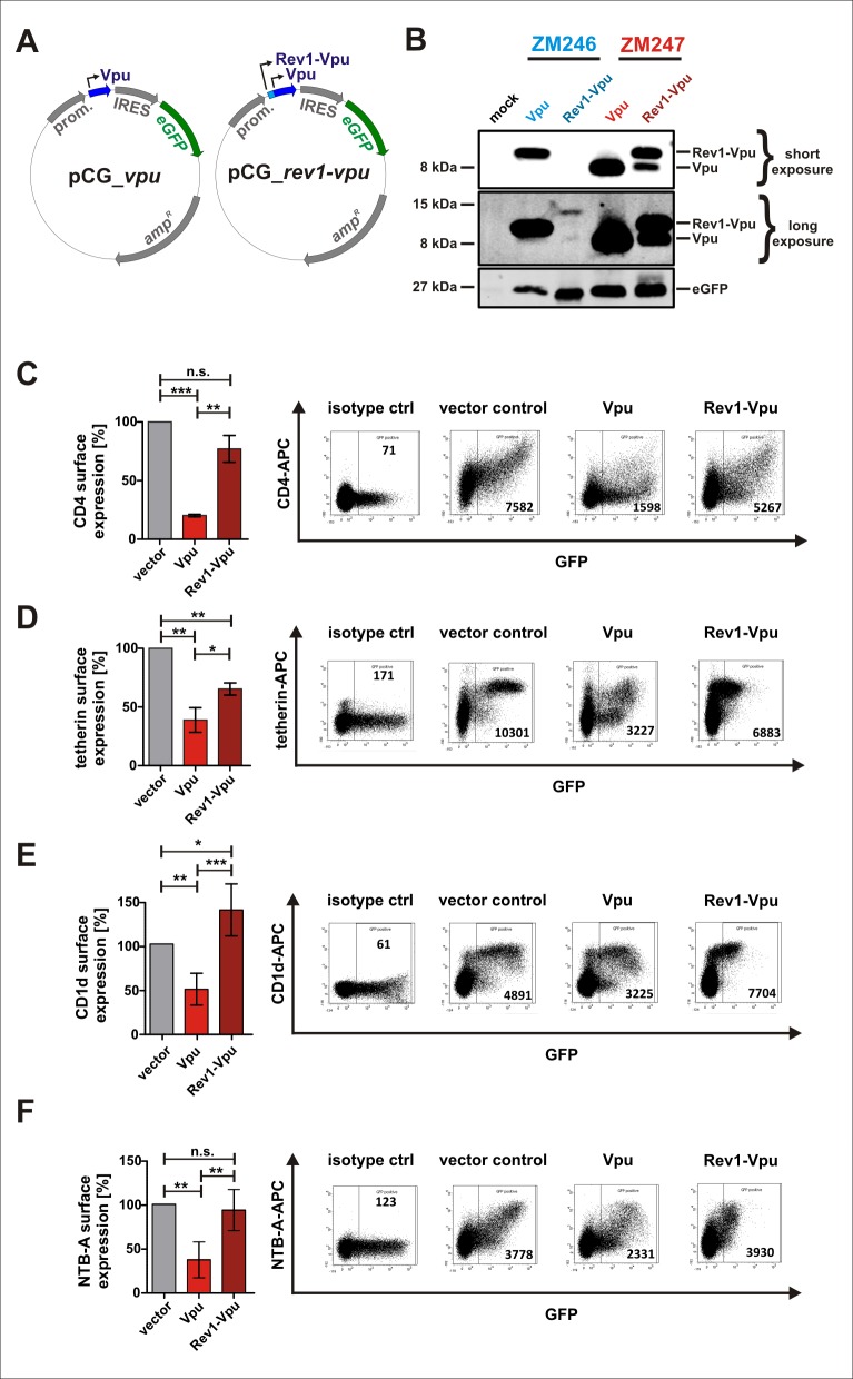
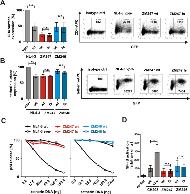
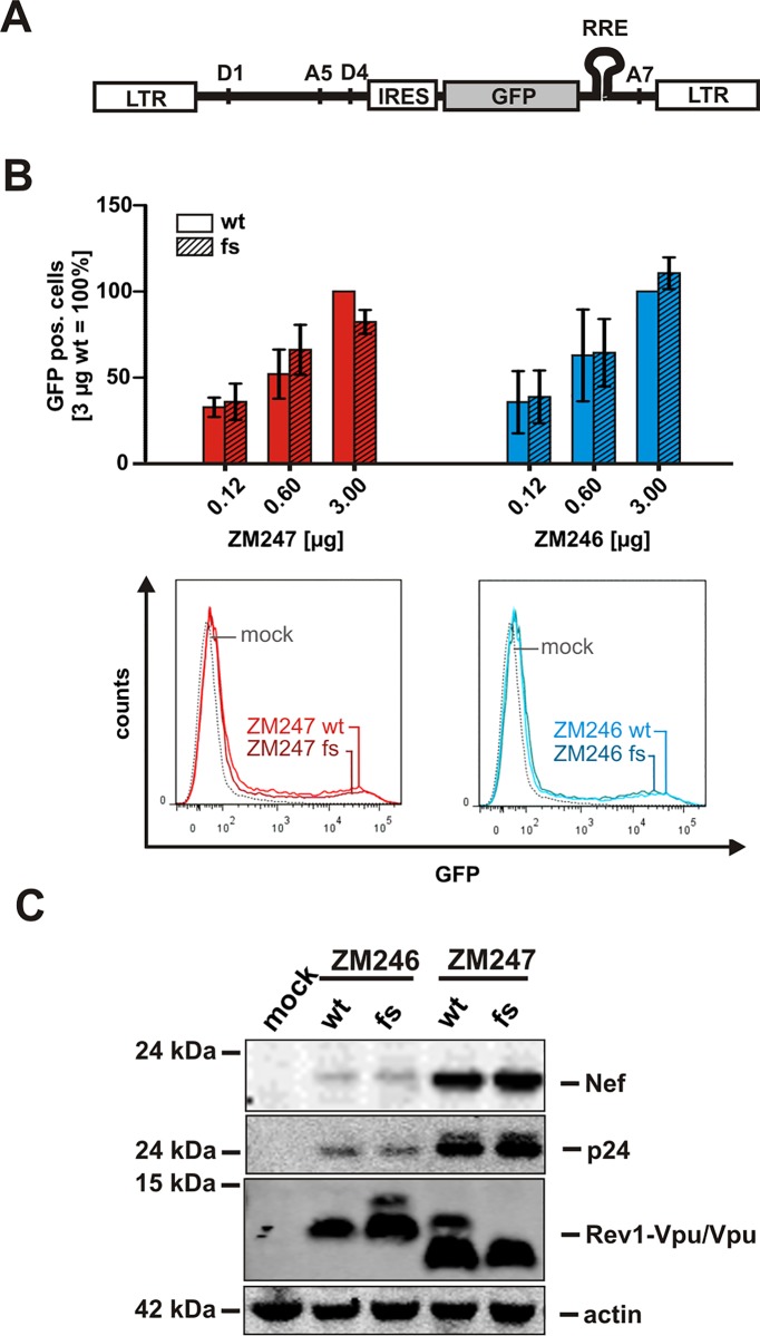
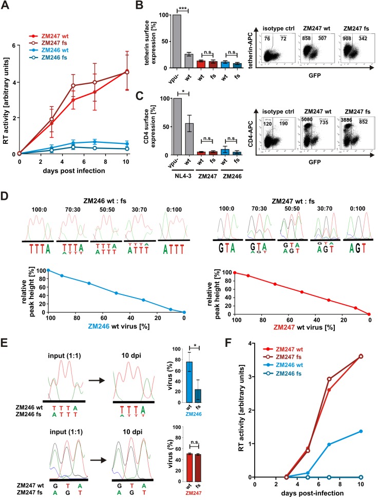
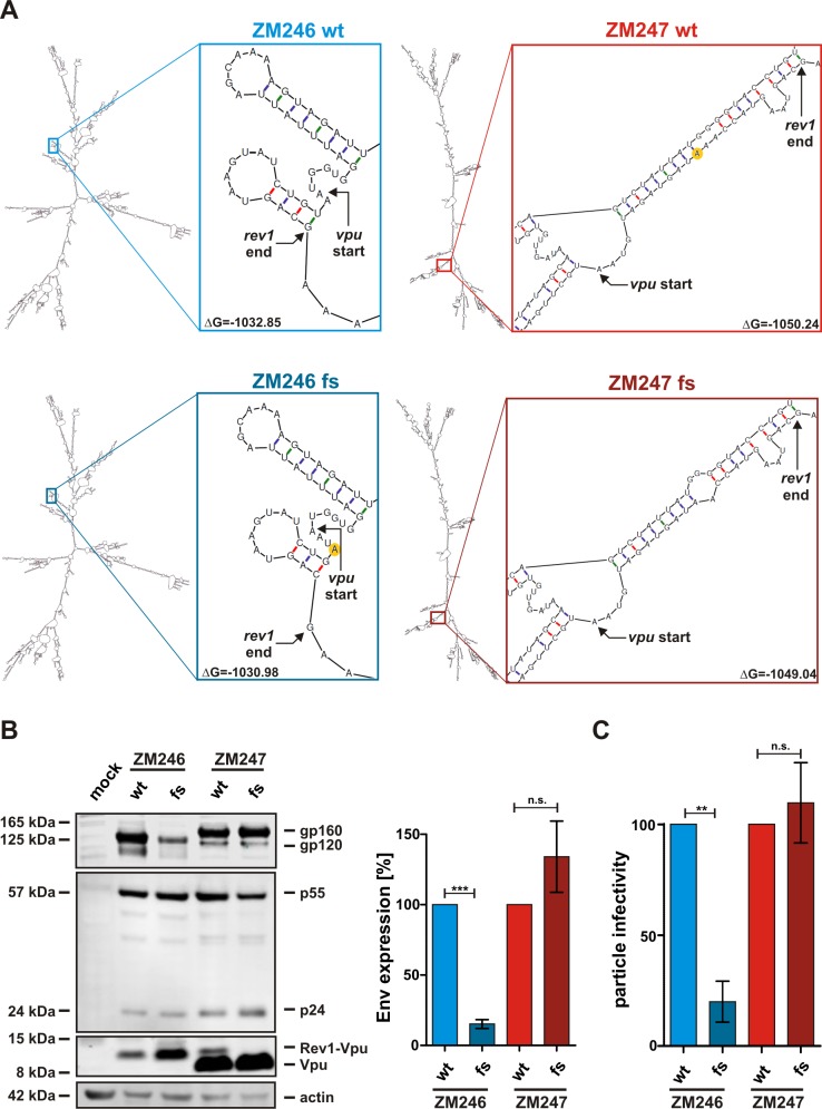
Results: Examining infectious molecular clones (IMCs) of HIV-1 that encode the rev1-vpu polymorphism, we show that a fusion protein is expressed in infected cells. Due to the splicing pattern of viral mRNA, however, these same IMCs also express a regular Vpu protein, which is produced at much higher levels. To investigate the function of the fusion gene, we characterized isogenic IMC pairs differing only in their ability to express a Rev1-Vpu protein. Analysis in transfected HEK293T and infected CD4+ T cells showed that all of these viruses were equally active in known Vpu functions, such as down-modulation of CD4 or counteraction of tetherin. Furthermore, the polymorphism did not affect Vpu-mediated inhibition of NF-кB activation or Rev-dependent nuclear export of incompletely spliced viral mRNAs. There was also no evidence for enhanced replication of Rev1-Vpu expressing viruses in primary PBMCs or ex vivo infected human lymphoid tissues. Finally, the frequency of HIV-1 quasispecies members that encoded a rev1-vpu fusion gene did not change in HIV-1 infected individuals over time.

Conclusions: Expression of a rev1-vpu fusion gene does not affect regular Rev and Vpu functions or alter HIV-1 replication in primary target cells. Since there is no evidence for increased replication fitness of rev1-vpu encoding viruses, this polymorphism likely emerged in the context of other mutations within and/or outside the rev1-vpu intergenic region, and may have a neutral phenotype.


Conflict of interest statement

Competing Interests: The authors have declared that no competing interests exist.

Figures

Fig 1
Fig 1. Varying genomic organization of HIV-1 in the intergenic region between rev1 and vpu.
(A) The relative position of genes within HIV-1 subtype C clones ZM246F-10 (upper panel) and ZM247Fv-1 (lower panel) is shown. Whereas ZM246 rev1 (green) and vpu (blue) lie within different reading frames and are separated by an intervening stop codon (*), ZM247 encodes a rev1-vpu fusion gene. Frameshift mutations that were introduced to generate or disrupt rev1-vpu in ZM246 and ZM247, respectively, are highlighted in yellow. (B) Putative topology of the Rev1-Vpu fusion protein and its parental proteins Rev (green) and Vpu (blue).
Fig 2
Fig 2. Expression of the Rev1-Vpu fusion protein.
(A) Western blot analysis of HEK293T cells co-transfected with the proviral clones described in Fig 1A or a vpu-deficient mutant thereof. Expression vectors containing rev1-vpu or vpu cassettes served as size controls. Vpu and Rev1-Vpu were detected with an antiserum raised against ZM247 Vpu. (B, C) Expression of Vpu and Rev1-Vpu in ZM247-infected PBMCs or SupD1 cells. Bands representing the Rev1-Vpu fusion protein are highlighted by red arrows. Detection of p55, p24 and actin served as internal controls.
Fig 3
Fig 3. Functional activity of Rev1-Vpu expressed from pCG expression plasmids.
(A) CMV-IE promoter-based pCG expression vectors containing vpu (left panel) or the rev1-vpu fusion gene (right panel). An enhanced version of the green fluorescent protein (eGFP) is co-expressed via an IRES. (B) Expression of Rev1-Vpu and Vpu in HEK293T cells transfected with the indicated pCG vectors. A Vpu-specific antiserum was used for detection. eGFP was detected to check transfection efficiencies. (C-F) FACS analysis of (C) CD4, (D) tetherin, (E) CD1d or (F) NTB-A receptor modulation by ZM247 Vpu and Rev1-Vpu. HEK293T cells were transfected with expression vectors for the respective surface receptor and Vpu or Rev1-Vpu. 40 h post transfection, surface receptor levels were monitored by two-color flow cytometry. Dot plots indicating the gating strategy are shown in the right panels. Bar diagrams summarizing three to five independent experiments +/- SD are shown on the left (***p<0.001; **p<0.01; *p<0.05; n.s. not significant).
Fig 4
Fig 4. Vpu function in isogenic viruses differing only in their ability to express Rev1-Vpu.
(A, B) FACS analysis of surface expression levels of (A) CD4 and (B) tetherin on HEK293T cells co-transfected with the indicated proviral constructs and expression vectors for the respective surface molecule. Dot plots indicating the gating strategy are shown in the right panels. Bar diagrams summarizing three to six independent experiments +/- SD are shown on the left. (C) p24 release from HEK293T cells co-transfected with the indicated proviral constructs and increasing amounts of a tetherin expression plasmid. 40 h post transfection, the amounts of cell-associated and cell-free p24 were analyzed by ELISA. Relative release was calculated as ratio of p24 in the supernatant to total p24. The means of at least three independent experiments are shown. (D) Activation of NF-кB by viruses harboring the fusion polymorphism or not. HEK293T cells were co-transfected with the indicated proviral clones, and an NF-кB-responsive firefly luciferase reporter construct. 40 h post transfection, firefly luciferase activity was determined and normalized to the activity of a Gaussia luciferase control plasmid. The means of three independent experiments +/- SD are shown (***p<0.001; **p<0.01; *p<0.05; n.s. not significant).
Fig 5
Fig 5. Rev function in isogenic viruses differing only in their ability to express Rev1-Vpu.
(A) Gene arrangement of a reporter construct expressing GFP in a Rev-dependent manner. The GFP ORF is flanked by splice donor site 4 (D4) and splice acceptor site 7 (A7). Rev mediates the export of intron-containing GFP expressing mRNA via binding to the RRE. (B) Rev-dependent gene expression was determined by co-transfection of HEK293T cells with increasing amounts of the indicated molecular clones of HIV-1, the GFP reporter construct and a BFP expressing control plasmid. 40 h post transfection, GFP expression levels of BFP positive cells were analyzed by flow cytometry. Examples of primary FACS data are shown in the lower panel. (C) Western blot, showing Rev-dependent expression of p24-capsid and Rev-independent expression of Nef in HEK293T cells transfected with the indicated proviral constructs.
Fig 6
Fig 6. Effect of Rev1-Vpu expression on HIV-1 replication in PBMCs and tonsillar explant cultures.
(A) PHA-stimulated PBMCs were infected with adjusted amounts of the indicated viruses. Virus replication was monitored by analyzing RT-activity in the supernatant. The means of three independent experiments +/- SEM are shown. (B, C) Surface expression levels of (B) tetherin and (C) CD4 were determined by flow cytometry at day 3 post infection. Infected cells were identified by intracellular p24 staining after surface staining of CD4 or tetherin. Dot plots indicating the gating strategy are shown in the right panels. Bar diagrams summarizing four to five independent experiments +/- SD are shown on the left. (D) Sequence analyses of viral mixtures. Wt and fs mutant virus stocks were normalized for infectivity, mixed at the indicated ratios, and the rev1-vpu region was sequenced after bulk amplification of cDNA. The lower panels show respective standard curves. The peak fluorescence of the T residue at position 1 (ZM246 wt) and the A residue at position 3 (ZM247 wt) is expressed as a fraction of the total fluorescence (relative peak height). (E) Sequence chromatograms of 1:1 input mixtures and viral cultures 10 days post infection (dpi). Percentages of wt and fs sequences displayed in the right panels were calculated from the standard curves shown in (D). (F) Human tonsil explant cultures were infected and analyzed as described in (A). One representative experiment for one of three independent donors is shown (***p<0.001; **p<0.01; *p<0.05; n.s. not significant).
Fig 7
Fig 7. Effect of rev1-vpu frameshift mutations on mRNA structure and Env expression.
(A) mRNA structures of ZM246 wt, ZM246 fs, ZM247 wt and ZM247 fs were predicted using the Mfold web server for nucleic acid folding and hybridization prediction [37]. The putative mRNA structures of full length rev/vpu/env enconding mRNAs (using splice sites D1 and A4c) are shown on the left of each panel. Close-ups of the rev1-vpu intergenic region and the minimum free energy ΔG are shown on the right. The nucleotides that are deleted in ZM247 wt and inserted in ZM246 fs are highlighted in yellow. (B) Western blot analysis of HEK293T cells co-transfected with the proviral clones described in Fig 1A. gp160 and gp120 were detected using an antiserum raised against HIV-1 M subtype C 96ZM651. A representative Western blot is shown on the left. Total Env expression (gp120+gp160) was normalized to total Gag expression (p55+p24). The mean values (+/- SEM) of four independent transfections are shown on the right. (C) Particle infectivity was determined by infecting TZM-bl reporter cells with equal amounts of p24. Three days post infection, β-galactosidase activity was measured. The mean values (+/- SEM) of triplicate infections from two to four independent transfections are shown (***p<0.001; **p<0.01; n.s. not significant).
Fig 8
Fig 8. Splice sites generating Rev1-Vpu encoding mRNA.
The HIV-1 genome (black) and nine ORFs encoding structural, regulatory and accessory proteins are depicted on top. Splice donor (D1-6) and acceptor (A1-8) sites are indicated by dotted lines. mRNAs encoding Rev1-Vpu, Vpu and Env are shown in grey. Depending on the cell type and time point post infection, 75–90% of Vpu and Env encoding mRNA species fail to express Rev1-Vpu since the usage of splice acceptor site A5 removes an intron containing the initiation codon of rev1. Only 10–25% of the Vpu encoding mRNAs have the potential to be translated into Rev1-Vpu since splice acceptor sites A4a, A4b and A4c retain the complete first exon of rev.

Similar articles

Cited by

References

    1. Ocwieja KE, Sherrill-Mix S, Mukherjee R, Custers-Allen R, David P, Brown M, et al. Dynamic regulation of HIV-1 mRNA populations analyzed by single-molecule enrichment and long-read sequencing. Nucleic Acids Res. 2012;40: 10345–10355. 10.1093/nar/gks753 - DOI - PMC - PubMed
    1. Salfeld J, Göttlinger HG, Sia RA, Park RE, Sodroski JG, Haseltine WA. A tripartite HIV-1 tat-env-rev fusion protein. EMBO J. 1990;9: 965–970. - PMC - PubMed
    1. Benko DM, Schwartz S, Pavlakis GN, Felber BK. A novel human immunodeficiency virus type 1 protein, tev, shares sequences with tat, env, and rev proteins. J Virol. 1990;64: 2505–2518. - PMC - PubMed
    1. Cohen EA, Lu Y, Göttlinger H, Dehni G, Jalinoos Y, Sodroski JG, et al. The T open reading frame of human immunodeficiency virus type 1. J Acquir Immune Defic Syndr. 1990;3: 601–608. - PubMed
    1. Göttlinger HG, Dorfman T, Cohen EA, Haseltine WA. The role of the tnv protein and tnv RNA splicing signals in replication of HIV-1 IIIB isolates. Virology. 1992;189: 618–628. - PubMed

Publication types

MeSH terms

Substances