. 2015 Nov 10:6:8839.
doi: 10.1038/ncomms9839.

Cerebrospinal fluid-derived circulating tumour DNA better represents the genomic alterations of brain tumours than plasma

Affiliations

Cerebrospinal fluid-derived circulating tumour DNA better represents the genomic alterations of brain tumours than plasma

Leticia De Mattos-Arruda et al. Nat Commun. .

Abstract

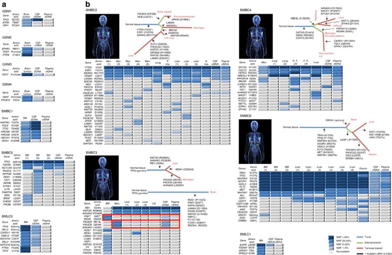
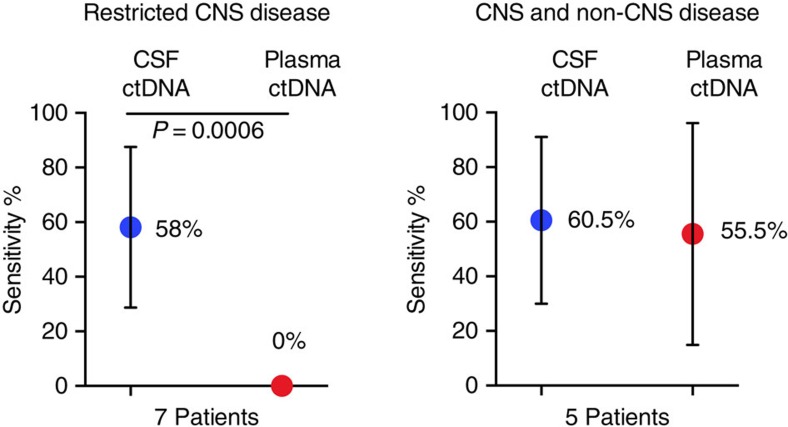
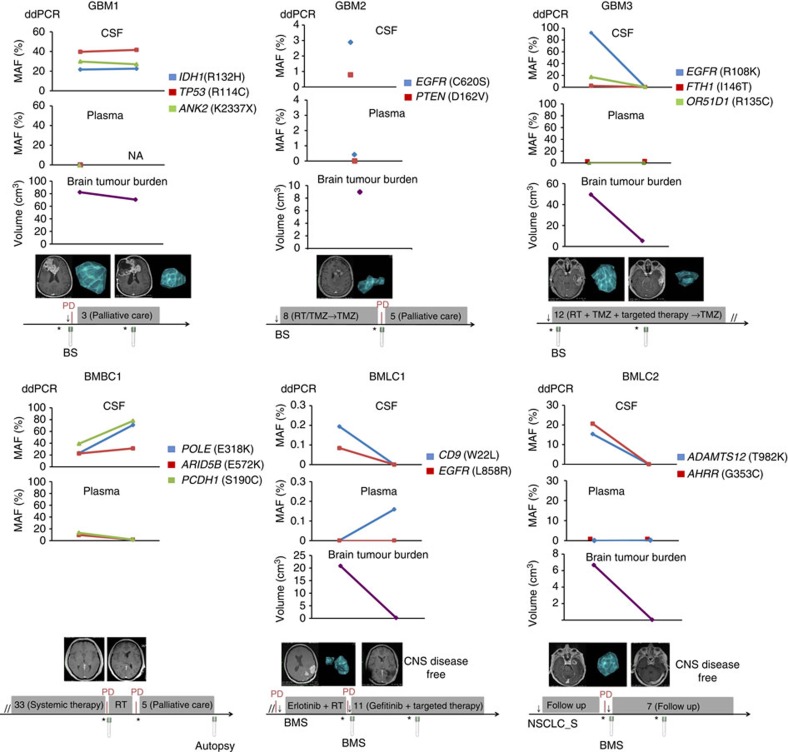
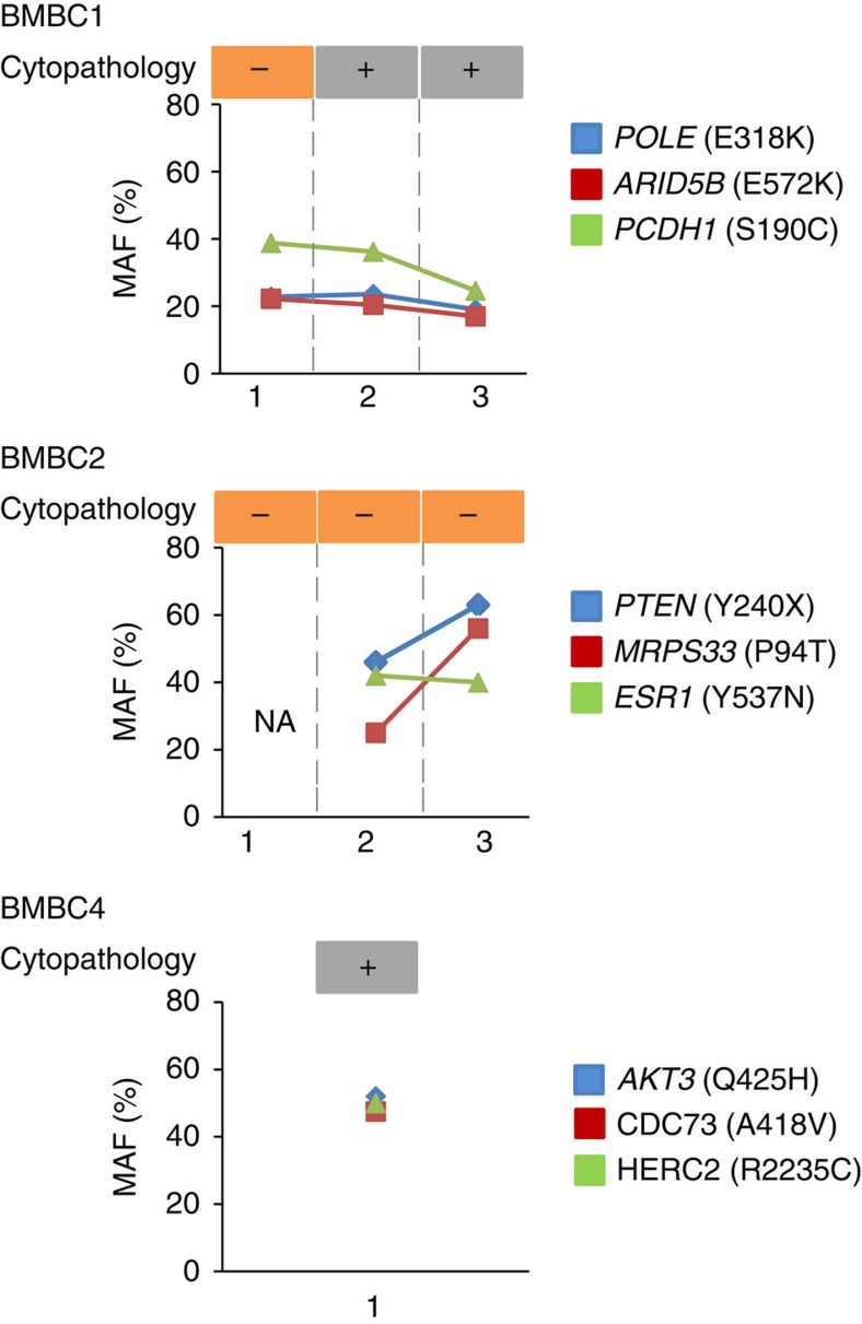
Cell-free circulating tumour DNA (ctDNA) in plasma has been shown to be informative of the genomic alterations present in tumours and has been used to monitor tumour progression and response to treatments. However, patients with brain tumours do not present with or present with low amounts of ctDNA in plasma precluding the genomic characterization of brain cancer through plasma ctDNA. Here we show that ctDNA derived from central nervous system tumours is more abundantly present in the cerebrospinal fluid (CSF) than in plasma. Massively parallel sequencing of CSF ctDNA more comprehensively characterizes the genomic alterations of brain tumours than plasma, allowing the identification of actionable brain tumour somatic mutations. We show that CSF ctDNA levels longitudinally fluctuate in time and follow the changes in brain tumour burden providing biomarkers to monitor brain malignancies. Moreover, CSF ctDNA is shown to facilitate and complement the diagnosis of leptomeningeal carcinomatosis.


Conflict of interest statement

The authors declare no competing financial interests.

Figures

Figure 1
Figure 1. CSF ctDNA better captures the genomic alterations in patients with brain tumours than plasma ctDNA.
(a,b) Analysis of CSF ctDNA, plasma ctDNA and primary brain tumour or metastatic lesions collected simultaneously. Heatmap of the non-silent genetic alterations from each of the twelve cases is shown and phylogenetic trees of the autopsied patients with brain metastasis from breast cancer (BMBC) are represented. Colour key for mutant allelic frequencies (MAFs) is shown. (a) Patients with restricted central nervous system (CNS) disease, glioblastoma (GBM), BMBC and brain metastasis from lung cancer (BMLC). (b) Patients with CNS and non-CNS disease. BM, brain metastasis; LN, lymph node; Men, meninges; P. Imp, pericardium implant; PT, para-tracheal; PP, peri-pancreatic.
Figure 2
Figure 2. Sensitivity analysis of CSF ctDNA and plasma ctDNA.
Sensitivity was inferred based on gene mutations detected in central nervous system (CNS) tumours, which were either identified in CSF or plasma ctDNA (Supplementary Table 5). Data were pooled and the mean with standard deviation error bars is shown. A Mann–Whitney test was used for the analysis and P value is shown.
Figure 3
Figure 3. Dynamic changes in CSF ctDNA recapitulate the treatment courses of patients with brain tumours.
Longitudinal monitoring of patients with GBM and brain metastases through CSF and plasma ctDNA and the analysis of brain tumour burden. Gene mutations were measured by ddPCR. Tumour volumes were calculated using computer aided planimetric analysis. Timelines reflect the most relevant clinical information for each patient. BS, brain surgery; BMS, brain metastasis surgery; CNS, central nervous system; NSCLC_S, non-small cell lung cancer surgery; PD, progressive disease; RT, radiotherapy; TMZ, temozolomide. Asterisk and arrow indicate time of magnetic resonance imaging and surgical procedure, respectively. Grey boxes indicate therapy or follow up, and their duration is provided in months.
Figure 4
Figure 4. Analysis of CSF ctDNA as a diagnostic tool for leptomeningeal carcinomatosis in three metastatic breast cancer patients.
The results of serial clinical cytopathology analyses are shown in the upper part of the graph. In the lower part, mutant allelic frequencies (MAFs) measured by ddPCR in the same CSF samples are depicted. NA, not available.

References

    1. Gerlinger M. et al. Intratumor heterogeneity and branched evolution revealed by multiregion sequencing. N. Engl. J. Med. 366, 883–892 (2012). - PMC - PubMed
    1. Sottoriva A. et al. Intratumor heterogeneity in human glioblastoma reflects cancer evolutionary dynamics. Proc. Natl Acad. Sci. USA 110, 4009–4014 (2013). - PMC - PubMed
    1. Omuro A. & DeAngelis L. M. Glioblastoma and other malignant gliomas: a clinical review. JAMA 310, 1842–1850 (2013). - PubMed
    1. Murtaza M. et al. Non-invasive analysis of acquired resistance to cancer therapy by sequencing of plasma DNA. Nature 497, 108–112 (2013). - PubMed
    1. Dawson S. J. et al. Analysis of circulating tumor DNA to monitor metastatic breast cancer. N. Engl. J. Med. 368, 1199–1209 (2013). - PubMed

Publication types

MeSH terms

Associated data