. 2015 Nov 9;35(3):281-94.
doi: 10.1016/j.devcel.2015.10.007.

An Engineered Minimal WASP-Myosin Fusion Protein Reveals Essential Functions for Endocytosis

Affiliations

An Engineered Minimal WASP-Myosin Fusion Protein Reveals Essential Functions for Endocytosis

Eric B Lewellyn et al. Dev Cell. .

Abstract

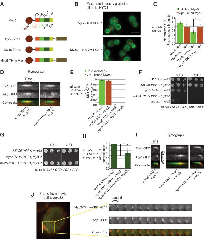
Actin polymerization powers membrane deformation during many processes, including clathrin-mediated endocytosis (CME). During CME in yeast, actin polymerization is triggered and coordinated by a six-protein WASP/Myosin complex that includes WASP, class I myosins (Myo3 and Myo5), WIP (Vrp1), and two other proteins. We show that a single engineered protein can replace this entire complex while still supporting CME. This engineered protein reveals that the WASP/Myosin complex has four essential activities: recruitment to endocytic sites, anchorage to the plasma membrane, Arp2/3 activation, and transient actin filament binding by the motor domain. The requirement for both membrane and F-actin binding reveals that myosin-mediated coupling between actin filaments and the base of endocytic sites is essential for allowing actin polymerization to drive membrane invagination.


Figures

Figure 1
Figure 1. Simplification of the WASP/Myosin complex
(A) A diagram of the components of the WASP/Myosin complex (circled with dotted line), the coat protein Sla1 (green) that is hypothesized to recruit Las17, and proteins of the Actin Module (red) that are directly or indirectly recruited by WASP/Myosin complex NPF activity. Black arrows indicate known or theorized physical interactions between complex components while gray lines indicate regulatory relationships. (B) Single frames from movies of wild-type (WT) cells and cells with various WASP/Myosin complex components deleted, all expressing Sla1-GFP and Abp1-RFP. A kymograph of a representative endocytic patch from each cell type is shown to the right of each image. (C) Proportion of Sla1-GFP patches that internalize more than 3 pixels (194 nm). At least 10 patches from the mother cell (or two similar cells if fewer than 10 patches could be counted for a cell) were scored for movement and an average was obtained for each cell. At least fifteen cells on three or more separate days of imaging were analyzed. Values represent the mean +/− SEM. *P<0.001 vs MYO5 (myo3Δ). (D) Growth of WASP/Myosin complex components on YPD plates. Starter cultures were diluted to an O.D600 of 0.02 and plated. Two additional 1:5 dilutions were also plated. Plates were grown at the indicated temperature for 48 hours and imaged. See also Figure S1, Movie S1, Tables S1 and S2.
Figure 2
Figure 2. Endocytic roles of the Myo5 TH2 and SH3 domains
(A) Diagram of the domains in Myo5 and each Myo5 variant analyzed in Figure 2. (B) Maximum intensity projection of Myo5-GFP and Myo5-Vrp1-GFP patches (Green) imaged over the course of a 120 second movie in the medial focal plane. Each culture also contains cells with a reference Myo5-GFP and Mtw1-mCherry for identification. A maximum intensity Z-projection of Mtw1-mCherry fluorescence is shown to identify the reference cells in each field (red dots). Scale bar is 5 μm. (C) Kymographs of representative endocytic patches from cells expressing Sla1-GFP and Abp1-RFP and the indicated Myo5 variant. (D) Growth of Myo5 variants on YPD plates, as described for Figure 1D. (E) Proportion of Sla1-GFP patches that internalize, as described in Figure 1C. *p<0.001 vs MYO5(myo3Δ); ** p<0.05 between bracketed variants; *** p<0.001 between bracketed variants. (F) Histogram of normalized fluorescence intensity peaks of the indicated GFP-tagged Myo5 variants. Values represent the mean +/− SEM. * p<0.01 vs Myo5-GFP; **p<0.05 between bracketed variants; ***p<0.001 between bracketed variants. See also Figure S2, Movie S2, Tables S1 and S2.
Figure 3
Figure 3. Endocytic roles of the Myo5 motor domain
(A) Diagram of each Myo5 variant analyzed in Figure 3. (B) Kymographs of representative endocytic patches from cells expressing Sla1-GFP and Abp1-RFP. (C) Proportion of Sla1-GFP patches that internalize, as described in Figure 1C. *P<0.001 vs MYO5 (myo3Δ). (D and E) Growth of Myo5 variants on YPD plates, as described for Figure 1D. (F) Proportion of Sla1-GFP patches that internalize, as described in Figure 1C. *P<0.001 vs MYO5 (myo3Δ). (G) Growth of Myo5 variants on YPD plates, as described for Figure 1D. (H) Proportion of Sla1-mCherry patches that internalize, as described in Figure 1C. *P<0.001 vs MYO5-GFP (MYO3). Myo5-GFP fluorescence was not imaged. (I) Single frame from movies of sla2Δ cells expressing Sla1-GFP and Abp1-RFP with the indicated motor domain variants indicated. Also, kymographs from each cell are shown to the right of each image to show the extent of Abp1-RFP internalization relative to the Myo5-GFP signal. Image scale bar represents 5 μm. Vertical scale bar in kymograph is 1.0 μm, horizontal scale bar in kymograph is 20 seconds. See also Figure S3, Movies S3, Tables S1 and S2.
Figure 4
Figure 4. Endocytic roles of the Myo5 TH1 domain
(A) Diagram of each Myo5 variant analyzed in Figure 4. (B) Maximum intensity projection of Myo5-TH1Δ-GFP and Myo5-TH1Δ-Vrp1-GFP patches mixed with reference Myo5-GFP cells; all as described for Figure 2B. Scale bar is 5 μm (C) Histogram of normalized fluorescence intensity peaks of the indicated GFP-tagged Myo5 variants. * p<0.01 vs Myo5-GFP; ***p<0.001 between bracketed variants. (D) Kymographs of representative endocytic patches from cells expressing Sla1-GFP and Abp1-RFP and the indicated Myo5 variant. (E) Proportion of Sla1-GFP patches that internalize, as described in Figure 1C. *P<0.001 vs MYO5 (myo3Δ). (F-G) Growth of Myo5 variants on YPD plates at the indicated temperatures, as described for Figure 1D. (H) Proportion of Sla1-GFP patches that internalize, as described in Figure 1C. *P<0.001 vs MYO5(myo3Δ); ***P<0.001 for bracketed variants. (I) Kymographs of representative endocytic patches from cells expressing Sla1-GFP and Abp1-RFP and the Myo5 variant indicated below. (J) Single frame from a movie of cells expressing Myo5-TH1Δ-Vrp1-GFP and Abp1-RFP. To the right is a montage showing the boxed from the image over several consecutive frames. The co-localization of Myo5-TH1Δ-Vrp1-GFP with the leading edge of Abp1-RFP tails is a feature seen in numerous cells with this genotype. See also Figure S4, Movie S4, Tables S1 and S2.
Figure 5
Figure 5. WASP/Myosin engineered protein can be simplified to contain only motor, TH1, PRR and NPF domains
(A) Diagram of all truncations of the Myo5-Las17 fusion protein analyzed in this Figure. (B) Growth of Myo5-Las17 fusion protein variants on YPD, as described in Figure 1D. (C) Kymographs of representative endocytic patches from cells expressing Sla1-GFP and Abp1-RFP and the indicated Myo5-Las17 variant indicated below. In all cells analyzed, the entire WASP/Myosin complex has been deleted and Myo5 has been replaced by the Myo5-Las17 fusion protein or fusion protein variant. (D) Proportion of Sla1-GFP patches that internalize, as described in Figure 1C. See also Figure S5.
Figure 6
Figure 6. WASP/Myosin complex functions as a single protein with only four activities
(A) Model: The WASP/Myosin complex has four activities that are necessary for CME and sufficient for a functional WASP/Myosin complex. (B) Theoretical outward-directed movement of individual, growing actin filaments that are tethered to the bud tip but not the base. (C) Model: The TH1 and motor domains constitute a membrane-actin linkage that tethers F-actin to the base of endocytic sites. While the F-actin at endocytic sites is a branched, cross-linked network, we have shown only two individual filaments for clarity of presentation. See also Movie S5.

References

    1. Aghamohammadzadeh S, Ayscough KR. Differential requirements for actin during yeast and mammalian endocytosis. Nat. Cell Biol. 2009;11:1039–1042. - PMC - PubMed
    1. Boettner DR, Friesen H, Andrews B, Lemmon SK. Clathrin light chain directs endocytosis by influencing the binding of the yeast Hip1R homologue, Sla2, to F-actin. Mol. Biol. Cell. 2011;22:3699–3714. - PMC - PubMed
    1. Boettner DR, Chi RJ, Lemmon SK. Lessons from yeast for clathrin-mediated endocytosis. Nat. Cell Biol. 2012;14:2–10. - PMC - PubMed
    1. Boulant S, Kural C, Zeeh J-C, Ubelmann F, Kirchhausen T. Actin dynamics counteract membrane tension during clathrin-mediated endocytosis. Nat. Cell Biol. 2012;13:1124–1131. - PMC - PubMed
    1. Burke T. a, Christensen JR, Barone E, Suarez C, Sirotkin V, Kovar DR. Homeostatic actin cytoskeleton networks are regulated by assembly factor competition for monomers. Curr. Biol. 2014;24:579–585. - PMC - PubMed

Publication types