. 2016 Jul;9(4):1039-1050.
doi: 10.1038/mi.2015.119. Epub 2015 Nov 11.

Pseudomonas aeruginosa pyocyanin modulates mucin glycosylation with sialyl-Lewis(x) to increase binding to airway epithelial cells

Affiliations

Pseudomonas aeruginosa pyocyanin modulates mucin glycosylation with sialyl-Lewis(x) to increase binding to airway epithelial cells

Jayme L Jeffries et al. Mucosal Immunol. 2016 Jul.

Abstract

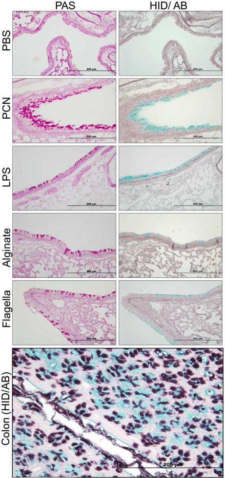
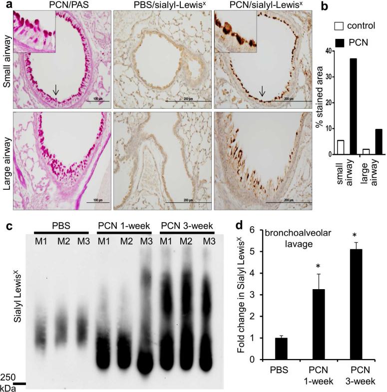
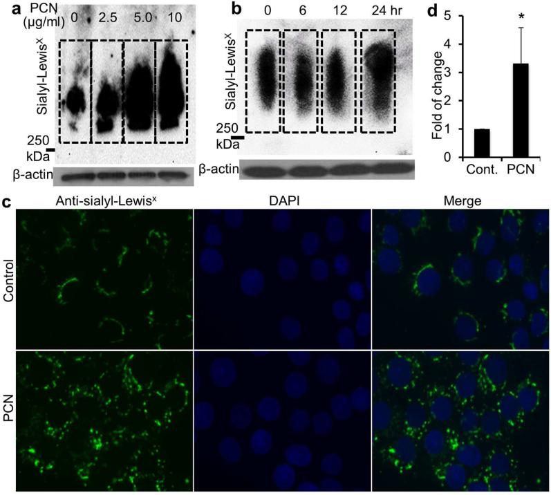
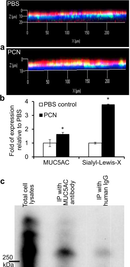
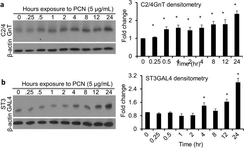
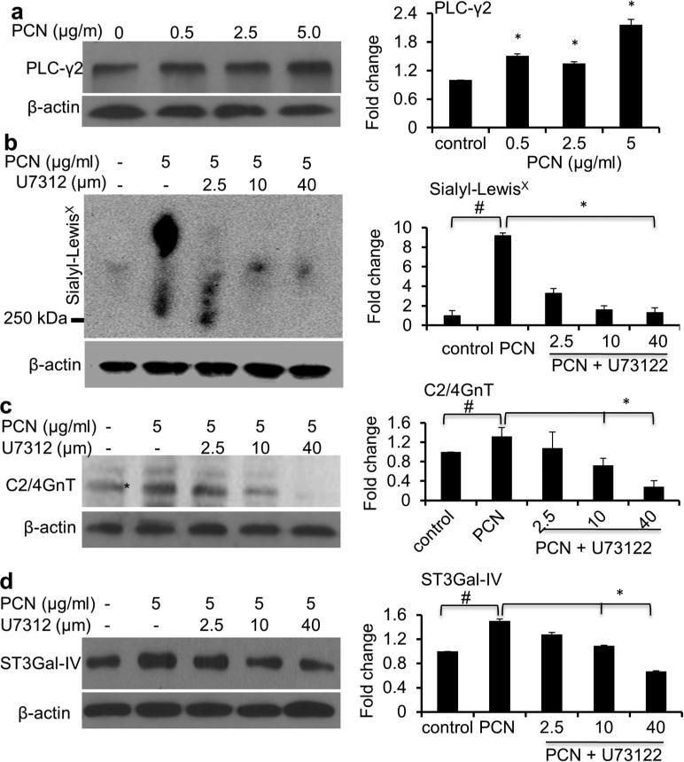
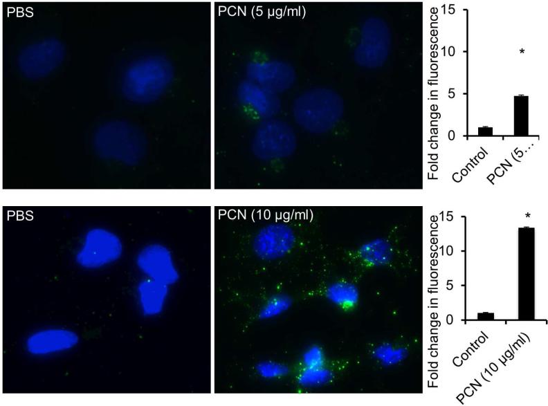
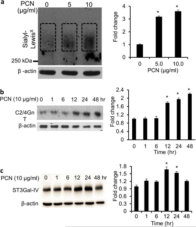
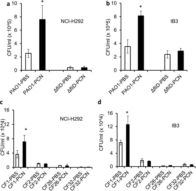
Cystic fibrosis (CF) patients battle life-long pulmonary infections with the respiratory pathogen Pseudomonas aeruginosa (PA). An overabundance of mucus in CF airways provides a favorable niche for PA growth. When compared with that of non-CF individuals, mucus of CF airways is enriched in sialyl-Lewis(x), a preferred binding receptor for PA. Notably, the levels of sialyl-Lewis(x) directly correlate with infection severity in CF patients. However, the mechanism by which PA causes increased sialylation remains uncharacterized. In this study, we examined the ability of PA virulence factors to modulate sialyl-Lewis(x) modification in airway mucins. We found pyocyanin (PCN) to be a potent inducer of sialyl-Lewis(x) in both mouse airways and in primary and immortalized CF and non-CF human airway epithelial cells. PCN increased the expression of C2/4GnT and ST3Gal-IV, two of the glycosyltransferases responsible for the stepwise biosynthesis of sialyl-Lewis(x), through a tumor necrosis factor (TNF)-α-mediated phosphoinositol-specific phospholipase C (PI-PLC)-dependent pathway. Furthermore, PA bound more efficiently to airway epithelial cells pre-exposed to PCN in a flagellar cap-dependent manner. Importantly, antibodies against sialyl-Lewis(x) and anti-TNF-α attenuated PA binding. These results indicate that PA secretes PCN to induce a favorable environment for chronic colonization of CF lungs by increasing the glycosylation of airway mucins with sialyl-Lewis(x).


Figures

Figure 1
Figure 1
PCN is a potent inducer of sialomucins. Serial sections of paraffin-embedded lungs from mice (n=10) exposed to PBS or various purified PA components were stained using PAS to detect goblet cells and high iron diamine/Alcian blue (HID/AB) to detect sialo- and sulfomucins. Sections of mouse colon were used as positive control tissues for the HID/ AB staining. PAS-stained goblet cells are pink. Sialomucins in colon stained blue. Sulfomucins stained brown.
Figure 2
Figure 2
PCN induces the expression of sialyl-Lewisx epitopes in mouse airways. (a) IHC analyses of mouse lung sections exposed to PCN or PBS stained with an anti-sialyl-Lewisx antibody. Serial PCN-exposed sections were stained with PAS for the presence of goblet cell hyperplasia and metaplasia. Magnified areas are indicated by arrows. (b) Sialyl-Lewisx stained areas in control versus PCN-treated large and small airways in C57BL6 mice were quantified using the ImageJ software. Average values large and small airways from 3 mice from each treatment, respectively, are shown. (c) Western blot analysis of sialyl-Lewisx in the BALF (15 μg total protein per lane) of mice exposed to PBS (once daily, 3 weeks) and or to 25 μg PCN (once daily, 1 week or 3 weeks. (d) Densitometry analysis of sialyl-LewisX expression in (c). Statistical significance comparisons among various time points were determined by using the one-way ANOVA analysis (p < 0.05). *p < 0.05 when compared against PBS control by using the Tukey's test.
Figure 3
Figure 3
PCN upregulates the expression of sialyl-Lewisx in time and concentration-dependent manners. (a and b) NCI-H292 cells were exposed to PBS (control) or indicated concentrations of PCN or at predetermined time intervals (with 5 μg/ml PCN). Total proteins were separated on an agarose-acrylamide gel. The expression of sialyl-Lewisx was analyzed using specific antibody by Western blots. β-actin was used as loading controls. Densitometry analysis of the sialyl-LewisX expression can be found in Figure S1. (c) Separate sets of cells from above were stained for sialyl-LewisX with antibodies and visualized with Alexa Fluor®488-conjugated secondary antibody (green color). (d) Quantification of total fluorescence of control and PCN treated cells. The mean ± standard error of total florescence in 10 representative high power fields from each treatment group in one typical experiment is shown. *p < 0.05 when compared against PBS control using the parametric Student's t test.
Figure 4
Figure 4
PCN induces sialylation of MUC5AC mucin in ALI culture of NHBE cells and in immortalized airway epithelial cells. (a) Differentiated NHBE cells were exposed to PBS or PCN (12.5 μg/ml) for 24 hr. MUC5AC mucin and sialyl-LewisX were stained with specific primary antibodies, and visualized with the Alexa Fluor®488-conjugated secondary antibody (green color, MUC5AC) or Alexa Fluor®647-conjugated secondary antibody (red color, sialyl-Lewisx). Nuclei were stained with DAPI (blue color). Three independent experiments showed similar results. Typical strips of cells are shown. (b) Expression of MUC5AC and sialyl-Lewisx from 10 representative strips were quantified using the AxioVision Rel. 4.8 software. *p < 0.05 when compared against PBS control using the parametric Student's t test. (c) Sialylation of MUC5AC in NCI-H292 cells. After exposure to PCN (24 hr, 5 μg/ml), total protein extracts (600 μg/ml) were immunoprecipitated with anti-MUC5AC antibody, or with human IgG as negative controls. Proteins were separated on an agarose-acrylamide gel and the expression of sialyl-Lewisx was analyzed using specific antibody by Western blotting. Experiments were repeated independently three times with similar results. A typical Western blot is shown.
Figure 5
Figure 5
PCN induces the expression of sialyl-Lewisx glycosyltransferases. NCI-H292 cells were exposed to 5 μg/ml PCN for the indicated time intervals. (a, b) The expression of C2/4GnT and ST3Gal-IV was analyzed by western blots. β-actin was used as loading controls. Representative western blots from one of the three independent experiments are shown. Densitometry analyses of C2/4GnT and ST3Gal-IV represent the mean ± standard error from three independent western blot experiments. Statistical significance comparisons among various time points were determined by using the one-way ANOVA analysis (p < 0.05). *p < 0.05 when compared against Time 0 hr by using the using the Tukey's test.
Figure 6
Figure 6
PCN upregulates sialyl-Lewisx through the PI-PLC pathway. (a) PCN induced the expression of PI-PLC signaling pathway effector PLC-γ2 in NCI-H292 cells exposed to increasing concentrations of PCN. (b-d) NCI-H292 cells were pre-exposed to PI-PLC inhibitor U73122 for 40 minutes before the addition of PBS or PCN (5 μg/ml) for 24 hr. Total cell lysates were used in Western blot analysis using antibodies against (a) PLC-γ2, (b) sialyl-LewisX, (c) C2/4GnT or (d) ST3Gal-IV. For sialyl-LewisX western blot (b), proteins were separated on an agarose-acrylamide gel. β-actin was used for loading controls. Experiments were repeated three times. Representative western blots are shown. Densitometry analyses represent the mean ± standard error of western blots from three independent experiments. *p < 0.05 against PBS control, or in the presence or absence of U73122 by using the one-way ANOVA analysis. #p < 0.05 against PBS control by using the Tukey's test.
Figure 7
Figure 7
PCN upregulates the expression of sialyl-Lewisx in immortalized CF epithelial cells. IB3-1 cells were exposed to PBS (control) or indicated concentrations of PCN for 48 hr. Sialyl-LewisX was stained with antibodies and visualized with Alexa Fluor®488-conjugated secondary antibody (green color). Quantification of total fluorescence of control and PCN treated cells. The mean ± standard error of total florescence in 10 representative high power fields from each treatment group in one typical experiment is shown. *p < 0.05 when compared against PBS control by parametric Student's t-test.
Figure 8
Figure 8
PCN upregulates the expression of sialyl-Lewisx in CF airway epithelial cells. IB3-1 cells were exposed to PBS (control) or indicated concentrations of PCN or time intervals (with 5 μg/ml PCN). (a) The expression of sialyl-Lewisx was analyzed using specific antibody by Western blots. Total protein extracts were separated on an agarose-acrylamide gel. (b and c) The expression of C2/4GnT (b) and ST3Gal-IV (c) was analyzed by western blots. β-actin was used as loading controls. Representative western blots from one of the three independent experiments are shown. Densitometry analyses represent the mean ± standard error from three independent western blot experiments. *p < 0.05 when compared sialyl-Lewisx (a) and C2/4GnT (b) and ST3Gal-IV (c) expression among the group by using the one-way ANOVA analysis. *p < 0.05 when compared each sample against PBS control (a) or Time 0 hr (b) by using the Tukey's test.
Figure 9
Figure 9
Pyocyanin increases P. aeruginosa binding efficiency to NCI-H292 cells. PA strain PAO1 was added in a 1:1 ratio to NCI-H292 cells (n = 12 wells) pretreated with PBS or PCN (5. μg/ml) for 24 hr. After 1 hr, cells were washed to remove unattached PAO1, lysed, serially diluted, and plated on Pseudomonas isolation agar. Data represents CFU recovered from each group. *p < 0.05 when compared bacterial binding among various treatments by using the one-way ANOVA analysis. *p < 0.05 when compared bacterial binding in each sample against PBS control by using the Tukey's test.
Figure 10
Figure 10
Antibodies against sialyl-LewisX and TNF-α attenuate the binding of P. aeruginosa to airway epithelial cells pre-exposed to PCN. NCI-H292 and IB3-1 cells were serum-starved for 24 hr before exposure to PCN (5 μg/ml for 24 hr or 10 μg/ml for 48 hr, respectively). To block sialomucins, anti-sialyl-LewisX antibodies (15 μg/well) were added 2 hr before the binding assay. To block TNF-α, anti-TNF-α antibodies (15 μg/well) were added simultaneously with PCN for the same duration. Human IgG (15 μg/well) was used as controls. PA strains PAO1 or CF1 were added in a 1:1 ratio to NCI-H292 or IB3-1 cells (n = 12 wells). After 1 hr, cells were washed to remove unattached PAO1, lysed, serially diluted, and plated on Pseudomonas isolation agar. Data represents CFU recovered from each group. *p < 0.05 when compared bacterial binding among various treatments by using the one-way ANOVA analysis. *p < 0.05 when compared bacterial binding in each sample against human IgG controls by using the Tukey's test. PAO1-IgG and CF1-IgG: binding of bacteria to airway cells pre-blocked with human IgG. PAO1-α-SLX and CF1-α-SLX: binding of bacteria to airway cells pre-blocked with anti-sialyl-LewisX antibodies. PAO1-α-TNF-α and CF1-α-TNF-α: binding of bacteria to airway cells pre-blocked with anti- TNF-α antibodies.

References

    1. Boucher RC. An overview of the pathogenesis of cystic fibrosis lung disease. Adv. Drug Deliver. Rev. 2002;54:1359–1371. - PubMed
    1. Lyczak JB, Cannon CL, Pier GB. Lung infections associated with cystic fibrosis. Clin. Microbiol. Rev. 2002;15:194–222. - PMC - PubMed
    1. Burns JL, et al. Longitudinal assessment of Pseudomonas aeruginosa in young children with cystic fibrosis. J. Infect. Dis. 2001;183:444–452. - PubMed
    1. Carnoy C, Scharfman A, Van Brussel E, Lamblin G, Ramphal R, Roussel P. Pseudomonas aeruginosa outer membrane adhesins for human respiratory mucus glycoproteins. Infect. Immun. 1994;62:1896–1900. - PMC - PubMed
    1. Lamblin G, et al. Human airway mucin glycosylation: a combinatory of carbohydrate determinants which vary in cystic fibrosis. Glycoconjugate J. 2001;18:661–684. - PubMed

MeSH terms