

Disruption of Ah Receptor Signaling during Mouse Development Leads to Abnormal Cardiac Structure and Function in the Adult
- PMID: 26555816
- PMCID: PMC4640841
- DOI: 10.1371/journal.pone.0142440
Disruption of Ah Receptor Signaling during Mouse Development Leads to Abnormal Cardiac Structure and Function in the Adult
Abstract
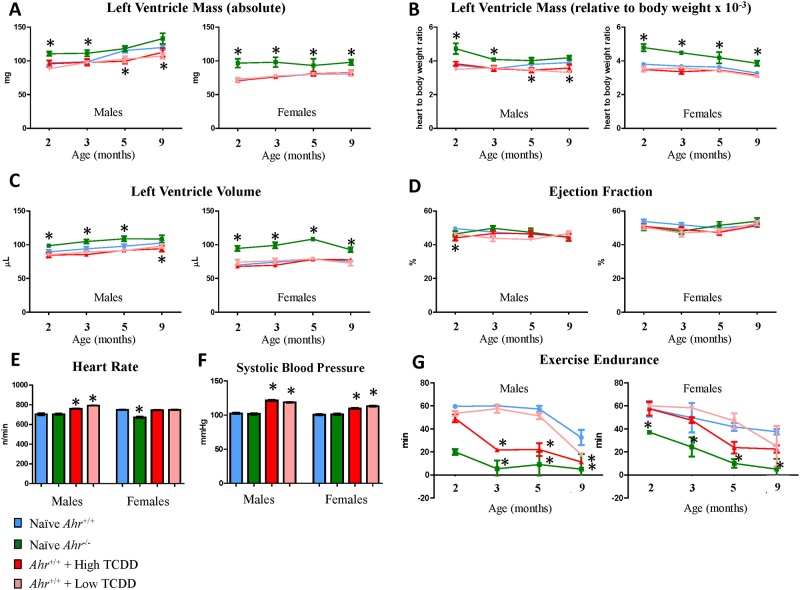
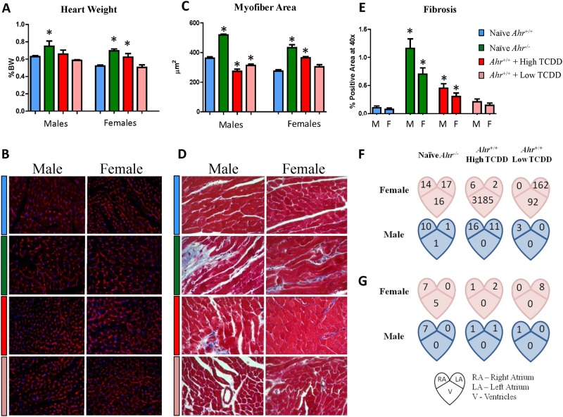
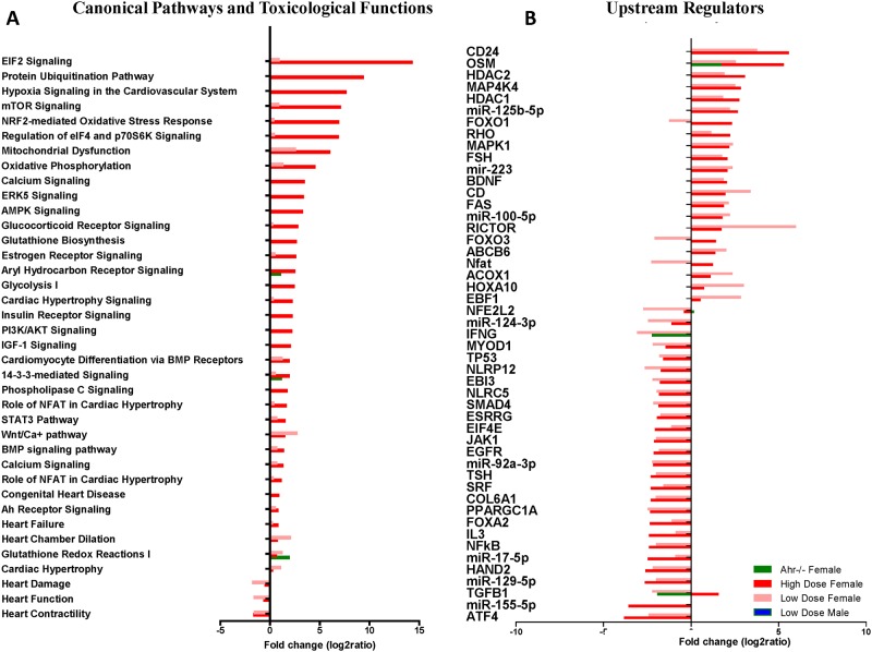
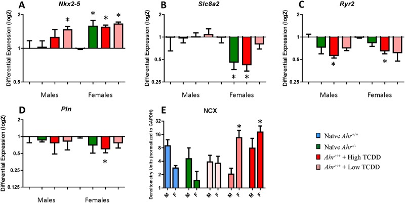
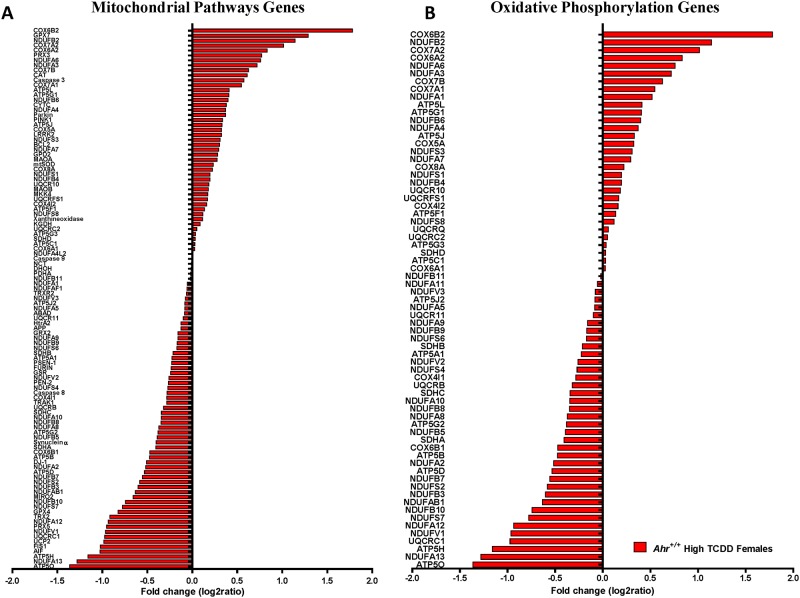
The Developmental Origins of Health and Disease (DOHaD) Theory proposes that the environment encountered during fetal life and infancy permanently shapes tissue physiology and homeostasis such that damage resulting from maternal stress, poor nutrition or exposure to environmental agents may be at the heart of adult onset disease. Interference with endogenous developmental functions of the aryl hydrocarbon receptor (AHR), either by gene ablation or by exposure in utero to 2,3,7,8-tetrachlorodibenzo-p-dioxin (TCDD), a potent AHR ligand, causes structural, molecular and functional cardiac abnormalities and altered heart physiology in mouse embryos. To test if embryonic effects progress into an adult phenotype, we investigated whether Ahr ablation or TCDD exposure in utero resulted in cardiac abnormalities in adult mice long after removal of the agent. Ten-months old adult Ahr-/- and in utero TCDD-exposed Ahr+/+ mice showed sexually dimorphic abnormal cardiovascular phenotypes characterized by echocardiographic findings of hypertrophy, ventricular dilation and increased heart weight, resting heart rate and systolic and mean blood pressure, and decreased exercise tolerance. Underlying these effects, genes in signaling networks related to cardiac hypertrophy and mitochondrial function were differentially expressed. Cardiac dysfunction in mouse embryos resulting from AHR signaling disruption seems to progress into abnormal cardiac structure and function that predispose adults to cardiac disease, but while embryonic dysfunction is equally robust in males and females, the adult abnormalities are more prevalent in females, with the highest severity in Ahr-/- females. The findings reported here underscore the conclusion that AHR signaling in the developing heart is one potential target of environmental factors associated with cardiovascular disease.
Conflict of interest statement
Figures
Similar articles
-
Developmental and lifelong dioxin exposure induces measurable changes in cardiac structure and function in adulthood.Sci Rep. 2021 May 17;11(1):10378. doi: 10.1038/s41598-021-89825-w. Sci Rep. 2021. PMID: 34001975 Free PMC article.
-
Ah receptor expression in cardiomyocytes protects adult female mice from heart dysfunction induced by TCDD exposure.Toxicology. 2016 Apr 29;355-356:9-20. doi: 10.1016/j.tox.2016.05.006. Epub 2016 May 6. Toxicology. 2016. PMID: 27163630
-
Aryl Hydrocarbon Receptor Ablation in Cardiomyocytes Protects Male Mice From Heart Dysfunction Induced by NKX2.5 Haploinsufficiency.Toxicol Sci. 2017 Nov 1;160(1):74-82. doi: 10.1093/toxsci/kfx164. Toxicol Sci. 2017. PMID: 28973413 Free PMC article.
-
The aryl hydrocarbon receptor: studies using the AHR-null mice.Drug Metab Dispos. 1998 Dec;26(12):1194-8. Drug Metab Dispos. 1998. PMID: 9860927 Review.
-
The Impact of the Aryl Hydrocarbon Receptor on Antenatal Chemical Exposure-Induced Cardiovascular-Kidney-Metabolic Programming.Int J Mol Sci. 2024 Apr 23;25(9):4599. doi: 10.3390/ijms25094599. Int J Mol Sci. 2024. PMID: 38731818 Free PMC article. Review.
Cited by
-
Design, Synthesis, and In Vitro Evaluation of Novel Indolyl DiHydropyrazole Derivatives as Potential Anticancer Agents.Molecules. 2021 Aug 29;26(17):5235. doi: 10.3390/molecules26175235. Molecules. 2021. PMID: 34500672 Free PMC article.
-
Fine particulate matter‑induced cardiac developmental toxicity (Review).Exp Ther Med. 2024 Oct 29;29(1):6. doi: 10.3892/etm.2024.12756. eCollection 2025 Jan. Exp Ther Med. 2024. PMID: 39534282 Free PMC article. Review.
-
Developmental and lifelong dioxin exposure induces measurable changes in cardiac structure and function in adulthood.Sci Rep. 2021 May 17;11(1):10378. doi: 10.1038/s41598-021-89825-w. Sci Rep. 2021. PMID: 34001975 Free PMC article.
-
Dioxin and AHR impairs mesoderm gene expression and cardiac differentiation in human embryonic stem cells.Sci Total Environ. 2019 Feb 15;651(Pt 1):1038-1046. doi: 10.1016/j.scitotenv.2018.09.247. Epub 2018 Sep 20. Sci Total Environ. 2019. PMID: 30266049 Free PMC article.
-
Interferon regulatory factor 8 regulates caspase-1 expression to facilitate Epstein-Barr virus reactivation in response to B cell receptor stimulation and chemical induction.PLoS Pathog. 2018 Jan 22;14(1):e1006868. doi: 10.1371/journal.ppat.1006868. eCollection 2018 Jan. PLoS Pathog. 2018. PMID: 29357389 Free PMC article.
References
Publication types
MeSH terms
Substances
Associated data
- Actions
Grants and funding
LinkOut - more resources
Full Text Sources
Other Literature Sources
Medical
Molecular Biology Databases
