. 2015 Nov 4;16(11):26333-46.
doi: 10.3390/ijms161125958.

Isolation and Culture of Pig Spermatogonial Stem Cells and Their in Vitro Differentiation into Neuron-Like Cells and Adipocytes

Affiliations

Isolation and Culture of Pig Spermatogonial Stem Cells and Their in Vitro Differentiation into Neuron-Like Cells and Adipocytes

Xiaoyan Wang et al. Int J Mol Sci. .

Abstract

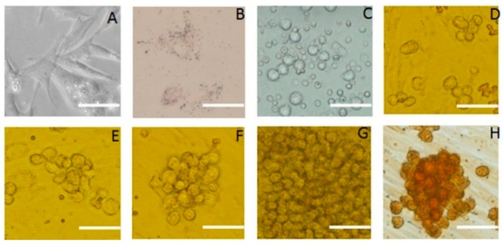
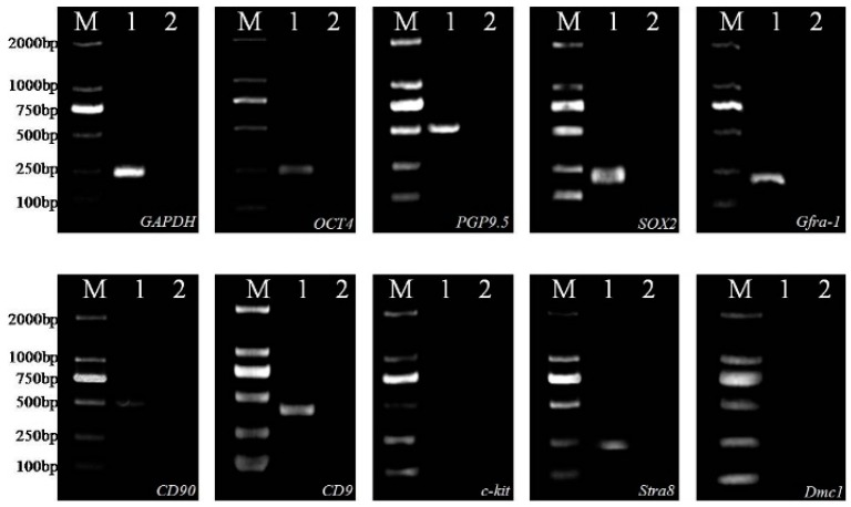
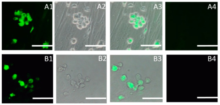
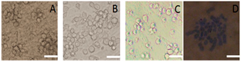
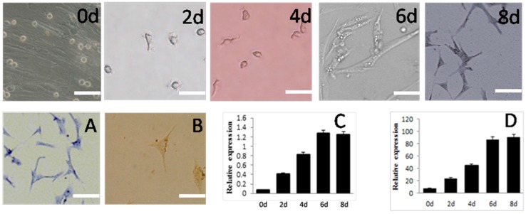
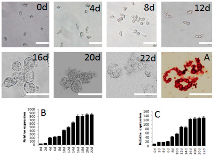
Spermatogonial stem cells (SSCs) renew themselves throughout the life of an organism and also differentiate into sperm in the adult. They are multipopent and therefore, can be induced to differentiate into many cells types in vitro. SSCs from pigs, considered an ideal animal model, are used in studies of male infertility, regenerative medicine, and preparation of transgenic animals. Here, we report on a culture system for porcine SSCs and the differentiation of these cells into neuron-like cells and adipocytes. SSCs and Sertoli cells were isolated from neonatal piglet testis by differential adhesion and SSCs were cultured on a feeder layer of Sertoli cells. Third-generation SSCs were induced to differentiate into neuron-like cells by addition of retinoic acid, β-mercaptoethanol, and 3-isobutyl-1-methylxanthine (IBMX) to the induction media and into adipocytes by the addition of hexadecadrol, insulin, and IBMX to the induction media. The differentiated cells were characterized by biochemical staining, qRT-PCR, and immunocytochemistry. The cells were positive for SSC markers, including alkaline phosphatase and SSC-specific genes, consistent with the cells being undifferentiated. The isolated SSCs survived on the Sertoli cells for 15 generations. Karyotyping confirmed that the chromosomal number of the SSCs were normal for pig (2n = 38, n = 19). Pig SSCs were successfully induced into neuron-like cells eight days after induction and into adipocytes 22 days after induction as determined by biochemical and immunocytochemical staining. qPCR results also support this conclusion. The nervous tissue markers genes, Nestin and β-tubulin, were expressed in the neuron-like cells and the adipocyte marker genes, PPARγ and C/EBPα, were expressed in the adipocytes.

Keywords: SSCs; adipocytes; in vitro culture; induction; neuron-like cells.


Figures

Figure 1
Figure 1
Morphology of SSCs and Sertoli cells in culture. Appearance under the microscope and various staining properties were used to identify the cells. (A) Sertoli cells derived from porcine testicular tissue; (B) Oil red-o staining of Sertoli cells; (C) Primary SSCs derived from porcine testicular tissue; (DG) Colony of SSCs grown on feeder cells for 3, 5, 7, 12 days; (H) SSCs clone cells stained for alkaline phosphatase activity; the image indicates strong alkaline phosphatase activity. Scale bar: 5 μm.
Figure 2
Figure 2
Expression of SSC-specific genes in cultured cells as determined by RT-PCR. M, DL2000 Marker used as a molecular size indicator; Lane 1, specific genes expression with cDNA of cultured cells; Lane 2, negative control with RNA of cultured cells; GAPDH used as positive control in cultured cells.
Figure 3
Figure 3
Immunocytochemical staining of SSCs using antibodies (FITC-conjugated secondary antibody, green) against SOX2 and SSEA-1 considered markers of pluripotency. (A1) SOX2, dark field; (A2) SOX2, bright field; (A3) Merged with dark field and bright; (A4) Negative control; (B1) SSEA-1, dark field; (B2) SSEA-1, bright field; (B3) Merged with dark field and bright; (B4) Negative control. Scale bar: 5 μm.
Figure 4
Figure 4
Images of cultured porcine SSCs after repeated subculture and their karyotype. (A) Cells in the fifth subculture; (B) Cells in the 10th subculture; (C) Cells in the15th subculture; (D) Karyotype cells in the 15th subculture. Scale bar: 5 μm.
Figure 5
Figure 5
Morphology and gene expression of SSCs differentiated into neuron-like cells in vitro. Appearance under the microscope and various staining properties were used to confirm their identity.Schematic represents the strategy used for in vitro differentiation. 0d, 2d, 4d, 6d, 8d mean the same, 2th, 4th, 6th, 8th day after induction. (A) Toluidine blue staining of SSCs-derived cells; (B) Immunocytochemcal staining of SSCs-derived cells; (C) Expression of Nestin and (D) β-tubulin, both measured by mRNA levels during induction. Scale bar: 5 μm.
Figure 6
Figure 6
Morphology and gene expression of SSCs differentiated into adipocytes in vitro. Appearance under the microscope and various staining properties were used to confirm their identity. Schematic represents the strategy used for in vitro differentiation. 0d, 2d, 4d, 6d, 8d, 10d, 12d, 14d, 16d, 18d, 20d, 22d mean the same, 2th, 4th, 6th, 8th, 10th, 12th, 14th, 16th, 18th, 20th, 22th day after induction. (A) Oil red-O staining of adipocytes derived from SSCs; (B) Expression of PPARγ and (C) C/EBPα, both measured by mRNA levels, during induction. Scale bar: 5 μm.

References

    1. De Rooij D.G. Stem cells in the testis. Int. J. Exp. Pathol. 1998;79:67–80. doi: 10.1046/j.1365-2613.1998.00057.x. - DOI - PMC - PubMed
    1. Brinster R.L. Germline stem cell transplantation and transgenesis. Science. 2002;296:2174–2176. doi: 10.1126/science.1071607. - DOI - PMC - PubMed
    1. Waheeb R., Hofmann M.C. Human spermatogonial stem cells: A possible origin for spermatocytic seminoma. Int. J. Androl. 2011;34:296–305. doi: 10.1111/j.1365-2605.2011.01199.x. - DOI - PMC - PubMed
    1. Tegelenbosch R.A., de Rooij D.G. A quantitative study of spermatogonial multiplication and stem cell renewal in the C3H/101 F1 hybrid mouse. Mutat Res. 1993;290:193–200. doi: 10.1016/0027-5107(93)90159-D. - DOI - PubMed
    1. Kubota H., Brinster R.L. Technology insight: In vitro culture of spermatogonial stem cells and their potential therapeutic uses. Nat. Clin. Pract. Endocrinol. Metab. 2006;2:99–108. doi: 10.1038/ncpendmet0098. - DOI - PMC - PubMed

Publication types