Review
. 2014:2014:473134.
doi: 10.1155/2014/473134. Epub 2014 Jan 23.

Mesenchymal Conversion of Mesothelial Cells Is a Key Event in the Pathophysiology of the Peritoneum during Peritoneal Dialysis

Affiliations
Review

Mesenchymal Conversion of Mesothelial Cells Is a Key Event in the Pathophysiology of the Peritoneum during Peritoneal Dialysis

Manuel López-Cabrera. Adv Med. 2014.

Abstract

Peritoneal dialysis (PD) is a therapeutic option for the treatment of end-stage renal disease and is based on the use of the peritoneum as a semipermeable membrane for the exchange of toxic solutes and water. Long-term exposure of the peritoneal membrane to hyperosmotic PD fluids causes inflammation, loss of the mesothelial cells monolayer, fibrosis, vasculopathy, and angiogenesis, which may lead to peritoneal functional decline. Peritonitis may further exacerbate the injury of the peritoneal membrane. In parallel with these peritoneal alterations, mesothelial cells undergo an epithelial to mesenchymal transition (EMT), which has been associated with peritoneal deterioration. Factors contributing to the bioincompatibility of classical PD fluids include the high content of glucose/glucose degradation products (GDPs) and their acidic pH. New generation low-GDPs-neutral pH fluids have improved biocompatibility resulting in better preservation of the peritoneum. However, standard glucose-based fluids are still needed, as biocompatible solutions are expensive for many potential users. An alternative approach to preserve the peritoneal membrane, complementary to the efforts to improve fluid biocompatibility, is the use of pharmacological agents protecting the mesothelium. This paper provides a comprehensive review of recent advances that point to the EMT of mesothelial cells as a potential therapeutic target to preserve membrane function.


Figures
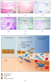
Figure 1
Figure 1
Structural alteration of the peritoneal membrane during PD. (a) Normal peritoneal tissue from a healthy donor stained with Haematoxylin-eosin (H&E) shows a preserved MCs monolayer that lines a compact zone of connective tissue (A). Peritoneal membrane from a PD patient stained with H&E shows the loss of the MCs monolayer and increased thickness of the compact zone (B). Magnification ×200. Staining of the peritoneal vessels with anti-CD31 antibody demonstrates an intense angiogenesis in peritoneal membrane from PD patient (C). Hyalinizing vasculopathy can be observed in the peritoneal tissue from PD patient (D). Immunohistochemical analysis of the peritoneal membrane from PD patient reveals the presence of fibroblast-like cells embedded in the fibrotic stroma expressing the mesothelial markers cytokeratins and calretinin (E) and (F). Magnification ×150. (b) Schematic representation of the progressive alterations of the peritoneal membrane in the time course of PD.
Figure 2
Figure 2
Multiple origins of myofibroblasts have been proposed in tissue fibrosis. Myofibroblasts may derive from at least five different sources through various mechanisms: phenotypic activation from interstitial fibroblasts; differentiation from vascular pericytes; recruitment from circulating fibrocytes; capillary endothelial-mesenchymal transition (EndMT); and epithelial-mesenchymal transition (EMT). The relative contribution of each source to the myofibroblast pool in peritoneal fibrosis still requires further studies.
Figure 3
Figure 3
Schematic illustration of the key events during MMT. Mesothelial to mesenchymal transition (MMT) occurs when mesothelial cells lose their epithelial-like characteristics, including dissolution of cell-cell junctions, that is, tight junctions, adherens junctions and desmosomes, and loss of apical-basolateral polarity, and acquire a mesenchymal phenotype, characterized by actin reorganization and stress fiber formation, migration, and invasion. The diagram shows four key steps essential for the completion of entire MMT, the most commonly used mesothelial and mesenchymal markers, and the molecules and signal transduction pathways that act either as inducer or modulator of the MMT process. See text for details.
Figure 4
Figure 4
Smad-dependent signaling pathways of TGF-β1 and BMP-7. The binding of TGF-β1 and BMP-7 to their primary receptors (receptors type II) allows the recruitment, transphosphorylation, and activation of the signaling receptors (receptors type I). The receptor type I of TGF-β1 phosphorylate Smad2 and Smad3. The receptor type I of BMP-7 phosphorylate Smad1, Smad5, and Smad8. These receptor-activated Smads form heterodimers with Smad4. The resulting Smad complexes are then translocated into the nucleus where they activate target genes involved either in the mesenchymal conversion of MCs (MMT) in the case of Smads2/3 or in the blocking/reversion of the mesenchymal transition (rMMT) in the case of Smads1/5/8. Smad6 and Smad7 control BMP-7- and TGF-β1-triggered Smad signaling by preventing the phosphorylation and/or nuclear translocation and by inducing receptor complex degradation through the recruitment of ubiquitin ligases. Extracellular regulation of TGF-β1 and BMP-7 is achieved by various molecules. CTGF inhibits BMP-7 and activates TGF-β1 signals by direct binding in the extracellular space. BMP-7 signaling might also be influenced by other BMP-7 modulators such as gremlin-1, kielin/chordin-like protein (KCP), or uterine sensitization-associated gene 1 (USAG-1).
Figure 5
Figure 5
Non-Smad signaling in response to TGF-β1 and BMP-7. TGF-β1 activates MAP kinases JNK and p38 signaling and NF-κB through the activation of TAK1 by receptor-associated TRAF6. TGF-β1 also activates MAP kinase ERK 1/2 signaling through recruitment and phosphorylation of Shc by the TGF-β1 type I receptor. In MCs the p38-mediated pathway acts as modulator of the mesenchymal conversion by a feedback mechanism based on the downregulation of ERKs 1/2, NF-κB, and TAK-1 activities. Interestingly, BMP-7 activates p38 signaling by receptor-associated XIAB, which may contribute to the maintaining epithelial-like phenotype. TGF-β1 also induces PI3-K/Akt pathway leading to the activation of mTOR and the stabilization of β-catenin and snail through the inactivation of GSK-3β. As a result, β-catenin localizes to the nucleus, where it feeds into the Wnt signaling pathway by interacting with lymphoid enhancer factor-1/T-cell factor (LEF1/TCF) and contributes to the transcription of mesenchymal-related genes. In addition, the nuclear translocation of snail promotes the transcriptional repression of E-cadherin and other intercellular adhesion molecules.
Figure 6
Figure 6
Therapeutic strategies for peritoneal membrane failure based on MMT. MMT in vivo results from integrated signals induced by multiple stimuli. These include high concentration of glucose and glucose degradation products (GDPs) in the PD fluids, which contribute to the formation of advanced glycation-end products (AGEs) and stimulate the mesenchymal conversion of MCs. The low pH of the dialysates and the mechanical injury during PD fluid exchanges may cause tissue irritation and contribute to chronic inflammation of the peritoneum, which promote MMT. Episodes of bacterial or fungal infections or hemoperitoneum cause acute inflammation and upregulation of cytokines and growth factors such as TGF-β, IL-1, TNF-α, and Angiotensin II, among others, which are strong inducers of MMT. The therapeutic strategies may be designed either to prevent or reverse the MMT itself, to decrease the MMT-promoting stimuli, or to treat MMT-associated effects such as the invasion capacity to avoid their accumulation in the compact zone. The diagram illustrates aspects related with the MMT process that can be clinically managed, alone or in combination, in order to prevent peritoneal membrane failure. See text for details.

Similar articles

Cited by

References

    1. Chaimovitz C. Peritoneal dialysis. Kidney International. 1994;45(4):1226–1240. - PubMed
    1. Devuyst O., Margetts P. J., Topley N. The pathophysiology of the peritoneal membrane. Journal of the American Society of Nephrology. 2010;21(7):1077–1085. doi: 10.1681/ASN.2009070694. - DOI - PubMed
    1. Krediet R. T. The peritoneal membrane in chronic peritoneal dialysis. Kidney International. 1999;55(1):341–356. doi: 10.1046/j.1523-1755.1999.00264.x. - DOI - PubMed
    1. Breborowicz A., Oreopoulos D. G. Biocompatibility of peritoneal dialysis solutions. American Journal of Kidney Diseases. 1996;27(5):738–743. - PubMed
    1. Wieczorowska-Tobis K., Polubinska A., Wisniewska J., et al. Multidirectional approach to study peritoneal dialysis fluid biocompatibility in a chronic peritoneal dialysis model in the rat. Nephrology Dialysis Transplantation. 2001;16(3):655–656. - PubMed

LinkOut - more resources