. 2016 Jan 12;7(2):1262-75.
doi: 10.18632/oncotarget.6014.

Leptin as a mediator of tumor-stromal interactions promotes breast cancer stem cell activity

Affiliations

Leptin as a mediator of tumor-stromal interactions promotes breast cancer stem cell activity

Cinzia Giordano et al. Oncotarget. .

Abstract

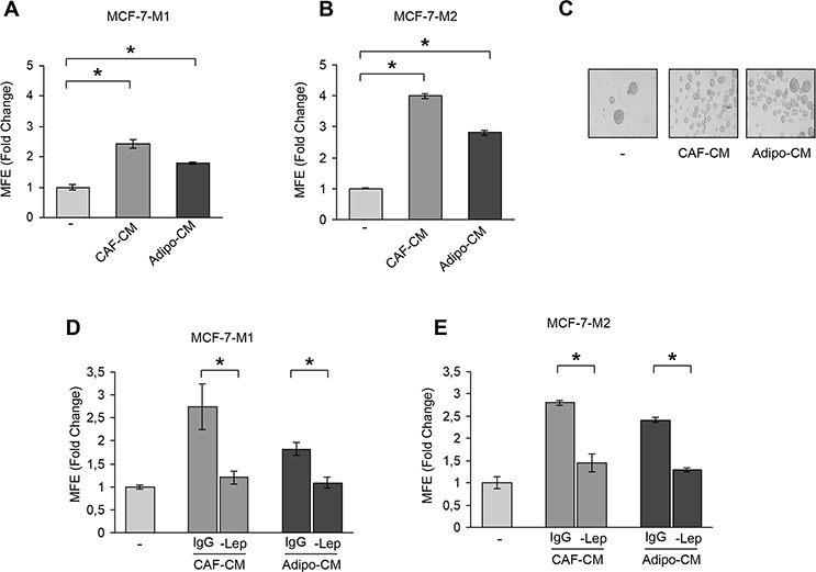
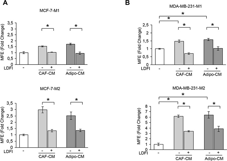
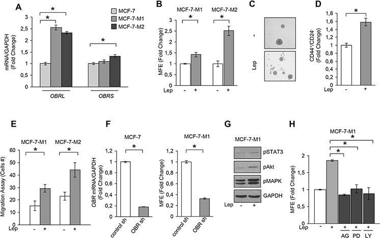
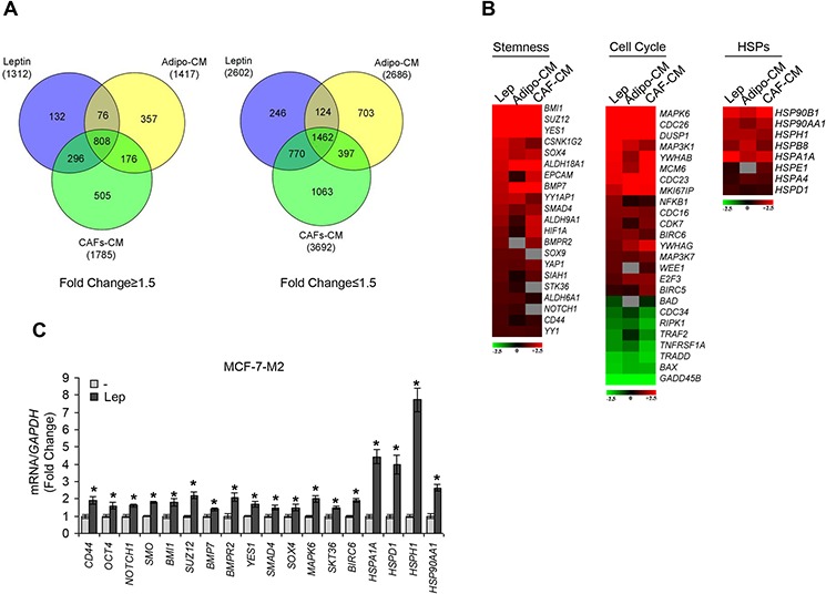
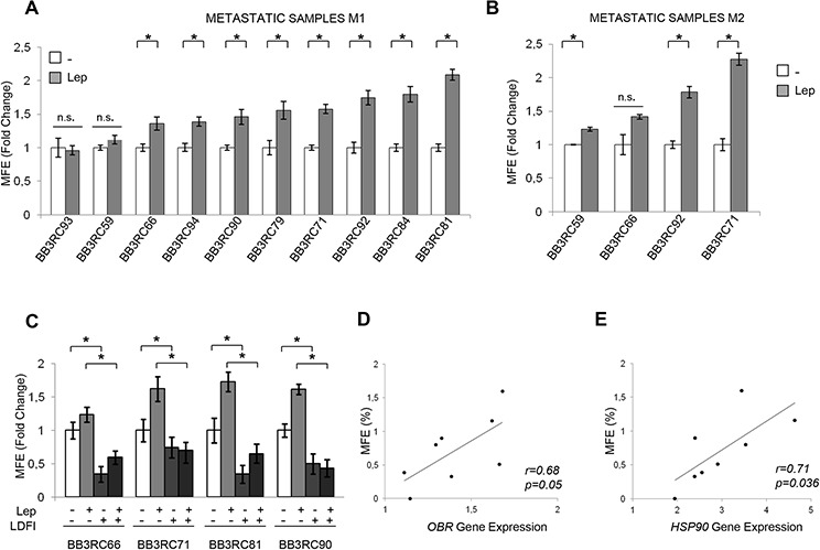
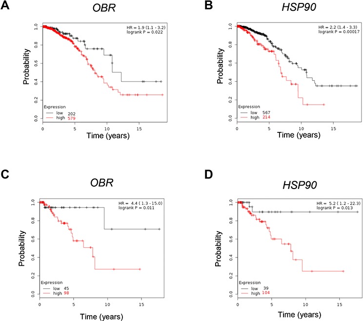
Breast cancer stem cells (BCSCs) play crucial roles in tumor initiation, metastasis and therapeutic resistance. A strict dependency between BCSCs and stromal cell components of tumor microenvironment exists. Thus, novel therapeutic strategies aimed to target the crosstalk between activated microenvironment and BCSCs have the potential to improve clinical outcome. Here, we investigated how leptin, as a mediator of tumor-stromal interactions, may affect BCSC activity using patient-derived samples (n = 16) and breast cancer cell lines, and determined the potential benefit of targeting leptin signaling in these model systems. Conditioned media (CM) from cancer-associated fibroblasts and breast adipocytes significantly increased mammosphere formation in breast cancer cells and depletion of leptin from CM completely abrogated this effect. Mammosphere cultures exhibited increased leptin receptor (OBR) expression and leptin exposure enhanced mammosphere formation. Microarray analyses revealed a similar expression profile of genes involved in stem cell biology among mammospheres treated with CM and leptin. Interestingly, leptin increased mammosphere formation in metastatic breast cancers and expression of OBR as well as HSP90, a target of leptin signaling, were directly correlated with mammosphere formation in metastatic samples (r = 0.68/p = 0.05; r = 0.71/p = 0.036, respectively). Kaplan-Meier survival curves indicated that OBR and HSP90 expression were associated with reduced overall survival in breast cancer patients (HR = 1.9/p = 0.022; HR = 2.2/p = 0.00017, respectively). Furthermore, blocking leptin signaling by using a full leptin receptor antagonist significantly reduced mammosphere formation in breast cancer cell lines and patient-derived samples. Our results suggest that leptin/leptin receptor signaling may represent a potential therapeutic target that can block the stromal-tumor interactions driving BCSC-mediated disease progression.

Keywords: CAFs; breast cancer; breast cancer stem cells; leptin; microenvironment.


Conflict of interest statement

CONFLICTS OF INTEREST

The authors declare they have no conflict of interest.

Figures

Figure 1
Figure 1. Leptin mediates the effects of stromal cell-CM on breast cancer cell mammosphere formation
Mammosphere Forming Efficiency (MFE) evaluated in MCF-7-M1 (A) and MCF-7-M2 (B) in the presence or absence (−) of CAF- and Adipocyte-derived Conditioned Media (CAF-CM and Adipo-CM, respectively). MFE was calculated by dividing the number of mammospheres (colonies > 50 μm) formed by the number of the cells plated and expressed as fold change compared to untreated cells (−). (C) Representative phase-contrast images of mammospheres treated as in panel (B) are shown. MFE evaluated in MCF-7-M1 (D) and MCF-7-M2 (E) in the presence or absence (−) of leptin-immunodepleted CAF-CM and Adipo-CM (-Lep). IgG: CM immunodepleted with nonspecific antibody. The values represent the means ± s.d. of three different experiments each performed in triplicate. *p < 0.05.
Figure 2
Figure 2. Effects of a selective leptin receptor antagonist on breast cancer stem cell activity
MFE evaluated in MCF-7-M1 and MCF-7-M2 (A) and in MDA-MB-231-M1 and MDA-MB-231-M2 (B) treated with CAF-CM and Adipo-CM with/without peptide LDFI (1 μg/ml). The values represent the means ± s.d. of three different experiments each performed in triplicate. *p < 0.05.
Figure 3
Figure 3. Leptin induces MFE in breast cancer cells
A. Leptin receptor long (OBRL) and short (OBRS) isoform mRNA levels, evaluated by real time RT-PCR, in MCF-7, MCF-7-M1 and MCF-7-M2 cells. Each sample was normalized to its GAPDH mRNA content. B. MFE in MCF-7-M1 and MCF-7-M2 in the presence or absence (−) of leptin 500 ng/ml (Lep). C. Representative phase-contrast images of mammospheres treated as in panel (B) are shown. D. CD44+/CD24 population in MCF-7-M2 cells treated or not (−) with Lep. E. Transmigration assays in MCF-7-M1 and MCF-7-M2-derived cells treated or not (−) with Lep. F. MCF-7 cells were stably transfected with either a scrambled shRNA (control-sh) or OBR shRNA (OBR-sh). OBRL mRNA content was evaluated by real time RT-PCR (left panel). Each sample was normalized to its GAPDH mRNA content. MFE in MCF-7-M1 derived from either control-sh or OBR-sh clones (right panel). G. Immunoblotting of phosphorylated (p), STAT3 (Tyr705), Akt (Ser473), and MAPK (Thr202/Tyr204) at the indicated residues measured in cellular extracts from MCF-7-M1 cells treated or not (−) with Lep. GAPDH, loading control. H. MFE in MCF-7-M1 treated with Lep and AG490 (AG-20 μmol/L), PD98059 (PD-10 μmol/L) or LY294002 (LY-10 μmol/L). The values represent the means ± s.d. of three different experiments each performed in triplicate. *p < 0.05.
Figure 4
Figure 4. Gene expression profiling in mammosphere cultures treated with stromal cell-CM or leptin
A. Venn diagram of up-(left panel) and down-(right panel) regulated transcript identified by microarray analysis in MCF-7-M2 cells treated with CAF-CM, Adipo-CM or Lep compared to untreated samples. B. Heat-maps of stemness related-genes, cell cycle related-genes and HSP family genes from microarray data. Gene expression changes were calculated in treated cells with respect to the untreated controls. Transcript showing a DiffScore ≤ − 30 and ≥ 30, corresponding to a p-value of 0.001, and significant fold change in treated vs untreated ≥ 1.5 were considered. C. Real-time RT-PCR validation of a subset of genes in MCF-7-M2 cells treated or not (−) with Lep. Each sample was normalized to its GAPDH mRNA content. The values represent the means ± s.d. of three different experiments each performed in triplicate. *p < 0.05 vs untreated (−) sample.
Figure 5
Figure 5. Leptin enhances mammospheres formation/self-renewal activity in patient-derived metastatic cells
10 metastatic fluid samples obtained from breast cancer patients (BB3RC59/BB3RC66/BB3RC71–94) undergoing palliative drainage of symptomatic ascites or pleural effusions were used (Table 1). MFE in metastatic patient-derived cells grown as primary (Metastatic samples M1) (A) or secondary (Metastatic samples M2) (B) mammospheres in the presence or absence (−) of Lep. (C) MFE in 4 Metastatic sample M1 untreated (−) or treated with Lep, peptide LDFI (1 μg/ml), and Lep+LDFI. The values represent the means ± s.d. of three different experiments each performed in triplicate. *p < 0.05. n.s.:nonsignificant. Correlation between OBR (D) or HSP90 mRNA expression (E) in cells of the metastatic fluids and MFE (8 patients/BB3RC29–70) (Pearson correlation coefficient, r = 0.68, p = 0.05; r = 0.71, p = 0.036, respectively).
Figure 6
Figure 6. Correlation between OBR and HSP90 mRNA levels and overall survival in breast cancer
Kaplan–Meier survival analysis in breast carcinoma patients (n = 781) with high and low OBR (A) or HSP90 (B) expression analyzed as described in Materials and Methods. Kaplan–Meier survival analysis in basal breast cancer patients (n = 143) with high and low OBR (C) or HSP90 (D) expression. Kaplan-Meier survival graph, and hazard ratio (HR) with 95% confidence intervals and logrank P value.

References

    1. Jemal A, Siegel R, Xu J, Ward E. Cancer statistics, 2010. CA Cancer J Clin. 2010;60:277–300. - PubMed
    1. Visvader JE, Lindeman GJ. Cancer stem cells in solid tumours: accumulating evidence and unresolved questions. Nat Rev Cancer. 2008;8:755–768. - PubMed
    1. Clarke MF, Dick JE, Dirks PB, Eaves CJ, Jamieson CH, Jones DL, Visvader J, Weissman IL, Wahl GM. Cancer stem cells—perspectives on current status and future directions: AACR Workshop on cancer stem cells. Cancer Res. 2006;66:9339–9344. - PubMed
    1. Li X, Lewis MT, Huang J, Gutierrez C, Osborne CK, Wu MF, Hilsenbeck SG, Pavlick A, Zhang X, Chamness GC, Wong H, Rosen J, Chang JC. Intrinsic resistance of tumorigenic breast cancer cells to chemotherapy. J Natl Cancer Inst. 2008;100:672–679. - PubMed
    1. Phillips TM, McBride WH, Pajonk F. The response of CD24(-/low)/CD44+ breast cancer-initiating cells to radiation. J Natl Cancer Inst. 2006;98:1777–1785. - PubMed

Publication types

MeSH terms