Review
. 2015 Nov 7;21(41):11502-21.
doi: 10.3748/wjg.v21.i41.11502.

Cirrhotic cardiomyopathy

Affiliations
Review

Cirrhotic cardiomyopathy

Luis Ruiz-del-Árbol et al. World J Gastroenterol. .

Abstract

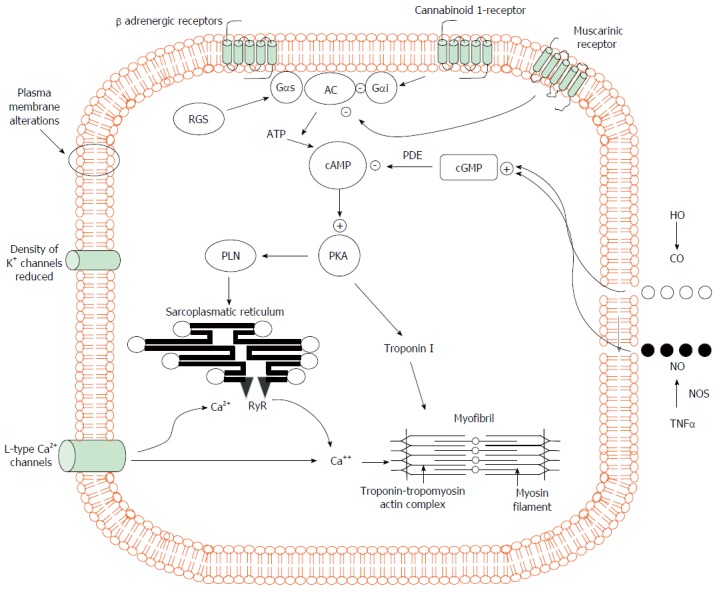
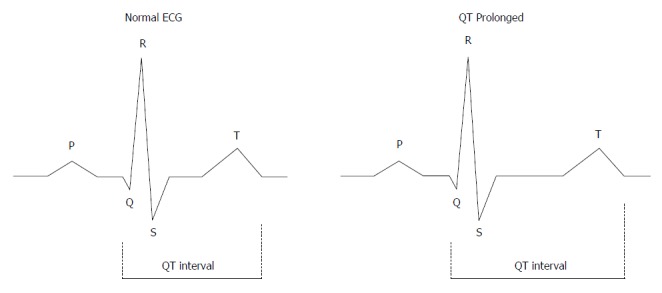
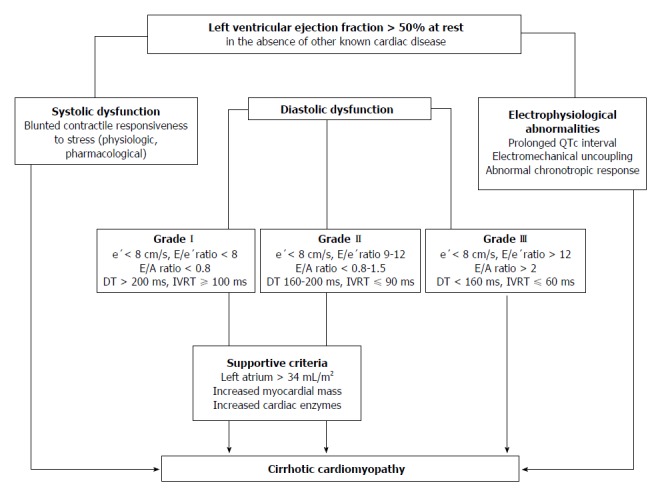
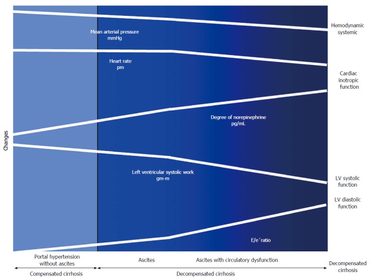
During the course of cirrhosis, there is a progressive deterioration of cardiac function manifested by the disappearance of the hyperdynamic circulation due to a failure in heart function with decreased cardiac output. This is due to a deterioration in inotropic and chronotropic function which takes place in parallel with a diastolic dysfunction and cardiac hypertrophy in the absence of other known cardiac disease. Other findings of this specific cardiomyopathy include impaired contractile responsiveness to stress stimuli and electrophysiological abnormalities with prolonged QT interval. The pathogenic mechanisms of cirrhotic cardiomyopathy include impairment of the b-adrenergic receptor signalling, abnormal cardiomyocyte membrane lipid composition and biophysical properties, ion channel defects and overactivity of humoral cardiodepressant factors. Cirrhotic cardiomyopathy may be difficult to determine due to the lack of a specific diagnosis test. However, an echocardiogram allows the detection of the diastolic dysfunction and the E/e' ratio may be used in the follow-up progression of the illness. Cirrhotic cardiomyopathy plays an important role in the pathogenesis of the impairment of effective arterial blood volume and correlates with the degree of liver failure. A clinical consequence of cardiac dysfunction is an inadequate cardiac response in the setting of vascular stress that may result in renal hypoperfusion leading to renal failure. The prognosis is difficult to establish but the severity of diastolic dysfunction may be a marker of mortality risk. Treatment is non-specific and liver transplantation may normalize the cardiac function.

Keywords: Arterial blood volume; Cirrhosis; Cirrhotic cardiomyopathy; E/e’ ratio; Hepatorenal syndrome; Inotropic heart dysfunction; Left ventricular diastolic dysfunction; Liver failure.


Figures

Figure 1
Figure 1
Pathogenic mechanisms of cardiomyocyte contraction in experimental cirrhosis. Cardiomyocytes of cirrhotic animals showed reduced contractility via impairment of the βAR signalling (down-regulation, desensitization) which in turn leads to a reduction in βAR density, Gs proteins, and AC activity with resultant decreased cAMP generation. This second messenger cAMP, phosphorylates several proteins leading to intracellular calcium fluxes, calcium release and myocyte contraction. Postreceptor signaling through cAMP is severely impaired. In addition, the decreased fluidity of cardiomyocyte membrane interferes with the function of βAR, L-type calcium and potassium channels and impair stimulation of muscarinic acetylcholine receptors. The activation of CB1 inhibiting L-type calcium channels and AC together with increased NO and CO levels via a cGMP-dependent mechanism also contribute to decreased calcium ion influx or release from the sarcoplasmic reticulum with a related fall of calcium ion content and contractility. +: stimulatory influence; -: inhibitory influence. βAR: β-adrenergic receptors; CB1: Cannabinoid 1-receptor; AC: Adenylcyclase; cAMP: Cyclic adenosine monophosphate; Gαi: Inhibitory G protein; Gαs: Stimulatory G protein; RGS: Regulator of G-protein signaling; PLN: Phosphoslamban; PKA: Protein kinase A; cGMP: Cyclic guanosine monophosphate; PDE: Phosphosdiesterase; RyR: Ryanodine receptor; HO: Haemoxygenase; CO: Carbon monoxide; NO: Nitric oxide; NOS: Nitric oxide synthase; TNFα: Tumour necrosis factor α.
Figure 2
Figure 2
The QT interval. The QT interval is a measure of the time between the start of the Q wave and the end of the T wave in the heart during electrical cycle. The QT interval is dependent on the heart rate and needs to be corrected by heart rate. The corrected QT interval (QTc) estimates the QT interval at a heart rate of 60 bpm. The standard clinical correction in cirrhosis uses Fredericia’s formula: QTc = QT/RR1/3. The RR interval is given in seconds (RR interval = 60/heart rate). QTc is prolonged if > 450 ms in men or > 470 ms in women. QTc interval prolongation is due to altered repolarization.
Figure 3
Figure 3
Algorithm for the diagnosis of cirrhotic cardiomyopathy. Three ways to diagnose CCM with normal EF at rest have been displayed: (1) Systolic function. Patients have documented blunted responsiveness to volume and postural challenge, exercise, or pharmacological infusion but the manoeuvres that can bring systolic dysfunction have not been standardized yet; (2) Diastolic function. The diagnosis of LVDD can be obtained by TDI (E/e′ > 15). If TDI yields an E/e′ ratio (8 < E/e′ < 15) other echocardiographic investigations such as blood flow Doppler of mitral valve or pulmonary veins, LV mass index or left atrial volume index are required for diagnostic of LVDD; (3) Electrophysiological abnormalities. a, the prolongation of the electrocardiographic corrected QT interval is common in cirrhosis; b, electromechanical uncoupling is a dyssynchrony between electrical and mechanical systole. The electrical systole is longer in patients with cirrhosis; c, chronotropic incompetence is the inability of the heart to proportionally increase HR in response to stimuli (exercise, tilting, paracentesis, infections and pharmacological agents). CCM: Cirrhotic cardiomyopathy; EF: Ejection fraction; LVDD: Left ventricular diastolic dysfunction; TDI: Tissue Doppler imaging; e’: Peak early diastolic velocity at the basal part of the septal and lateral corner of the mitral annulus; E/e’ratio: Peak E-wave transmitral/early diastolic mitral annular velocity; E/A ratio: Early diastolic mitral inflow velocity/late diastolic velocity; DT: Deceleration time; IVRT: Isovolumic relaxation time.
Figure 4
Figure 4
Cardiac and circulatory dysfunction according to presence of compensated and decompensated cirrhosis. There is a progressive splanchnic arterial vasodilatation due to the overproduction of vasodilator molecules. Initially, circulatory homeostasis is maintained by the development of a hyperdynamic circulation. Later during the course of decompensated cirrhosis, patients develop an activation of the vasoconstrictor systems to maintain arterial pressure. In subsequent stages, the progressive decrease in cardiac afterload is not followed by an increase in HR and CO due to a decrease in cardiac function. Finally, in the advanced phase, when impairment of effective arterial blood volume is extreme, patients showed increased activity of NE concentration, arterial hypotension and reduced LVSF (CO and LV stroke work), and cardiac chronotropic function, and a higher degree in LVDD (E/e’) as compared to those with ascites, but normal NE, or without ascites. E/e’ratio: Peak E-wave transmitral/Peak early diastolic mitral annular velocity.

Similar articles

  • Cirrhotic cardiomyopathy: a cardiologist's perspective.
    Gassanov N, Caglayan E, Semmo N, Massenkeil G, Er F. Gassanov N, et al. World J Gastroenterol. 2014 Nov 14;20(42):15492-8. doi: 10.3748/wjg.v20.i42.15492. World J Gastroenterol. 2014. PMID: 25400434 Free PMC article. Review.
  • Cirrhotic cardiomyopathy.
    Baik SK, Fouad TR, Lee SS. Baik SK, et al. Orphanet J Rare Dis. 2007 Mar 27;2:15. doi: 10.1186/1750-1172-2-15. Orphanet J Rare Dis. 2007. PMID: 17389039 Free PMC article. Review.
  • [Cirrhotic cardiomyopathy].
    Kim MY, Baik SK. Kim MY, et al. Korean J Hepatol. 2007 Mar;13(1):20-6. Korean J Hepatol. 2007. PMID: 17380071 Review. Korean.
  • Recent advances in cirrhotic cardiomyopathy.
    Karagiannakis DS, Papatheodoridis G, Vlachogiannakos J. Karagiannakis DS, et al. Dig Dis Sci. 2015 May;60(5):1141-51. doi: 10.1007/s10620-014-3432-8. Epub 2014 Nov 18. Dig Dis Sci. 2015. PMID: 25404411 Review.
  • Pathophysiological and clinical approach to cirrhotic cardiomyopathy.
    Páll A, Czifra A, Vitális Z, Papp M, Paragh G, Szabó Z. Páll A, et al. J Gastrointestin Liver Dis. 2014 Sep;23(3):301-10. doi: 10.15403/jgld.2014.1121.233.apac. J Gastrointestin Liver Dis. 2014. PMID: 25267959 Review.

Cited by

References

    1. Kowalski HJ, Abelmann WH. The cardiac output at rest in Laennec’s cirrhosis. J Clin Invest. 1953;32:1025–1033. - PMC - PubMed
    1. Murray JF, Dawson AM, Sherlock S. Circulatory changes in chronic liver disease. Am J Med. 1958;24:358–367. - PubMed
    1. Gould L, Shariff M, Zahir M, Di Lieto M. Cardiac hemodynamics in alcoholic patients with chronic liver disease and a presystolic gallop. J Clin Invest. 1969;48:860–868. - PMC - PubMed
    1. Lee SS. Cardiac abnormalities in liver cirrhosis. West J Med. 1989;151:530–535. - PMC - PubMed
    1. Bernardi M, Rubboli A, Trevisani F, Cancellieri C, Ligabue A, Baraldini M, Gasbarrini G. Reduced cardiovascular responsiveness to exercise-induced sympathoadrenergic stimulation in patients with cirrhosis. J Hepatol. 1991;12:207–216. - PubMed

Publication types