

Endosonography guided management of pancreatic fluid collections
- PMID: 26557008
- PMCID: PMC4631982
- DOI: 10.3748/wjg.v21.i41.11842
Endosonography guided management of pancreatic fluid collections
Abstract
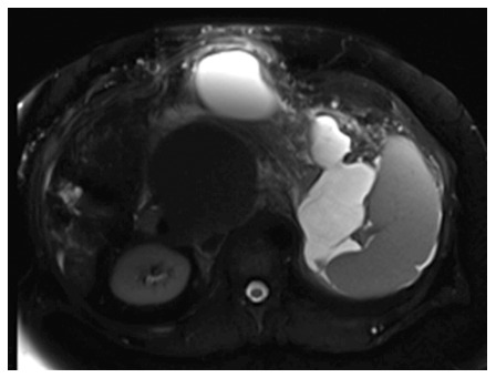
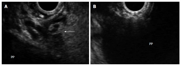
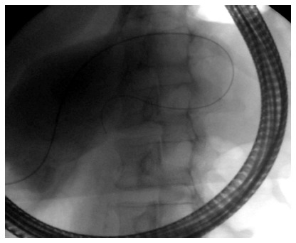
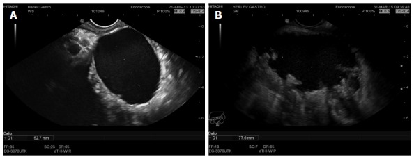
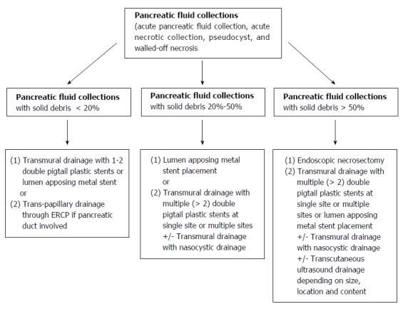
The revised Atlanta classification of acute pancreatitis was adopted by international consensus, and is based on actual local and systemic determinants of disease severity. The local determinant is pancreatic necrosis (sterile or infected), and the systemic determinant is organ failure. Local complications of pancreatitis can include acute peri-pancreatic fluid collection, acute necrotic collection, pseudocyst formation, and walled-off necrosis. Interventional endoscopic ultrasound (EUS) has been increasing utilized in managing these local complications. After performing a PubMed search, the authors manually applied pre-defined inclusion criteria or a filter to identify publications relevant to EUS and pancreatic collections (PFCs). The authors then reviewed the utility, efficacy, and risks associated with using therapeutic EUS and involved EUS devices in treating PFCs. Due to the development and regulatory approval of improved and novel endoscopic devices specifically designed for transmural drainage of fluid and necrotic debris (access and patency devices), the authors predict continuing evolution in the management of PFCs. We believe that EUS will become an indispensable part of procedures used to diagnose PFCs and perform image-guided interventions. After draining a PFC, the amount of tissue necrosis is the most important predictor of a successful outcome. Hence, it seems logical to classify these collections based on their percentage of necrotic component or debris present when viewed by imaging methods or EUS. Finally, the authors propose an algorithm for managing fluid collections based on their size, location, associated symptoms, internal echogenic patterns, and content.
Keywords: Abscess; Drainage; Endoscopic ultrasound; Pancreas; Pancreatic fluid collection; Patency device; Pseudocyst; Walled of necrosis.
Figures

References
-
- Banks PA, Bollen TL, Dervenis C, Gooszen HG, Johnson CD, Sarr MG, Tsiotos GG, Vege SS. Classification of acute pancreatitis--2012: revision of the Atlanta classification and definitions by international consensus. Gut. 2013;62:102–111. - PubMed
-
- Dellinger EP, Forsmark CE, Layer P, Lévy P, Maraví-Poma E, Petrov MS, Shimosegawa T, Siriwardena AK, Uomo G, Whitcomb DC, et al. Determinant-based classification of acute pancreatitis severity: an international multidisciplinary consultation. Ann Surg. 2012;256:875–880. - PubMed
-
- Thoeni RF. The revised Atlanta classification of acute pancreatitis: its importance for the radiologist and its effect on treatment. Radiology. 2012;262:751–764. - PubMed
-
- Kaul V, Adler DG, Conway JD, Farraye FA, Kantsevoy SV, Kethu SR, Kwon RS, Mamula P, Pedrosa MC, Rodriguez SA, et al. Interventional EUS. Gastrointest Endosc. 2010;72:1–4. - PubMed
-
- Alvarez-Sánchez MV, Jenssen C, Faiss S, Napoléon B. Interventional endoscopic ultrasonography: an overview of safety and complications. Surg Endosc. 2014;28:712–734. - PubMed
Publication types
MeSH terms
LinkOut - more resources
Full Text Sources
Other Literature Sources
Medical
Miscellaneous
