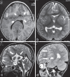
Middle interhemispheric variant of holoprosencephaly: A rare midline malformation
- PMID: 26557166
- PMCID: PMC4611894
- DOI: 10.4103/1817-1745.165678
Middle interhemispheric variant of holoprosencephaly: A rare midline malformation
Abstract
Middle interhemispheric variant (MIH) of holoprosencephaly (HPE) or syntelencephaly is a rare variant of HPE characterized by abnormal midline union of the posterior frontal and parietal lobes with variable fusion of thalami. It varies from classic HPE in embryopathogenesis, severity of fusion of brain structures, associated craniofacial anomalies and clinical presentation. We report a case of MIH in a 5-year-old girl, who presented with severe developmental delay and discuss the features differentiating it from other more common forms of HPE.
Keywords: Holoprosencephaly; middle interhemispheric variant; syntelencephaly.
Figures


Similar articles
-
The middle interhemispheric variant of holoprosencephaly.AJNR Am J Neuroradiol. 2002 Jan;23(1):151-6. AJNR Am J Neuroradiol. 2002. PMID: 11827888 Free PMC article.
-
Holoprosencephaly: a survey of the entity, with embryology and fetal imaging.Radiographics. 2015 Jan-Feb;35(1):275-90. doi: 10.1148/rg.351140040. Radiographics. 2015. PMID: 25590404 Review.
-
Holoprosencephaly.2024 Jun 7. In: StatPearls [Internet]. Treasure Island (FL): StatPearls Publishing; 2025 Jan–. 2024 Jun 7. In: StatPearls [Internet]. Treasure Island (FL): StatPearls Publishing; 2025 Jan–. PMID: 32809696 Free Books & Documents.
-
Syntelencephaly in an infant of a diabetic mother.Am J Med Genet. 1996 Dec 30;66(4):433-7. doi: 10.1002/(SICI)1096-8628(19961230)66:4<433::AID-AJMG9>3.0.CO;2-L. Am J Med Genet. 1996. PMID: 8989462
-
Prenatal diagnosis of middle interhemispheric variant of holoprosencephaly: review of literature and prenatal case series.J Matern Fetal Neonatal Med. 2022 Dec;35(25):4976-4984. doi: 10.1080/14767058.2021.1873942. Epub 2021 Jan 17. J Matern Fetal Neonatal Med. 2022. PMID: 33455493 Review.
Cited by
-
Holoprosencephaly spectrum: an up-to-date overview of classification, genetics and neuroimaging.Jpn J Radiol. 2025 Jan;43(1):13-31. doi: 10.1007/s11604-024-01655-8. Epub 2024 Sep 11. Jpn J Radiol. 2025. PMID: 39259418 Review.
References
-
- DeMyer W. Holoprosencephaly. In: Vinker P, Bruyn J, editors. Handbook of Clinical Neurology. Amsterdam: North Holland; pp. 431–78.
-
- Lewis AJ, Simon EM, Barkovich AJ, Clegg NJ, Delgado MR, Levey E, et al. Middle interhemispheric variant of holoprosencephaly: A distinct cliniconeuroradiologic subtype. Neurology. 2002;59:1860–5. - PubMed
-
- Lee KJ, Dietrich P, Jessell TM. Genetic ablation reveals that the roof plate is essential for dorsal interneuron specification. Nature. 2000;403:734–40. - PubMed
-
- Hahn JS, Barnes PD. Neuroimaging advances in holoprosencephaly: Refining the spectrum of the midline malformation. Am J Med Genet C Semin Med Genet. 2010;154C:120–32. - PubMed
Publication types
LinkOut - more resources
Full Text Sources
Other Literature Sources

