

Fatal cerebellar hemorrhage as an initial presentation of medulloblastoma in a child
- PMID: 26557180
- PMCID: PMC4611908
- DOI: 10.4103/1817-1745.165727
Fatal cerebellar hemorrhage as an initial presentation of medulloblastoma in a child
Abstract
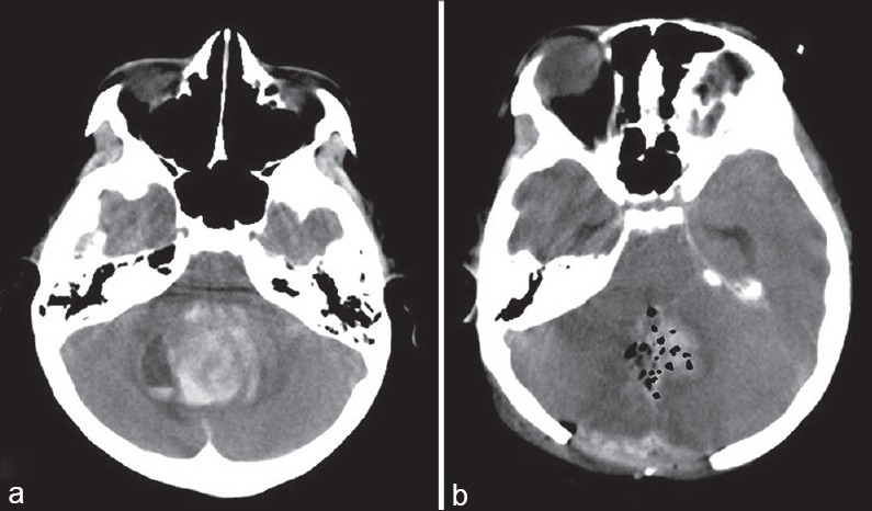
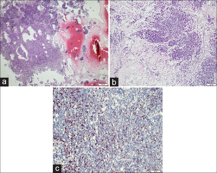
Children with medulloblastomas most commonly present with signs and symptoms of elevated intracranial pressure due to obstructive hydrocephalus, especially headaches and vomiting. However, some pediatric patients present with sudden neurological deterioration due to intracerebellar hemorrhage associated with medulloblastoma, although very few reports exist that document this phenomenon. An 8-year-old girl was admitted to our emergency department who presented with sudden loss of consciousness, vomiting, and bradycardia. The neuroradiological evaluation revealed a hemorrhagic mass lesion in the posterior fossa. Urgent evacuation of the hematoma was performed. The postoperative course was uneventful, and the postoperative histopathological examination revealed the lesion to be a medulloblastoma. This report presents an unusual case of a medulloblastoma presenting with fatal intracranial hemorrhage in a child. The clinical features and intraoperative and pathologic findings of the case are discussed.
Keywords: Cerebellar hemorrhage; childhood; medulloblastoma.
Figures
Similar articles
-
[Pediatric medulloblastoma presenting as cerebellar hemorrhage: a case report].No Shinkei Geka. 2014 Jun;42(6):545-51. No Shinkei Geka. 2014. PMID: 24920742 Japanese.
-
Multiple aneurysms of the distal posterior inferior cerebellar artery: two case reports.Minim Invasive Neurosurg. 2008 Oct;51(5):249-52. doi: 10.1055/s-0028-1082302. Epub 2008 Oct 14. Minim Invasive Neurosurg. 2008. PMID: 18855286
-
Supratentorial primitive neuroectodermal tumor presenting with intracranial hemorrhage in adult.J Neurosci Rural Pract. 2014 Apr;5(2):176-9. doi: 10.4103/0976-3147.131672. J Neurosci Rural Pract. 2014. PMID: 24966562 Free PMC article.
-
Undiagnosed medulloblastoma presenting as fatal hemorrhage in a 14-year-old boy: case report and review of the literature.Childs Nerv Syst. 2007 Jul;23(7):799-805. doi: 10.1007/s00381-006-0290-5. Epub 2007 Feb 6. Childs Nerv Syst. 2007. PMID: 17279429 Review.
-
Intraoperative hemorrhage in medulloblastoma: a case report and review of the literature.Childs Nerv Syst. 2002 Jul;18(6-7):356-60. doi: 10.1007/s00381-002-0601-4. Epub 2002 May 23. Childs Nerv Syst. 2002. PMID: 12172947 Review.
Cited by
-
Anomalous vascularization in a Wnt medulloblastoma: a case report.BMC Neurol. 2016 Jul 15;16:103. doi: 10.1186/s12883-016-0632-1. BMC Neurol. 2016. PMID: 27416922 Free PMC article.
-
H3 K27-altered diffuse midline glioma presenting as massive cerebellopontine hemorrhage.Childs Nerv Syst. 2023 Aug;39(8):2229-2232. doi: 10.1007/s00381-023-05904-5. Epub 2023 Mar 3. Childs Nerv Syst. 2023. PMID: 36867241
-
Pediatric WNT medulloblastoma predisposition in intraoperative blood loss: a retrospective observational cohort study.Front Neurol. 2024 Jul 2;15:1386121. doi: 10.3389/fneur.2024.1386121. eCollection 2024. Front Neurol. 2024. PMID: 39015321 Free PMC article.
-
Detecting Tumor-Associated Intracranial Hemorrhage Using Proton Magnetic Resonance Spectroscopy.Neurol Int. 2024 Dec 17;16(6):1856-1877. doi: 10.3390/neurolint16060133. Neurol Int. 2024. PMID: 39728759 Free PMC article. Review.
-
Clinical features of intracerebral hemorrhage in patients with colorectal cancer and its underlying pathogenesis.World J Gastrointest Oncol. 2021 Dec 15;13(12):2180-2189. doi: 10.4251/wjgo.v13.i12.2180. World J Gastrointest Oncol. 2021. PMID: 35070050 Free PMC article.
References
-
- Illinois, USA: Central Brain Tumor Registry of the United States; 2004-2005. CBTRUS. Statistical Report: (2004-2005), Primary Brain Tumors in the United States, 1997-2001.
-
- Santi M, Kadom N, Vezina G, Rushing EJ. Undiagnosed medulloblastoma presenting as fatal hemorrhage in a 14-year-old boy: Case report and review of the literature. Childs Nerv Syst. 2007;23:799–805. - PubMed
-
- Furuhata M, Aihara Y, Eguchi S, Horiba A, Tanaka M, Komori T, et al. Pediatric medulloblastoma presenting as cerebellar hemorrhage: A case report. No Shinkei Geka. 2014;42:545–51. - PubMed
-
- Elgamal EA, Richards PG, Patel UJ. Fatal haemorrhage in medulloblastoma following ventricular drainage. Case report and review of the literature. Pediatr Neurosurg. 2006;42:45–8. - PubMed
-
- Chugani HT, Rosemblat AM, Lavenstein BL, Palumbo FM, Luessenhop AJ, Manz HJ. Childhood medulloblastoma presenting with hemorrhage. Childs Brain. 1984;11:135–40. - PubMed
Publication types
LinkOut - more resources
Full Text Sources
Other Literature Sources
