

Evaluation of Optical Coherence Tomography as a Means of Identifying Earlier Stage Basal Cell Carcinomas while Reducing the Use of Diagnostic Biopsy
- PMID: 26557214
- PMCID: PMC4633207
Evaluation of Optical Coherence Tomography as a Means of Identifying Earlier Stage Basal Cell Carcinomas while Reducing the Use of Diagnostic Biopsy
Abstract
Objective: To determine the diagnostic accuracy of optical coherence tomography for basal cell carcinoma and the proportion of biopsies that could be avoided if optical coherence tomography is used to rule-in surgery.
Design: Multicenter, prospective, observational study.
Setting: Dermatology clinics.
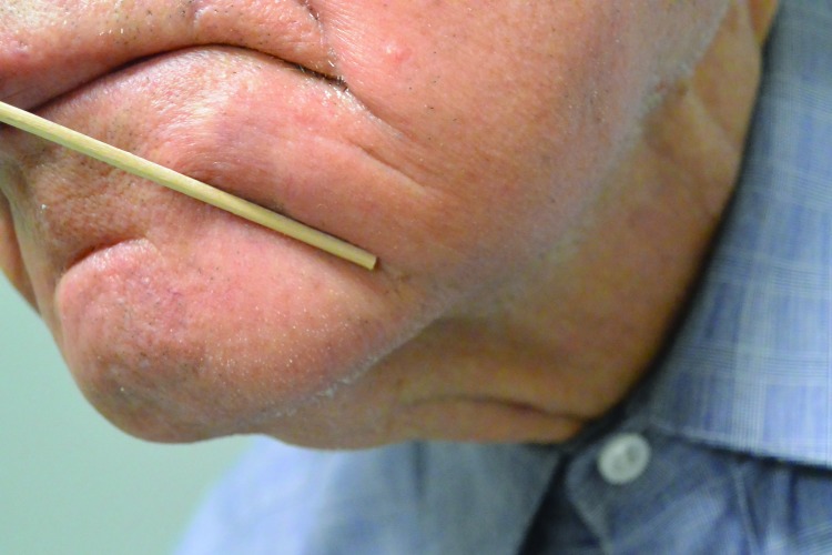
Participants: Consecutive patients with clinically challenging pink lesions suspicious for basal cell carcinoma.
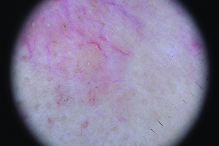
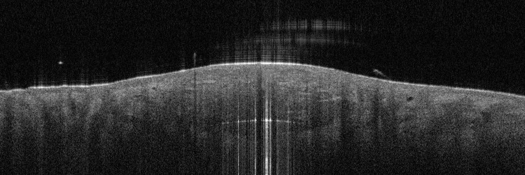
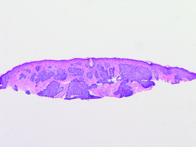
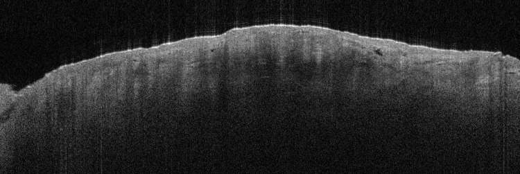
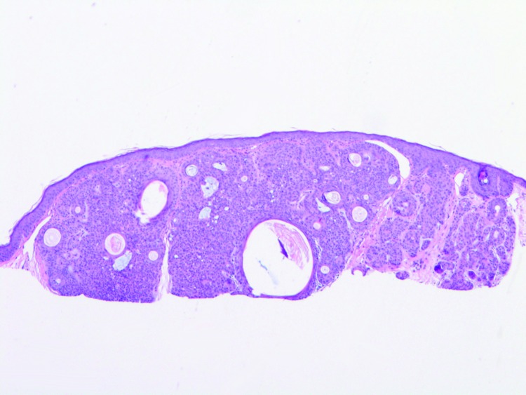
Measurements: Clinical, dermoscopic, and optical coherence tomography images were obtained for all subjects. At each stage, the clinician made a diagnosis (pathology + subtype if applicable), and assessed his/her own confidence in the diagnosis.
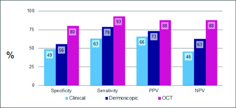
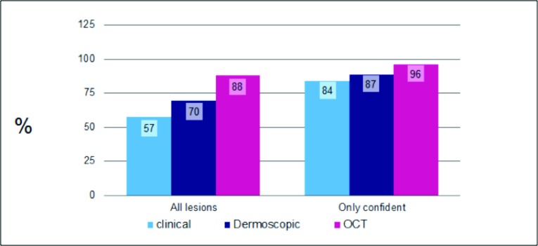
Results: Optical coherence tomography significantly (p<0.01) improved sensitivity and specificity over clinical or dermoscopic evaluation. The percentage of correct diagnoses was 57.4 percent (clinical), 69.6 percent (dermoscopy), and 87.8 percent (optical coherence tomography). Optical coherence tomography significantly increased the certainty of diagnosis; clinicians indicated they were certain (>95% confident) in 17 percent of lesions examined clinically, in 38.6 percent examined with dermoscopy, and in 70 percent examined with optical coherence tomography. With the use of optical coherence tomography in the diagnosis of basal cell carcinoma, more than 1 in 3 patients could avoid a diagnostic biopsy.
Conclusion: In a population of clinically challenging lesions, optical coherence tomography improved diagnostic certainty by a factor of four over clinical examination alone and improved diagnostic accuracy by 50 percent (57-88%). The addition of optical coherence tomography to other standard assessments can improve the false-positive rate and give a high degree of certainty for ruling in a positive diagnosis for basal cell carcinoma. A reduction of 36 percent in overall biopsies could be achieved by sending high certainty basal cell carcinoma positive optical coherence tomography diagnoses straight to surgery.
Figures


References
-
- Debski T, Lembas L, Jethon J. Basal cell carcinoma. In: Agullo F, editor. Current Concepts in Plastic Surgery. Rijeka, Croatia: InTech; 2012.
-
- Lomas A, Leonardi-Bee J, Bath-Hextall F. A systematic review of worldwide incidence of nonmelanoma skin cancer. Br J Dermatol. 2012;166:1069–1080. - PubMed
-
- Rogers HW, Weinstock MA, Feldman SR, et al. Incidence estimate of nonmelanoma skin cancer (keratinocyte carcinomas) in the US population, 2012. J Am Acad Dermatol. 2015 Apr 30. Epub. - PubMed
-
- Rogers HW, Weinstock MA, Harris AR, et al. Incidence estimate of nonmelanoma skin cancer in the United States, 2006. Arch Dermatol. 2010;146:283–287. - PubMed
LinkOut - more resources
Full Text Sources