

The Impact of Multispectral Digital Skin Lesion Analysis on German Dermatologist Decisions to Biopsy Atypical Pigmented Lesions with Clinical Characteristics of Melanoma
- PMID: 26557216
- PMCID: PMC4633209
The Impact of Multispectral Digital Skin Lesion Analysis on German Dermatologist Decisions to Biopsy Atypical Pigmented Lesions with Clinical Characteristics of Melanoma
Abstract
Objective: To determine the impact of multispectral digital skin lesion analysis on German dermatologist biopsy decisions of atypical pigmented skin lesions.
Design: Participants were shown high-resolution clinical images of 12 atypical pigmented skin lesions previously analyzed by multispectral digital skin lesion analysis. Participants were asked if they would biopsy the lesion based on clinical images and high-resolution dermoscopy images and again when subsequently shown multispectral digital skin lesion analysis probability information.
Setting/participants: Forty-one dermatologists at a skin cancer conference in Germany in September 2014.
Measurements: Sensitivity, specificity, diagnostic accuracy, percent biopsying all melanomas, and overall biopsy rates.
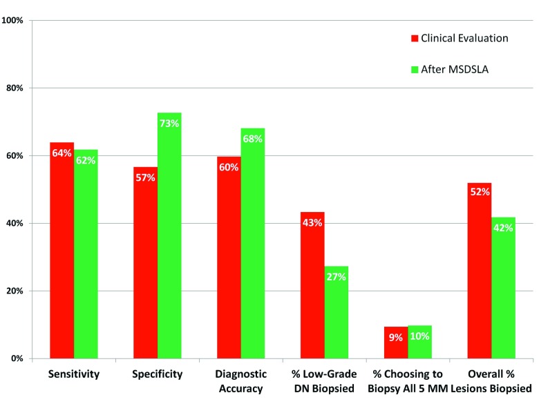
Results: Sensitivity for the detection of melanoma following clinical evaluation was 64 percent. After receipt of multispectral digital skin lesion analysis probability information, sensitivity decreased nonsignificantly to 62 percent. Specificity with clinical evaluation was 57 percent and increased to 73 percent using multispectral digital skin lesion analysis. Overall biopsy accuracy increased from 60 percent with clinical evaluation to 68 percent with multispectral digital skin lesion analysis. The percentage of low-grade dysplastic nevi chosen for biopsy decreased from 43 percent after clinical evaluation to 27 percent with multispectral digital skin lesion analysis. Finally, the overall percentage of lesions biopsied decreased from 52 percent with clinical evaluation to 42 percent after multispectral digital skin lesion analysis.
Conclusion: Multispectral digital skin lesion analysis can be used reliably to detect melanoma as well as clinical evaluation. Dermatologists can confidently use multispectral digital skin lesion analysis to significantly improve specificity and reduce their overall number of biopsies while increasing overall diagnostic accuracy.
Figures
References
-
- Rigel DS, Russak J, Friedman R. The evolution of melanoma diagnosis: 25 years beyond the ABCDs. CA Cancer J Clin. 2010;60(5):301–316. - PubMed
-
- Elbaum M, Kopf AW, Rabinovitz HS, et al. Automatic differentiation of melanoma and melanocytic nevi with multispectral digital dermoscopy: a feasibility study. J Am Acad Dermatol. 2001;44(2):207–218. - PubMed
-
- Gutkowicz-Krusin D, Elbaum M, Jacobs A, et al. Precision of automatic measurements of pigmented skin lesion parameters with a MelaFind(TM) multispectral digital dermoscope. Melanoma Res. 2000;10(6):563–570. - PubMed
-
- Monheit G, Cognetta AB, Ferris L, et al. The performance of MelaFind: a prospective multicenter study. Arch Dermatol. 2011;147(2):188–194. - PubMed
-
- Hauschild A, Chen SC, Weichenthal M, et al. To excise or not: impact of MelaFind on German dermatologists’ decisions to biopsy atypical lesions. J Dtsch Dermatol Ges. 2014;12(7):606–614. - PubMed
LinkOut - more resources
Full Text Sources