

Ocular Inflammatory Myofibroblastic Tumor in the Left Eye with Phthisis Right Eye: A Rare Occurrence in a Child
- PMID: 26557400
- PMCID: PMC4628688
- DOI: 10.1155/2015/281528
Ocular Inflammatory Myofibroblastic Tumor in the Left Eye with Phthisis Right Eye: A Rare Occurrence in a Child
Abstract
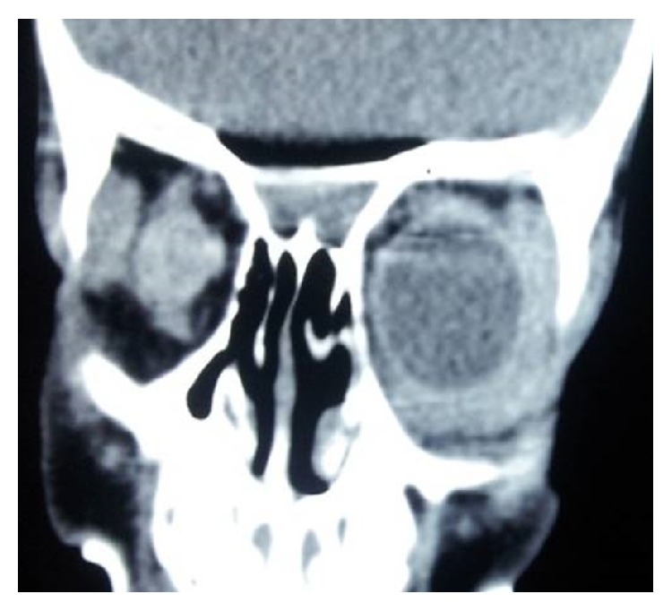
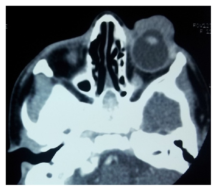
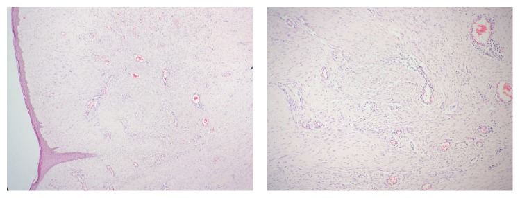
Inflammatory myofibroblastic tumor (IMT) is a benign pseudoneoplastic inflammatory condition with the potential for persistent local growth and recurrence that rarely affects the orbit. We report a very rare case of anterior orbital IMT in a child who presented with gradually progressive mass in left eye for 16 months. Ocular examination showed a cauliflower like exophytic mass at 360 degrees of the perilimbal area covering the entire cornea and obscuring the visualization of anterior and posterior segments. The right eye was phthisical. CT scan showed a lobulated exophytic soft tissue mass in the preseptal region and along the anterior portion of the left globe extending from medial canthus to the lateral canthus. Enucleation of the left eye was performed and the histopathological examination confirmed the diagnosis of IMT. This report aims to raise awareness about this rare ocular entity and emphasizes its early treatment as delay can result in loss of the eye.
Figures


Similar articles
-
A Rare Presentation of Inflammatory Myofibroblastic Tumor in the Nasolabial Fold.Case Rep Otolaryngol. 2019 Jan 23;2019:3257697. doi: 10.1155/2019/3257697. eCollection 2019. Case Rep Otolaryngol. 2019. PMID: 30809407 Free PMC article.
-
Idiopathic Orbital Inflammatory Pseudotumor With Bone Erosion.J Craniofac Surg. 2016 Oct;27(7):e607-e608. doi: 10.1097/SCS.0000000000002921. J Craniofac Surg. 2016. PMID: 27464561
-
Inflammatory Myofibroblastic Tumor of the Breast Mimicking Malignancy in an Elderly Male.Ochsner J. 2017 Fall;17(3):277-279. Ochsner J. 2017. PMID: 29026362 Free PMC article.
-
Inflammatory myofibroblastic tumor in children: diagnosis and treatment.J Pediatr Surg. 2001 Jun;36(6):908-12. doi: 10.1053/jpsu.2001.23970. J Pediatr Surg. 2001. PMID: 11381424 Review.
-
Orbital Compartment Syndrome without Evidence of Orbital Mass or Ocular Compression After Pterional Craniotomy for Removal of Meningioma of the Frontal Lobe: A Case Report and Literature Review.World Neurosurg. 2020 Jul;139:588-591. doi: 10.1016/j.wneu.2020.04.094. Epub 2020 Apr 25. World Neurosurg. 2020. PMID: 32344145 Review.
Cited by
-
Distribution of phthisis bulbi and status of fellow eyes at a tertiary eye-care centre in Nigeria: a ten-year review.Afr Health Sci. 2021 Mar;21(1):437-444. doi: 10.4314/ahs.v21i1.54. Afr Health Sci. 2021. PMID: 34394326 Free PMC article.
-
Inflammatory Myofibroblastic Tumor of the Orbit: A Case Series and Literature Review.J Inflamm Res. 2024 Dec 14;17:11029-11039. doi: 10.2147/JIR.S485499. eCollection 2024. J Inflamm Res. 2024. PMID: 39697794 Free PMC article.
-
A Rare Presentation of Inflammatory Myofibroblastic Tumor in the Nasolabial Fold.Case Rep Otolaryngol. 2019 Jan 23;2019:3257697. doi: 10.1155/2019/3257697. eCollection 2019. Case Rep Otolaryngol. 2019. PMID: 30809407 Free PMC article.
References
-
- Sa H. S., Ji J. Y., Suh Y. L., Kim Y. D. Inflammatory myofibroblastic tumor of the orbit presenting as a subconjunctival mass. Ophthalmic Plastic and Reconstructive Surgery. 2005;21(3):211–215. - PubMed
-
- Coffin C. M., Humphrey P. A., Dehner L. P. Extrapulmonary inflammatory myofibroblastic tumor: a clinical and pathological survey. Seminars in Diagnostic Pathology. 1998;15(2):85–101. - PubMed
-
- Coffin C. M., Dehner L. P., Meis-Kindblom J. M. Inflammatory myofibroblastic tumor, inflammatory fibrosarcoma, and related lesions: an historical review with differential diagnostic considerations. Seminars in Diagnostic Pathology. 1998;15(2):102–110. - PubMed
LinkOut - more resources
Full Text Sources
Other Literature Sources
