. 2015 Jul-Sep;5(3):170-8.
doi: 10.4103/2229-5151.164940.

Central line complications

Affiliations

Central line complications

Craig Kornbau et al. Int J Crit Illn Inj Sci. 2015 Jul-Sep.

Abstract

Central venous access is a common procedure performed in many clinical settings for a variety of indications. Central lines are not without risk, and there are a multitude of complications that are associated with their placement. Complications can present in an immediate or delayed fashion and vary based on type of central venous access. Significant morbidity and mortality can result from complications related to central venous access. These complications can cause a significant healthcare burden in cost, hospital days, and patient quality of life. Advances in imaging, access technique, and medical devices have reduced and altered the types of complications encountered in clinical practice; but most complications still center around vascular injury, infection, and misplacement. Recognition and management of central line complications is important when caring for patients with vascular access, but prevention is the ultimate goal. This article discusses common and rare complications associated with central venous access, as well as techniques to recognize, manage, and prevent complications.

Keywords: Bleeding; central line; complications; infection; pneumothorax.


Figures

Figure 1
Figure 1
Chest X-ray demonstrating a pulmonary artery catheter inadvertently placed via the right carotid artery into the thoracic aorta. Despite pressure waveform monitoring, erroneous placement was not noticed until a post-procedure chest was obtained. The catheter was removed immediately. Follow-up carotid ultrasound was unremarkable for arteriovenous (AV) fistula or pseudoaneurysm. This patient had no residual complications as this complication was immediately recognized and managed
Figure 2
Figure 2
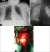
(a) Vascular injury following dialysis catheter placement. Initial chest X-ray after reported uneventful place of a tunneled right internal jugular dialysis line in a 67-year-old female with end-stage renal disease. (b) After initial attempts to access catheter demonstrated that fluid was easily flushed, but not aspirated out of the catheter, attempts to change the “malfunctioning” catheter over a wire suggested that the wire was in the pleural space, and hence the catheter tip was extravascular. (c) Due to concerns of a vascular injury, removal under direct visualization was recommended. Following median sternotomy, the intraoperative findings demonstrated the catheter had punctured the confluence of the right subclavian vein as it entered into the superior vena cava. The catheter was in the right pleural space. The pericardial space had not been violated. Under direct visualization the catheter was removed and the residual hole was repaired with a purse-string and pledgeted suture. Blood loss was minimal and the postoperative recovery was uneventful. Repeat line placement several days later, under fluoroscopic guidance, was uneventful
Figure 3
Figure 3
Contralateral pneumothorax. Pneumothorax following failed chronic port placement on contralateral side
Figure 4
Figure 4
Ipsilateral pneumothorax. Pneumothorax following left-sided subclavian line placement
Figure 5
Figure 5
Fractured catheters following chronic port removal. An elderly lady who underwent combined chemo and radiation therapy for breast cancer presented for elective removal of her chronic intravascular port (top image). Upon attempted removal of the port in the operating room, the distal half fractured (bottom image). As it appeared to be immobile and potentially adherent to the vascular wall, the initial recommendation was to follow it closely for signs of migration with serial chest X-rays. Patient concern for embolization prompted attempted extraction by interventional catheter-directed techniques by interventional radiology. Such attempts were unsuccessful as the catheter appeared, as suspected, to be adherent to the innominate vein. Due to patient frailty and concerns for causing a major injury, further attempts at removal were abandoned. The patient remains asymptomatic
Figure 6
Figure 6
Chronic catheter fracture. Catheter fragment incidentally found on chest X-ray 4 years following chronic port removal. The patient was asymptomatic and opted for no intervention

Similar articles

Cited by

References

    1. McGee DC, Gould MK. Preventing complications of central venous catheterization. N Engl J Med. 2003;348:1123–33. - PubMed
    1. O’Grady NP, Alexander M, Burns LA, Dellinger EP, Garland J, Heard SO, et al. Healthcare Infection Control Practices Advisory Committee (HICPAC) (Appendix 1). Summary of recommendations: Guidelines for the prevention of intravascular catheter-related infections. Clin Infect Dis. 2011;52:1087–99. - PMC - PubMed
    1. Kusminsky RE. Complications of central venous catheterization. J Am Coll Surg. 2007;204:681–96. - PubMed
    1. Sznajder JI, Zveibil FR, Bitterman H, Weiner P, Bursztein S. Central vein catheterization. Failure and complication rates by three percutaneous approaches. Arch Intern Med. 1986;146:259–61. - PubMed
    1. Bhutta ST, Culp WC. Evaluation and management of central venous access complications. Tech Vasc Interv Radiol. 2011;14:217–24. - PubMed