

Analysis of Retinal Peripapillary Segmentation in Early Alzheimer's Disease Patients
- PMID: 26557684
- PMCID: PMC4628738
- DOI: 10.1155/2015/636548
Analysis of Retinal Peripapillary Segmentation in Early Alzheimer's Disease Patients
Abstract
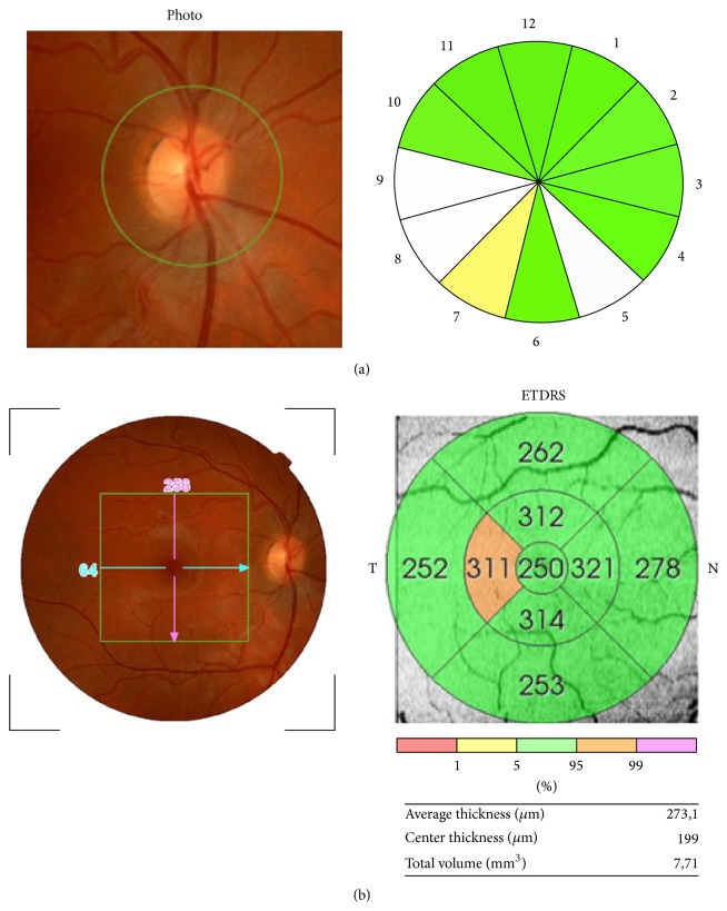
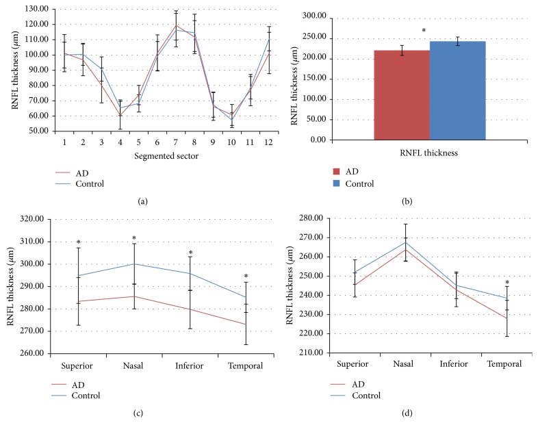
Decreased thickness of the retinal nerve fiber layer (RNFL) may reflect retinal neuronal-ganglion cell death. A decrease in the RNFL has been demonstrated in Alzheimer's disease (AD) in addition to aging by optical coherence tomography (OCT). Twenty-three mild-AD patients and 28 age-matched control subjects with mean Mini-Mental State Examination 23.3 and 28.2, respectively, with no ocular disease or systemic disorders affecting vision, were considered for study. OCT peripapillary and macular segmentation thickness were examined in the right eye of each patient. Compared to controls, eyes of patients with mild-AD patients showed no statistical difference in peripapillary RNFL thickness (P > 0.05); however, sectors 2, 3, 4, 8, 9, and 11 of the papilla showed thinning, while in sectors 1, 5, 6, 7, and 10 there was thickening. Total macular volume and RNFL thickness of the fovea in all four inner quadrants and in the outer temporal quadrants proved to be significantly decreased (P < 0.01). Despite the fact that peripapillary RNFL thickness did not statistically differ in comparison to control eyes, the increase in peripapillary thickness in our mild-AD patients could correspond to an early neurodegeneration stage and may entail the existence of an inflammatory process that could lead to progressive peripapillary fiber damage.
Figures
Similar articles
-
OCT in Alzheimer's disease: thinning of the RNFL and superior hemiretina.Graefes Arch Clin Exp Ophthalmol. 2017 Sep;255(9):1827-1835. doi: 10.1007/s00417-017-3715-9. Epub 2017 Jun 22. Graefes Arch Clin Exp Ophthalmol. 2017. PMID: 28643042
-
Evaluation of retinal nerve fiber layer and ganglion cell layer thickness in Alzheimer's disease using spectral-domain optical coherence tomography.Invest Ophthalmol Vis Sci. 2013 Sep 5;54(9):5953-8. doi: 10.1167/iovs.13-12046. Invest Ophthalmol Vis Sci. 2013. PMID: 23920375
-
Macular Thickness Measurements with Frequency Domain-OCT for Quantification of Retinal Neural Loss and its Correlation with Cognitive Impairment in Alzheimer's Disease.PLoS One. 2016 Apr 22;11(4):e0153830. doi: 10.1371/journal.pone.0153830. eCollection 2016. PLoS One. 2016. PMID: 27104962 Free PMC article.
-
Spectral-Domain OCT Measurements in Alzheimer's Disease: A Systematic Review and Meta-analysis.Ophthalmology. 2019 Apr;126(4):497-510. doi: 10.1016/j.ophtha.2018.08.009. Epub 2018 Aug 13. Ophthalmology. 2019. PMID: 30114417 Free PMC article.
-
Alzheimer's disease: A review of its visual system neuropathology. Optical coherence tomography-a potential role as a study tool in vivo.Graefes Arch Clin Exp Ophthalmol. 2016 Nov;254(11):2079-2092. doi: 10.1007/s00417-016-3430-y. Epub 2016 Jul 4. Graefes Arch Clin Exp Ophthalmol. 2016. PMID: 27377656 Review.
Cited by
-
Glaucoma as a Tauopathy-Is It the Missing Piece in the Glaucoma Puzzle?J Clin Med. 2023 Nov 2;12(21):6900. doi: 10.3390/jcm12216900. J Clin Med. 2023. PMID: 37959365 Free PMC article. Review.
-
Evaluation of macular thickness and volume tested by optical coherence tomography as biomarkers for Alzheimer's disease in a memory clinic.Sci Rep. 2020 Jan 31;10(1):1580. doi: 10.1038/s41598-020-58399-4. Sci Rep. 2020. PMID: 32005868 Free PMC article.
-
Retinal Thickness Changes Over Time in a Murine AD Model APP NL-F/NL-F.Front Aging Neurosci. 2021 Jan 15;12:625642. doi: 10.3389/fnagi.2020.625642. eCollection 2020. Front Aging Neurosci. 2021. PMID: 33542683 Free PMC article.
-
Optical Coherence Tomography in Patients with Alzheimer's Disease: What Can It Tell Us?Eye Brain. 2021 Jan 8;13:1-20. doi: 10.2147/EB.S235238. eCollection 2021. Eye Brain. 2021. PMID: 33447120 Free PMC article. Review.
-
Ocular changes as potential biomarkers for early diagnosis of Alzheimer's disease.Alzheimers Dement. 2025 Aug;21(8):e70476. doi: 10.1002/alz.70476. Alzheimers Dement. 2025. PMID: 40851106 Free PMC article. Review.
References
-
- Brookmeyer R., Johnson E., Ziegler-Graham K., Arrighi H. M. Forecasting the global burden of Alzheimer's disease. Alzheimer's and Dementia. 2007;3(3):186–191. - PubMed
-
- Cummings J. L., Vinters H. V., Cole G. M., Khachaturian Z. S. Alzheimer's disease. Neurology. 1998;51(1, supplement 1):S2–S17. - PubMed
-
- Small B. J., Gagnon E., Robinson B. Early identification of cognitive deficits: preclinical Alzheimer's disease and mild cognitive impairment. Geriatrics. 2007;62(4):19–23. - PubMed
Publication types
MeSH terms
LinkOut - more resources
Full Text Sources
Other Literature Sources
Medical
