

A huge cardiac hydatid cyst: An unusual cause of chest pain revealing multivisceral hydatidosis in a young woman
- PMID: 26557748
- PMCID: PMC4614895
- DOI: 10.1016/j.jsha.2015.04.003
A huge cardiac hydatid cyst: An unusual cause of chest pain revealing multivisceral hydatidosis in a young woman
Abstract
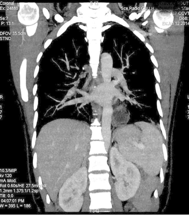
Hydatid disease remains endemic in some parts of the world. Cardiac hydatidosis with multivisceral involvement is uncommon but potentially fatal. We report the case of a 36-year-old Tunisian woman admitted with chest pain and T-wave inversion in the inferior leads on her electrocardiogram. Transthoracic echocardiography revealed a large hydatid cyst in the epicardium throughout the left ventricle. Thoraco-abdominal computerized tomography (CT) scan showed several hydatid cysts in the left lung, the liver, and in both breasts. After one week of albendazole treatment, surgical excision of the cardiac cyst on cardiopulmonary bypass was carried out as well as excision of the pulmonary and breast cysts. The postoperative course was uneventful and albendazole treatment was continued for six months. Though hydatid cardiac involvement is very rare, it should be considered in the differential diagnosis of atypical chest pain in young patients, especially those living in regions where hydatid disease is endemic.
Keywords: Cardiac hydatid cyst; Chest pain; Hydatid disease.
Figures


References
-
- Kireşi D.A., Karabacakoğlu A., Odev K., Karaköse S. Uncommon locations of hydatid cysts. Acta Radiol. 2003;44(6):622–636. - PubMed
-
- Grozavu C., Ilias M., Pantile D. Multivisceral echinococcosis: concept, diagnosis, management. Chirurgia (Bucur) 2014;109(6):758–768. - PubMed
-
- Ulgen M.S., Alan S., Karadede A., Aydinalp O., Toprak N. Cardiac hydatid cysts located in both the left ventricular apex and the intraventricular septum: case report. Heart Vessels. 2000;15(5):243–244. - PubMed
-
- Younis S.N., Faraj A.A. Cardiac hydatid disease, case report, and review of literature. Acta Clin Belg. 2014;69(1):66–68. - PubMed
LinkOut - more resources
Full Text Sources
Other Literature Sources
