Review
. 2015 Nov;8(6):384-94.
doi: 10.1177/1756283X15592584.

Therapeutic potential of ultrasound microbubbles in gastrointestinal oncology: recent advances and future prospects

Affiliations
Review

Therapeutic potential of ultrasound microbubbles in gastrointestinal oncology: recent advances and future prospects

Tatiana D Khokhlova et al. Therap Adv Gastroenterol. 2015 Nov.

Abstract

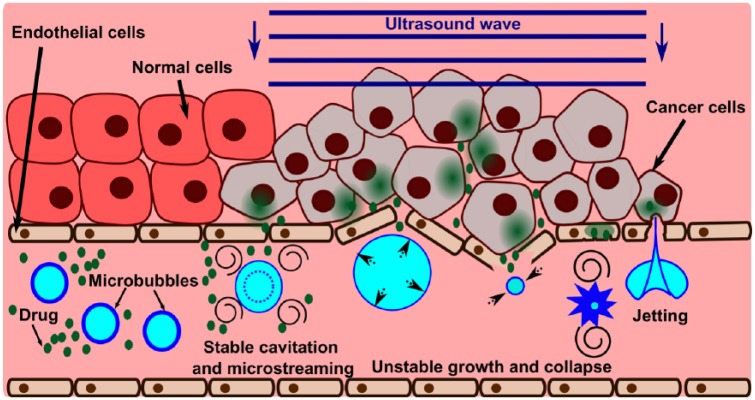
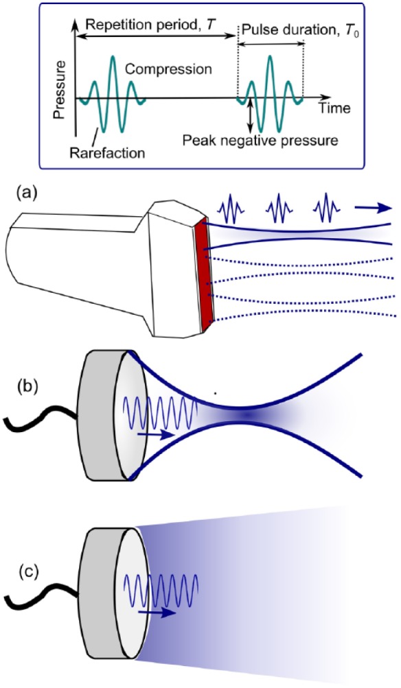
Microbubbles were initially invented as contrast agents for ultrasound imaging. However, lately more and more therapeutic applications of microbubbles are emerging, mostly related to drug and gene delivery. Ultrasound is a safe and noninvasive therapeutic modality which has the unique ability to interact with microbubbles and release their payload in situ in addition to permeabilizing the target tissues. The combination of drug-loaded microbubbles and ultrasound has been used in preclinical studies on blood-brain barrier opening, drug and gene delivery to solid tumors, and ablation of blood vessels. This review covers the basic principles of ultrasound-microbubble interaction, the types of microbubbles and the effect they have on tissue, and the preclinical and clinical experience with this approach to date in the field of gastrointestinal oncology.

Keywords: liver tumors; microbubbles; oncology; pancreatic cancer; review; ultrasound.


Conflict of interest statement

Conflict of interest statement: The authors declare no conflicts of interest in preparing this article.

Figures

Figure 1.
Figure 1.
Illustration of the physical mechanisms of microbubble-enhanced ultrasound drug delivery. At lower ultrasound intensities (e.g. at the edge of the beam), bubbles may undergo slight (up to about two-fold) oscillations in size – stable cavitation. This process causes fluid flow around the bubble (microstreaming) which may provide means for active convection in the vessel and increase the extravasation of the drug. At higher ultrasound intensities, the bubble undergoes an unstable growth followed by a rapid collapse that causes the distension and invagination of the vessel wall, respectively. These deformations are associated with high mechanical stress and damage to the endothelial lining of the vessel, thus allowing the drug to penetrate beyond the vessel wall. The bubble collapse is also accompanied by enhanced microstreaming and formation of liquid jets that may impinge on the vessel wall and disrupt cell membranes.
Figure 2.
Figure 2.
Ultrasound devices used for microbubble-enhanced drug and gene delivery. (a) Ultrasound imaging probe emits diagnostic level, short ultrasound pulses in a consecutive series of slightly focused beams to form an image. The ultrasound exposure parameters that influence the outcome of the treatment the most (top box) are peak negative (or peak rarefactional) pressure, ultrasound frequency, pulse duration T0, pulse repetition frequency, PRF=1/T and duty factor DF=(T0/T)·100%. (b) Spherically focused ultrasound transducers allow the achievement of the necessary pressure levels at large depth of tissue in a spatially localized focal area. (c) Plane ultrasound transducers are optimal for large, shallow targets.

References

    1. Adam R., De Gramont A., Figueras J., Guthrie A., Kokudo N., Kunstlinger F., et al. (2012) The oncosurgery approach to managing liver metastases from colorectal cancer: a multidisciplinary international consensus. Oncologist.17: 1225–1239. - PMC - PubMed
    1. Chen H., Kreider W., Brayman A., Bailey M., Matula T. (2011) Blood vessel deformations on microsecond time scales by ultrasonic cavitation. Phys Rev Lett 106: 034301. - PMC - PubMed
    1. Cochran M., Eisenbrey J., Soulen M., Schultz S., Ouma R., White S., et al. (2011) Disposition of ultrasound sensitive polymeric drug carrier in a rat hepatocellular carcinoma model. Acad Radiol 18: 1341–1348. - PMC - PubMed
    1. Dayton P., Morgan K., Klibanov A., Brandenburger G., Ferrara K. (1999) Optical and acoustical observations of the effects of ultrasound on contrast agents. IEEE Trans Ultrason Ferroelect Freq Control 46: 220–232. - PubMed
    1. Eisenbrey J., Burstein O., Kambhampati R., Forsberg F., Liu J., Wheatley M. (2010) Development and optimization of a doxorubicin loaded poly(lactic acid) contrast agent for ultrasound directed drug delivery. J Controlled Release 143: 38–44. - PMC - PubMed

LinkOut - more resources