. 2015 Aug 11:3:15021.
doi: 10.1038/boneres.2015.21. eCollection 2015.

Notch signaling controls chondrocyte hypertrophy via indirect regulation of Sox9

Affiliations

Notch signaling controls chondrocyte hypertrophy via indirect regulation of Sox9

Anat Kohn et al. Bone Res. .

Abstract

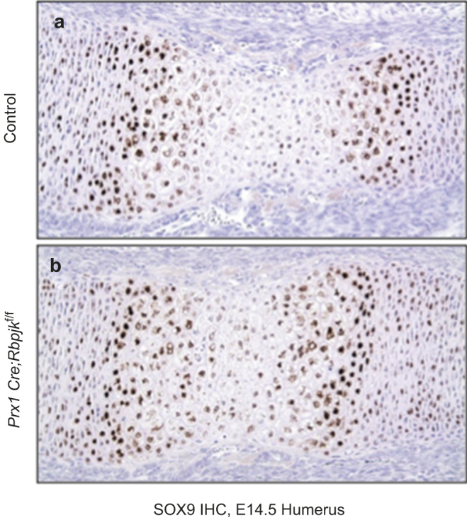
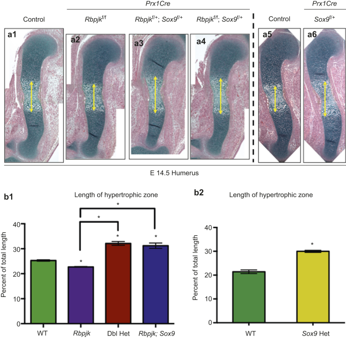
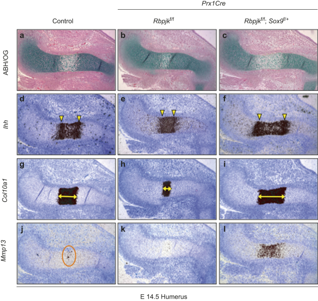
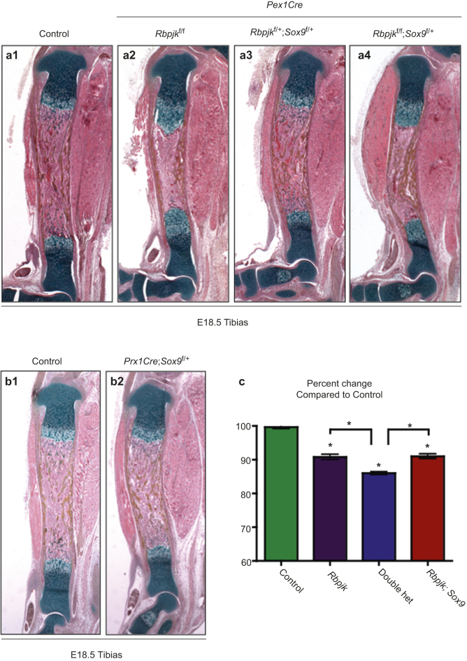
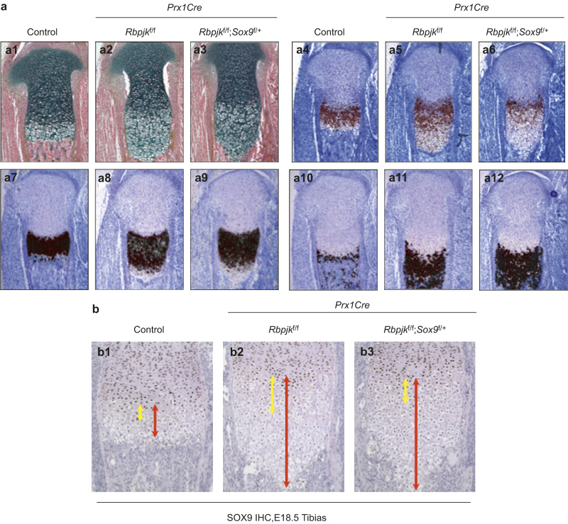
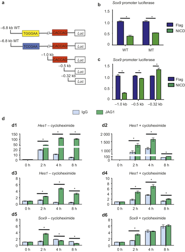
RBPjk-dependent Notch signaling regulates both the onset of chondrocyte hypertrophy and the progression to terminal chondrocyte maturation during endochondral ossification. It has been suggested that Notch signaling can regulate Sox9 transcription, although how this occurs at the molecular level in chondrocytes and whether this transcriptional regulation mediates Notch control of chondrocyte hypertrophy and cartilage development is unknown or controversial. Here we have provided conclusive genetic evidence linking RBPjk-dependent Notch signaling to the regulation of Sox9 expression and chondrocyte hypertrophy by examining tissue-specific Rbpjk mutant (Prx1Cre;Rbpjk(f/f) ), Rbpjk mutant/Sox9 haploinsufficient (Prx1Cre;Rbpjk(f/f);Sox9(f/+) ), and control embryos for alterations in SOX9 expression and chondrocyte hypertrophy during cartilage development. These studies demonstrate that Notch signaling regulates the onset of chondrocyte maturation in a SOX9-dependent manner, while Notch-mediated regulation of terminal chondrocyte maturation likely functions independently of SOX9. Furthermore, our in vitro molecular analyses of the Sox9 promoter and Notch-mediated regulation of Sox9 gene expression in chondrogenic cells identified the ability of Notch to induce Sox9 expression directly in the acute setting, but suppresses Sox9 transcription with prolonged Notch signaling that requires protein synthesis of secondary effectors.


Figures

Figure 1
Figure 1
Loss of RBPjk leads to inappropriate expression of SOX9 in hypertrophic chondrocytes. Immunohistochemistry for SOX9 in control (a) and Prx1Cre;Rbpjkf/f(b) E14.5 humerus sections.
Figure 2
Figure 2
Notch regulates the onset of chondrocyte hypertrophy via Sox9. (a) Histological analyses of control (a1), Prx1Cre;Rbpjkf/f (a2), Prx1Cre;Rbpjkf/+;Sox9f/+ (a3), Prx1Cre;Rbpjkf/f;Sox9f/+ (a4), as well as, control (a5) and Prx1Cre;Sox9f/+ embryonic tibia sections at E14.5. (b) Quantification of the lengths of the hypertrophic zone, expressed as a percentage of the total length of the element. (b1) WT wild-type, Rbpjk (Prx1Cre;Rbpjkf/f), Dbl Het (Prx1Cre;Rbpjkf/+;Sox9f/+), RBPJk; Sox9 (Prx1Cre;Rbpjkf/f;Sox9f/+). (b2) WT wild-type, Sox9 Het (Prx1Cre;Sox9f/+).
Figure 3
Figure 3
Notch regulates the onset of chondrocyte hypertrophy via Sox9. Histological and molecular analysis of control, Prx1Cre;Rbpjkf/f and Prx1Cre;Rbpjkf/f;Sox9f/+ embryonic tibia sections at E14.5. ABH/OG staining (a, b, c). In situ hybridization for markers of chondrocyte maturation – Indian Hedgehog (Ihh) (d, e, f), Collagen 10a1 (Col10a1) (g, h, i) and Matrix Metaloproteinase 13 (Mmp13) (j, k, l). Yellow arrowheads indicate primary Ihh expressing domains. Yellow double-headed arrows indicate Col10a1 expression domain. Orange circle highlights Mmp13 expression.
Figure 4
Figure 4
Cartilage element length reduction due to Sox9 haploinsufficiency is rescued by loss of RBPjk-dependent Notch signaling. (a) ABH/OG staining of control (a1), Prx1Cre;Rbpjkf/f (a2), Prx1Cre;Rbpjkf/+;Sox9f/+ (a3) and Prx1Cre;Rbpjkf/f;Sox9f/+ (a4) E18.5 full length tibia sections. (b) ABH/OG staining of control (b1) and Prx1Cre;Sox9f/+ (b2) E18.5 tibia sections. (c) Quantification of the size difference of E18.5 tibia sections.
Figure 5
Figure 5
Notch regulation of terminal chondrocyte maturation is not likely to be mediated via Sox9. (a) Histological and molecular analysis of control, Prx1Cre;RBPjkf/f, Prx1Cre;RBPjkf/f;Sox9f/+ embryonic tibia sections at E18.5. ABH/OG staining (a1a3). Markers of chondrocyte maturation – Indian Hedgehog (Ihh) (a4a6), Collagen 10a1 (Col10a1) (a7a9) and Matrix Metaloproteinase 13 (Mmp13) (a10a12) were analyzed by in situ hybridization. (b) Immunohistochemistry for SOX9 in control (b1), Prx1Cre;RBPjkf/f (b2), and Prx1Cre;RBPjkf/f;Sox9f/+ (b3) embryonic tibia sections at E18.5. Yellow double-headed arrows indicate the length of the SOX9 expressing hypertrophic chondrocyte domain. Red double-headed arrows indicate the total length of the hypertrophic zone.
Figure 6
Figure 6
Notch signaling inhibits Sox9 gene expression via secondary effectors. (a) Diagram for the localization of a RBPjk consensus binding site (yellow box is wild-type sequence and blue box is mutant sequence) and N-box consensus binding site (red box) in the Sox9 promoter and luciferase constructs. Core consensus sequences are listed in diagram. (b) ATDC5 cells were co-transfected with Flag or NICD1 over-expression vectors and either a wild-type 6.8 kb Sox9-Luciferase complex (6.8 kb WT) or 6.8 kb construct with a mutated RBPjk binding site (−6.8 kb MT). Luciferase levels were measured 24 hours after transfection. (c) ATDC5 cells were co-transfected with Flag or NICD1 over-expression vectors and either a 1 kb, 0.5 kb, or 0.32 kb Sox9-Luciferase deletion constructs. Luciferase levels were measured 24 hours after transfection. (d) Quantitative RT-PCR assessing Hes1 (d1, d2), Hey1 (d3, d4), and Sox9 (d5, d6) gene expression in ATDC5 cells at 0-, 2-, 4-, and 8-hour post-culture on IgG versus JAG1 coated plates in the absence (d1, d3, d5) or presence (d2, d4, d6) of cycloheximide.

Similar articles

Cited by

References

    1. Kronenberg HM. Developmental regulation of the growth plate. Nature 2003; 423: 332–336. - PubMed
    1. Akiyama H, Lefebvre V. Unraveling the transcriptional regulatory machinery in chondrogenesis. J Bone Miner Metab 2011; 29: 390–395. - PMC - PubMed
    1. Foster JW, Dominguez-Steglich MA, Guioli S et al. Campomelic dysplasia and autosomal sex reversal caused by mutations in an SRY-related gene. Nature 1994; 372: 525–530. - PubMed
    1. Wagner T, Wirth J, Meyer J et al. Autosomal sex reversal and campomelic dysplasia are caused by mutations in and around the SRY-related gene SOX9. Cell 1994; 79: 1111–1120. - PubMed
    1. Bi W, Huang W, Whitworth DJ et al. Haploinsufficiency of Sox9 results in defective cartilage primordia and premature skeletal mineralization. Proc Natl Acad Sci U S A 2001; 98: 6698–6703. - PMC - PubMed