

Congenital cystic adenomatoid malformation type I
- PMID: 26558243
- PMCID: PMC4636102
- DOI: 10.4322/acr.2015.019
Congenital cystic adenomatoid malformation type I
Abstract
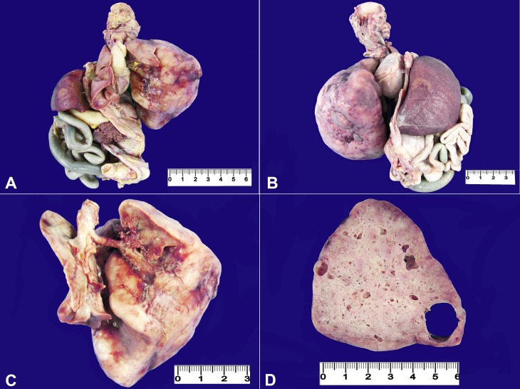
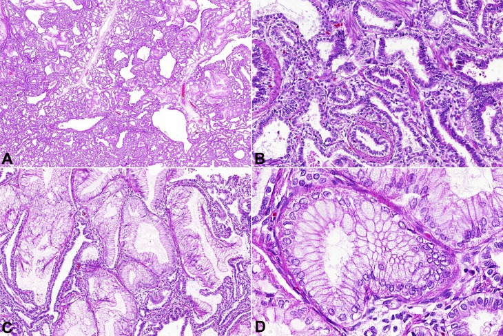
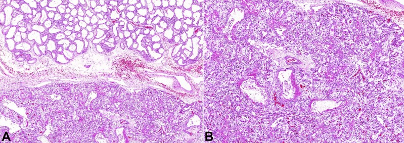
Congenital cystic adenomatoid malformation (CCAM) is an hamartomatous congenital pulmonary airway malformation with incidence ranging between 1:10,000 and 1:35,000 newborns. Currently CCAM is classified into five groups according to clinical and pathological features. The clinical outcome varies depending on the subtype and the extent of involvement. The authors report the case of a premature male newborn with the prenatal diagnosis of CCAM Type 1 associated with cardiac right axis deviation, who died 67 hours after birth due to respiratory failure. In addition to the autopsy report of this rare entity, the authors present its classification and prognosis.
Keywords: Autopsy; Classification; Cystic Adenomatoid Malformation of Lung, Congenital; Prognosis.
Conflict of interest statement
Conflict of interest: None
Figures
References
-
- Gilbert-Barness E, editor. Potter’s pathology of fetus and infant. St. Louis: Mosby; 1997. p. 508-509.
-
- Emery JL, Mithal A. The number of alveoli in the terminal respiratory unit of man during late intrauterine life and childhood. Arch Dis Child. 1960;35(184):544-7. http://dx.doi.org/10.1136/adc.35.184.544. PMid: - DOI - PMC - PubMed
-
- Stocker JT, Dehner LP, Husain AN. Stocker and Dehner’s pedriatric pathology. 3rd ed. Philadelphia: Lippincott Williams and Wilkins; 2001. p. 464-469.
-
- Durell J, Lakhoo K. Congenital cystic lesions of the lung. Early Hum Dev. 2014;90(12):935-9. http://dx.doi.org/10.1016/j.earlhumdev.2014.09.014. PMid: - DOI - PubMed
-
- Lakhoo K. Management of congenital cystic adenomatous malformations of the lung. Arch Dis Child Fetal Neonatal Ed. 2009;94(1):F73-6. http://dx.doi.org/10.1136/adc.2007.130542. PMid: - DOI - PubMed
Publication types
LinkOut - more resources
Full Text Sources
Other Literature Sources
