. 2015:2015:604028.
doi: 10.1155/2015/604028. Epub 2015 Oct 19.

No Evidence for Retinal Damage Evolving from Reduced Retinal Blood Flow in Carotid Artery Disease

Affiliations

No Evidence for Retinal Damage Evolving from Reduced Retinal Blood Flow in Carotid Artery Disease

Henning Heßler et al. Biomed Res Int. 2015.

Abstract

Introduction: Carotid artery disease (CAD) comprising high-grade internal carotid artery stenosis (CAS) or carotid artery occlusion (CAO) may lead to ipsilateral impaired cerebral blood flow and reduced retinal blood supply.

Objective: To examine the influence of chronic CAD on retinal blood flow, retinal morphology, and visual function.

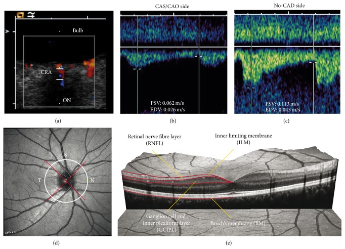
Methods: Patients with unilateral CAS ≥ 50% (ECST criteria) or CAO were grouped according to the grade of the stenosis and to the flow direction of the ophthalmic artery (OA). Retinal perfusion was measured by transorbital duplex ultrasound, assessing central retinal artery (CRA) blood flow velocities. In addition, optic nerve and optic nerve sheath diameter were measured. Optical coherence tomography (OCT) was performed to study retinal morphology. Visual function was assessed using high- and low-contrast visual paradigms.

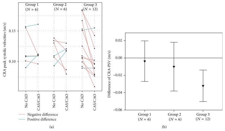
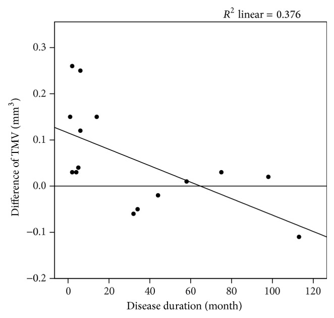
Results: Twenty-seven patients were enrolled. Eyes with CAS ≥ 80%/CAO and retrograde OA blood flow showed a significant reduction in CRA peak systolic velocity (no-CAD side: 0.130 ± 0.035 m/s, CAS/CAO side: 0.098 ± 0.028; p = 0.005; n = 12). OCT, optic nerve thicknesses, and visual functional parameters did not show a significant difference.

Conclusion: Despite assessable hemodynamic effects, chronic high-grade CAD does not lead to gaugeable morphological or functional changes of the retina.


Figures

Figure 1
Figure 1
(a)–(c) Transorbital ultrasound measurement of flow velocities in the central retinal artery (CRA) in a patient with carotid artery occlusion (CAO) and retrograde OA blood flow. (a) Transorbital B-mode scan of the optic nerve and color-mode imaging of the CRA. (b) Flow velocities ipsilateral to the CAO. Besides the reduced absolute flow velocity, there is a markedly delayed systolic flow rise. (c) Flow velocity of the no-CAD side. Note the normal steep systolic flow rise. (d) Exemplary peripapillary OCT ring scan with sectors: temporal (T), superior (S), inferior (I), and nasal (N). (e) Exemplary presentation of an OCT macular volume scan with segmentation of the retinal nerve fiber layer (RNFL) and combined ganglion cell and inner plexiform layer (GCIPL).
Figure 2
Figure 2
Results of peak systolic velocity (PSV) in the central retinal artery (CRA). (a) shows the individual measurements in stenosis subgroups. (b) shows PSV differences (PSV intereye difference = PSV CAS/CAO side − PSV no-CAD side) of stenosis subgroups. CAD: carotid artery disease; CAS: carotid artery stenosis; CAO: carotid artery occlusion, Group 1: CAS 50–79%; Group 2: CAS ≥ 80%/CAO with nonretrograde OA; Group 3: CAS ≥ 80%/CAO with retrograde OA.
Figure 3
Figure 3
Scatterplot describing the significant correlation (p = 0.005) between the disease duration of the CAS/CAO and the intereye difference of the total macular volume (TMV). Patients with initial CAS/CAO diagnosis in the last 15 months show thickening of the TMV. TMV intereye difference = TMV CAS/CAO side − TMV no-CAD side. CAD: carotid artery disease; CAS: carotid artery stenosis; CAO: carotid artery occlusion. Spearman-Rho test was used.

Similar articles

Cited by

References

    1. Lieb W. E., Flaharty P. M., Sergott R. C., et al. Color Doppler imaging provides accurate assessment of orbital blood flow in occlusive carotid artery disease. Ophthalmology. 1991;98(4):548–552. doi: 10.1016/S0161-6420(91)32257-7. - DOI - PubMed
    1. Ho A. C., Lieb W. E., Flaharty P. M., et al. Color Doppler imaging of the ocular ischemic syndrome. Ophthalmology. 1992;99(9):1453–1462. doi: 10.1016/S0161-6420(92)31784-1. - DOI - PubMed
    1. Hu H.-H., Sheng W.-Y., Yen M.-Y., Lai S.-T., Teng M. M.-H. Color doppler imaging of orbital arteries for detection of carotid occlusive disease. Stroke. 1993;24(8):1196–1203. doi: 10.1161/01.str.24.8.1196. - DOI - PubMed
    1. Costa V. P., Kuzniec S., Molnar L. J., Cerri G. G., Puech-Leao P., Carvalho C. A. Clinical findings and hemodynamic changes associated with severe occlusive carotid artery disease. Ophthalmology. 1997;104(12):1994–2002. doi: 10.1016/S0161-6420(97)30066-9. - DOI - PubMed
    1. Päivänsalo M., Riiheläinen K., Rissanen T., Suramo I., Laatikainen L. Effect of an internal carotid stenosis on orbital blood velocity. Acta Radiologica. 1999;40(3):270–275. doi: 10.3109/02841859909175553. - DOI - PubMed

Publication types

LinkOut - more resources