. 2015:2015:830315.
doi: 10.1155/2015/830315. Epub 2015 Oct 19.

CMP-Neu5Ac Hydroxylase Null Mice as a Model for Studying Metabolic Disorders Caused by the Evolutionary Loss of Neu5Gc in Humans

Affiliations

CMP-Neu5Ac Hydroxylase Null Mice as a Model for Studying Metabolic Disorders Caused by the Evolutionary Loss of Neu5Gc in Humans

Deug-Nam Kwon et al. Biomed Res Int. 2015.

Abstract

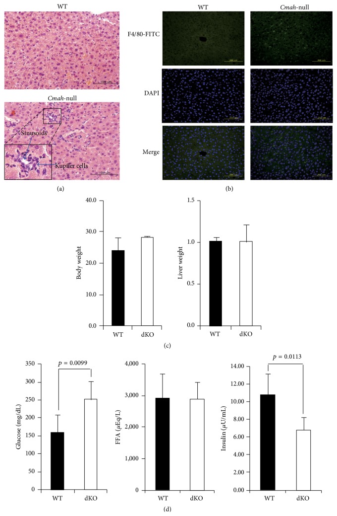
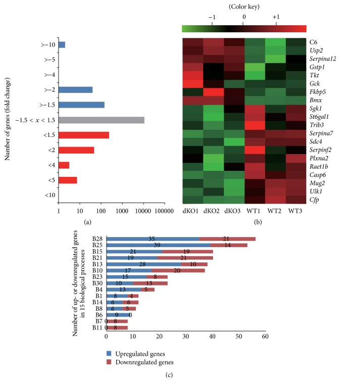
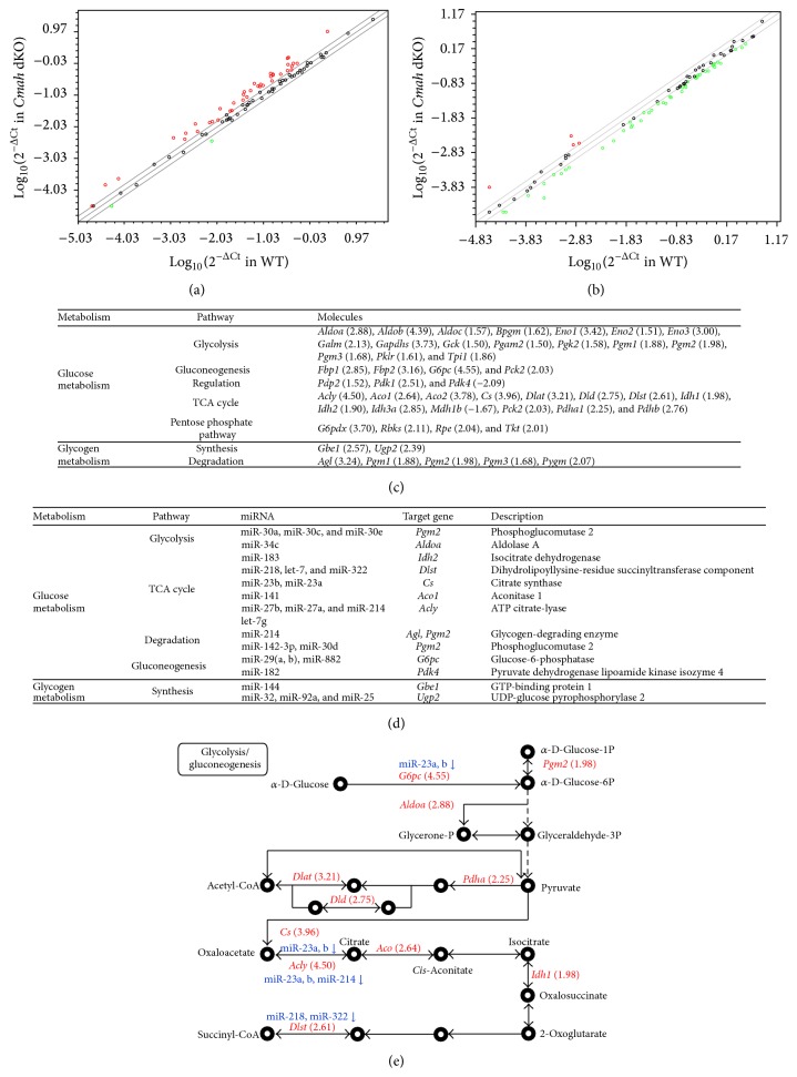
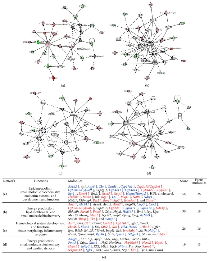
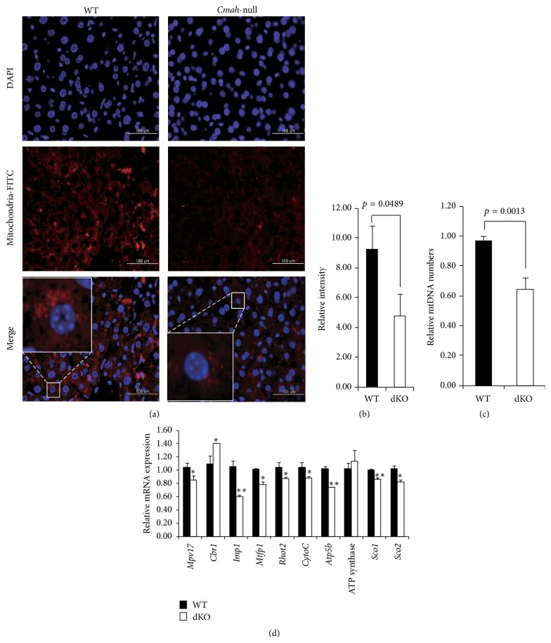
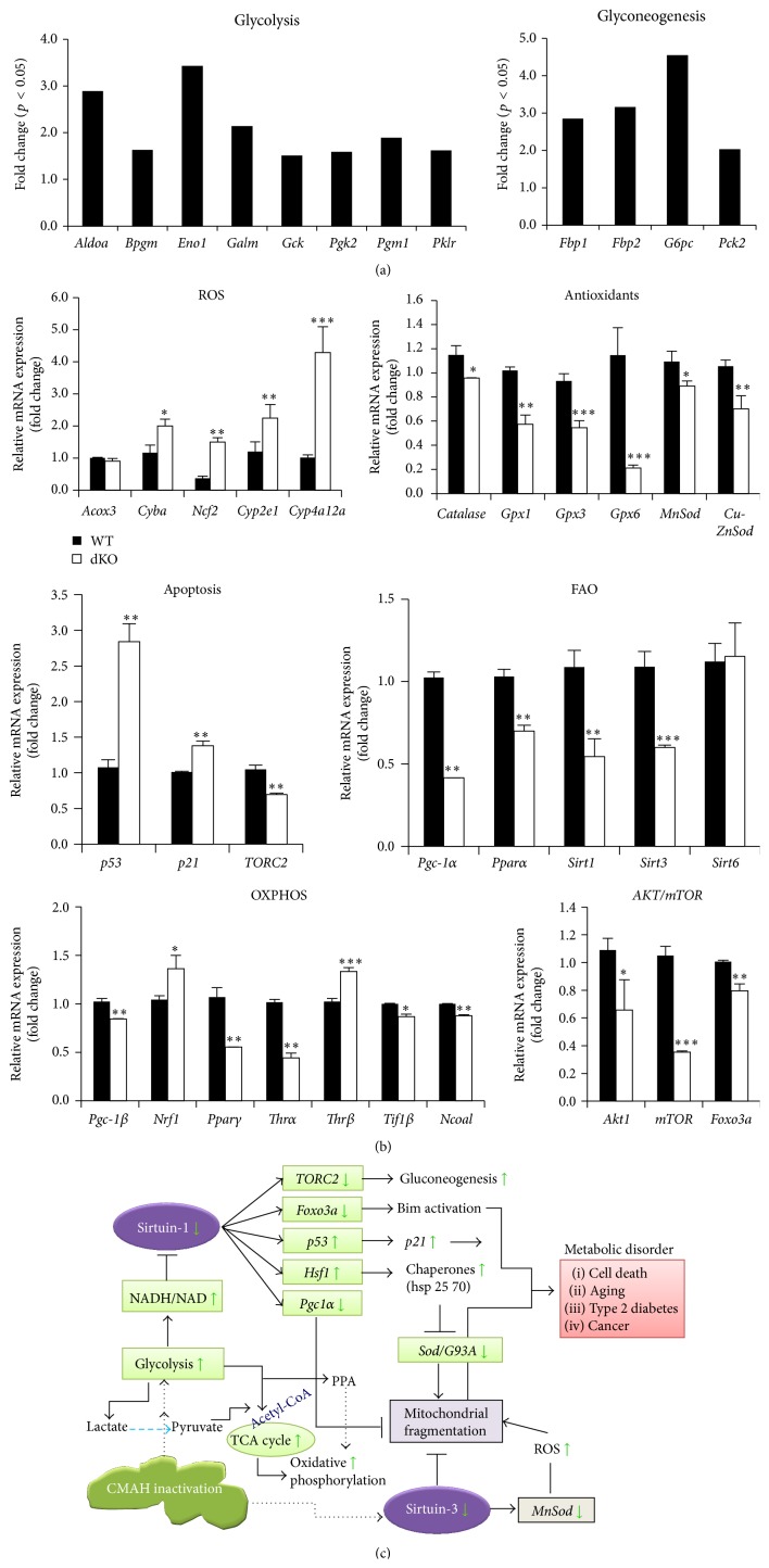
The purpose of this study was to identify the modification/turnover of gene products that are altered in humans due to evolutionary loss of Neu5Gc. CMP-Neu5Ac hydroxylase- (Cmah-) deficient mice show the infiltration of Kupffer cells within liver sinusoids, whereas body and liver weight develop normally. Pathway analysis by use of Illumina MouseRef-8 v2 Expression BeadChip provided evidence that a number of biological pathways, including the glycolysis, gluconeogenesis, TCA cycle, and pentose phosphate pathways, as well as glycogen metabolism-related gene expression, were significantly upregulated in Cmah-null mice. The intracellular glucose supply in Cmah-null mice resulted in mitochondrial dysfunction, oxidative stress, and the advanced glycation end products accumulation that could further induce oxidative stress. Finally, low sirtuin-1 and sirtuin-3 gene expressions due to higher NADH/NAD in Cmah-null mice decreased Foxo-1 and MnSOD gene expression, suggesting that oxidative stress may result in mitochondrial dysfunction in Cmah-null mouse. The present study suggests that mice with CMAH deficiency can be taken as an important model for studying metabolic disorders in humans.


Figures

Figure 1
Figure 1
Cmah-null mice show impaired insulin secretion in response to glucose. (a) Liver abnormality in Cmah-null mice. The number of Kupffer cells was significantly higher in Cmah-null mouse-derived liver tissues than in WT. Bar: 100 μm. (b) Immunofluorescence staining of Cmah-null-derived liver tissue to detect expression of Kupffer cells using F4/80 antibody. (c) Body and liver weights of WT and Cmah-null mice. (d) Quantity of glucose (mL/dL), FFA (μEq/L), and insulin (μU/mL) in sera of WT and Cmah-null mice. Significant differences are indicated by p value.
Figure 2
Figure 2
Alteration of gene expression in liver tissues of Cmah-null mice. (a) Histogram showing genes with a >1.5-fold difference in expression levels in liver tissues of Cmah-null mice. Blue and red bars indicate down- and upregulated genes, respectively. (b) Cluster analysis of 20 microarray pieces of data for WT and Cmah-null mice. (c) The differentially up- or downregulated genes were examined according to biological process. B1: amino acid metabolism, B2: apoptosis, B3: blood circulation and gas exchange, B4: carbohydrate metabolism, B5: cell adhesion, B6: cell cycle, B7: cell proliferation and differentiation, B8: cell structure and motility, B9: coenzyme and prosthetic group metabolism, B10: developmental processes, B11: electron transport, B12: homeostasis, B13: immunity and defense, B14: intracellular protein traffic, B15: lipid, fatty acid, and steroid metabolism, B16: miscellaneous, B17: muscle contraction, B18: neuronal activities, B19: nitrogen metabolism, B20: nonvertebrate processes, B21: nucleoside, nucleotide, and nucleic acid metabolism, B22: oncogenesis, B23: other metabolisms, B24: phosphate metabolism, B25: protein metabolism and modification, B26: protein targeting and localization, B27: sensory perception, B28: signal transduction, B29: sulfur metabolism, B30: transport, and B31: biological process unclassified. The bar graph includes up- and downregulated genes in the top 15 biological process categories.
Figure 3
Figure 3
Alteration of genes and miRNAs involved in metabolism-related pathways by CMAH disruption. (a) Comparison of the relative expressions of 84 genes involved in glucose metabolism between WT and Cmah-null mouse-derived liver tissues using a pathway-focused glucose metabolism PCR array. The figure depicts a scatter plot of the relative expression levels. Red and green colors indicate upregulation and downregulation of gene expression (>1.5-fold change), respectively. (b) Comparison of the relative expression levels of the 84 most abundantly expressed and best characterized miRNAs in miRBase using a miFinder miScript miRNA PCR Array. The figure depicts a scatter plot of the relative expression levels. Red and green colors indicate upregulation and downregulation of miRNA expression (>1.5-fold change). (c) Metabolism-related pathways and target molecules identified in liver tissues of Cmah-null mice. (d) miRNAs and target genes involved in glucose and glycogen metabolism. (e) Illustration of genes involved in glucose metabolism identified by the PCR array. Red and blue colors indicate upregulated genes and downregulated miRNAs, respectively.
Figure 4
Figure 4
Networks predicted by Ingenuity Pathway Analysis in Cmah-null mice. (a) Lipid metabolism, small molecule biochemistry, and endocrine system development and function. (b) Energy production, lipid metabolism, and small molecule biochemistry. (c) Hematological system development and function, tissue morphology, and inflammatory response. (d) Cell death and survival, embryonic development, and organ development, p < 0.05. Red and green denote upregulation and downregulation of genes, respectively. Solid lines and dotted lines indicate direct and indirect relationships, respectively. (e) List of genes involved in networks (a)–(d). This table includes their functions, molecules, score, and focus molecules. Red and blue indicate up- and downregulated genes, respectively, which are involved in the networks.
Figure 5
Figure 5
Characterization of mitochondrial dysfunction in liver of Cmah-null mice. (a) Reduction of mitochondrial activity in Cmah-null mice livers, stained using a mitochondrial antibody. (b) The intensity of the fluorescent signal indicates the level of mitochondrial activity. (c) The mtDNA/β-actin ratio, which represents the average copy number, was significantly decreased in Cmah-null mouse-derived livers. The CytB gene amplification level was normalized against the expression of nuclear β-actin. (d) Expression levels of genes involved in mitochondrial functional regulation were determined by RT-qPCR in RNA samples from the livers of WT and Cmah-null mice. Measurements were performed in triplicate, after which the mean expression was calculated and corrected using gapdh expression levels. Error bars indicate standard deviations. Significant differences are indicated by p < 0.05 and ∗∗ p < 0.01.
Figure 6
Figure 6
Molecular mechanisms of glucose-mediated regulation of sirtuins and oxidative stress in Cmah-null mice. (a) Expression of genes involved in glycolysis and glyconeogenesis, confirmed in WT and Cmah-null mouse-derived livers by a pathway-focused glucose metabolism PCR array. (b) Expression levels of genes involved in the molecular mechanisms of sirtuin and oxidative stress regulation were determined by RT-qPCR of RNA samples from liver tissues of WT and Cmah-null mice. Measurements were performed in triplicate, and the calculated mean expression was corrected using Gapdh expression levels. Error bars indicate standard deviations. Significant differences are indicated by p < 0.05 and ∗∗ p < 0.01, ∗∗∗ p < 0.001. (c) Glucose can directly or indirectly affect the main regulators of the aging process and sirtuin activity, as well as other contributors to aging such as oxidative stress. Increased glycolytic activity would tend to provoke an accumulation of NADH and lower the availability of NAD, resulting in decreased sirtuin activity. In addition, activation of the TCA cycle and β-oxidation of fatty acids distribute acetyl-CoA and acyl CoA, respectively, to the mitochondrial OXPHOS pathway. Therefore, lactate produced by glycolysis may be converted to pyruvate and ultimately enter the TCA cycle. Collectively, these results show that, via increased glycolysis, increased intracellular glucose levels can lead to (i) mitochondrial dysfunction and oxidative stress due to continuous ATP synthesis and (ii) accumulation of highly toxic advanced glycation end products (AGEs), which can provoke further oxidative stress. These observations suggest that the evolutionary loss of CMAH function may make humans more prone to diabetes or aging than other mammals.

Similar articles

Cited by

References

    1. Irie A., Koyamat S., Kozutsumi Y., Kawasaki T., Suzuki A. The molecular basis for the absence of N-glycolylneuraminic acid in humans. Journal of Biological Chemistry. 1998;273(25):15866–15871. doi: 10.1074/jbc.273.25.15866. - DOI - PubMed
    1. Hayakawa T., Satta Y., Gagneux P., Varki A., Takahata N. Alu-mediated inactivation of the human CMP-N-acetylneuraminic acid hydroxylase gene. Proceedings of the National Academy of Sciences of the United States of America. 2001;98(20):11399–11404. doi: 10.1073/pnas.191268198. - DOI - PMC - PubMed
    1. Brinkman-Van der Linden E. C. M., Sjoberg E. R., Juneja L. R., Crocker P. R., Varki N., Varki A. Loss of N-glycolylneuraminic acid in human evolution. Implications for sialic acid recognition by siglecs. The Journal of Biological Chemistry. 2000;275(12):8633–8640. doi: 10.1074/jbc.275.12.8633. - DOI - PubMed
    1. Varki A. Loss of N-glycolylneuraminic acid in humans: mechanisms, consequences, and implications for hominid evolution. American Journal of Physical Anthropology. 2001;116(supplement 33):54–69. doi: 10.1002/ajpa.10018. - DOI - PMC - PubMed
    1. Deng L., Song J., Gao X., et al. Host adaptation of a bacterial toxin from the human pathogen Salmonella Typhi. Cell. 2014;159(6):1290–1299. doi: 10.1016/j.cell.2014.10.057. - DOI - PMC - PubMed

Publication types

Substances