. 2015 Nov 12:6:8923.
doi: 10.1038/ncomms9923.

Guanine nucleotide binding to the Bateman domain mediates the allosteric inhibition of eukaryotic IMP dehydrogenases

Affiliations

Guanine nucleotide binding to the Bateman domain mediates the allosteric inhibition of eukaryotic IMP dehydrogenases

Rubén M Buey et al. Nat Commun. .

Abstract

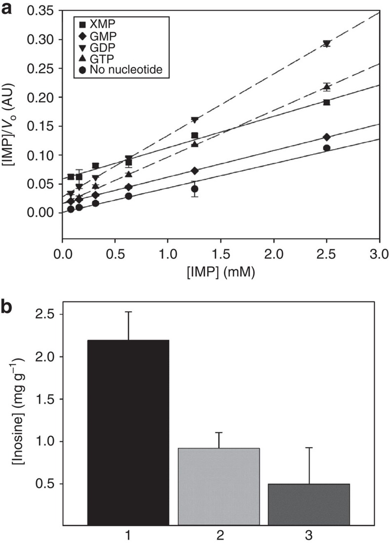
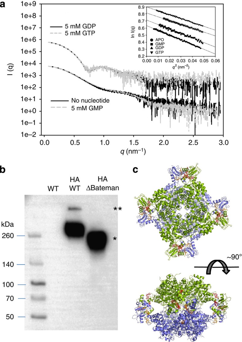
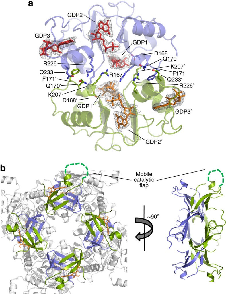
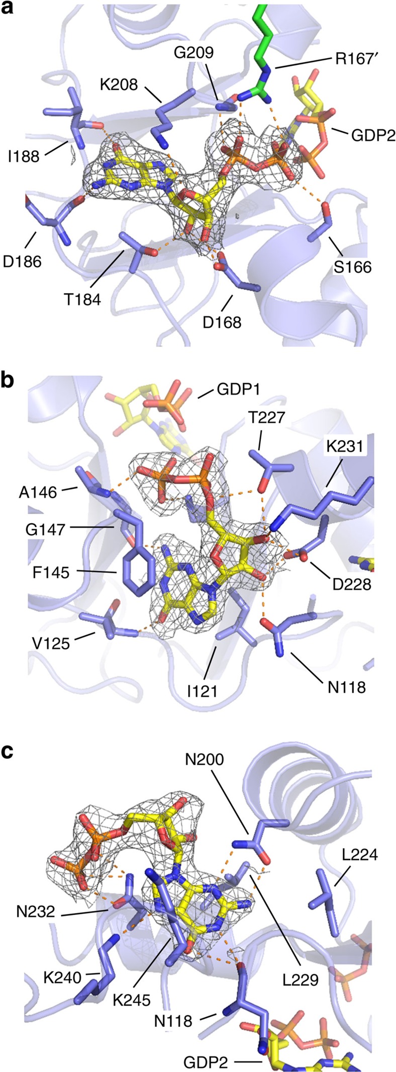
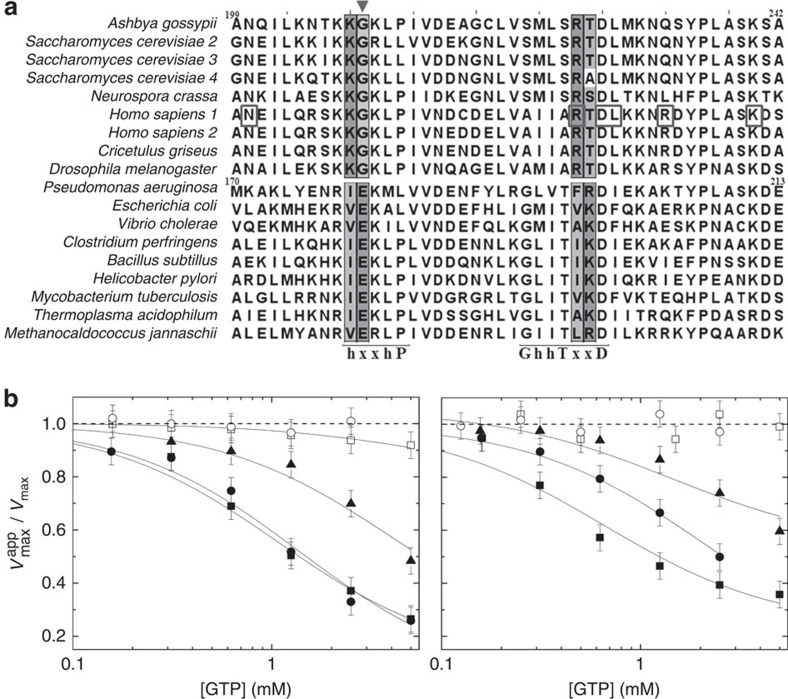
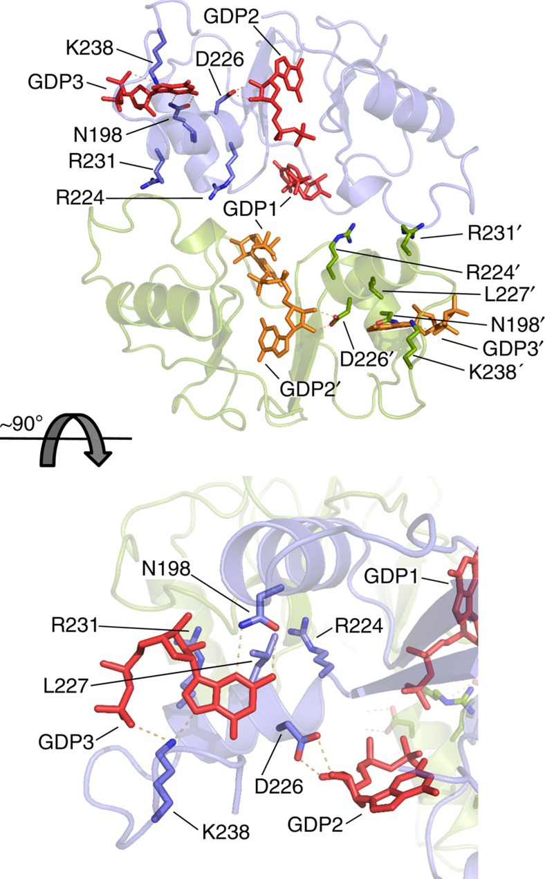
Inosine-5'-monophosphate dehydrogenase (IMPDH) plays key roles in purine nucleotide metabolism and cell proliferation. Although IMPDH is a widely studied therapeutic target, there is limited information about its physiological regulation. Using Ashbya gossypii as a model, we describe the molecular mechanism and the structural basis for the allosteric regulation of IMPDH by guanine nucleotides. We report that GTP and GDP bind to the regulatory Bateman domain, inducing octamers with compromised catalytic activity. Our data suggest that eukaryotic and prokaryotic IMPDHs might have developed different regulatory mechanisms, with GTP/GDP inhibiting only eukaryotic IMPDHs. Interestingly, mutations associated with human retinopathies map into the guanine nucleotide-binding sites including a previously undescribed non-canonical site and disrupt allosteric inhibition. Together, our results shed light on the mechanisms of the allosteric regulation of enzymes mediated by Bateman domains and provide a molecular basis for certain retinopathies, opening the door to new therapeutic approaches.


Figures

Figure 1
Figure 1. GTP and GDP allosterically inhibit AgIMPDH.
(a) Analysis of the enzyme kinetics in vitro data using the linear Hanes-Woolf plot. Initial velocities (Vo) were determined by fitting the time-course data to the Michaelis–Menten equation. Reactions contained 20 μg ml−1 AgIMPDH, 0.5 mM NAD+ and variable concentrations of IMP. Reaction buffer (100 mM Tris-HCl, 100 mM KCl, 2 mM DTT pH 8.0) did not contain Mg+2. (b) Milligrams of inosine excreted per gram of mycelium from different strains of A. gossypii (1: WT, 2: ΔBateman and 3: PGPD-ΔBateman) after 3 days of culture in MA2-rich medium at 28 °C with orbital shaking. Experiments were performed in triplicate. Error bars represent s.e.
Figure 2
Figure 2. GTP and GDP alter the quaternary structure of AgIMPDH.
(a) SAXS profiles of AgIMPDH in the presence of different nucleotides. The inset shows the Guinier plots for the different curves. The plots have been conveniently displaced along the y axis to facilitate visualization. (b) Anti-HA western blotting of cross-linked A. gossypii whole-cell extracts. The single and double asterisks show the bands unambiguously attributed to tetramers and octamers, respectively. (c) Different views of a cartoon representation of AgIMPDH octamers, obtained in the presence of GDP (red and orange sticks). The two tetramers that pile up tail-to-tail are coloured green and blue.
Figure 3
Figure 3. The bipartite interface of the AgIMPDH dimer of tetramers.
(a) Two Bateman domains from the upper and lower tetramers are shown in green and blue cartoons. GDP-binding protein residues and GDP molecules are shown in sticks. The grey meshes around GDP molecules represent the simulated annealing omit 2mFo−DFc electron density maps contoured at the 1σ level. (b) The finger domains from the upper and lower tetramers are shown in green and blue cartoons around the quaternary (left) and binary symmetry axes (right). GMP bound to the catalytic site is shown with orange sticks in the left panel. The catalytic mobile flap (not visible in our structure) is represented as a discontinuous green line in one of the monomers.
Figure 4
Figure 4. The Bateman domains of AgIMPDH bind guanine nucleotides.
Close-up views of the three GDP molecules bound to the Bateman domains of AgIMPDH (a) GDP1, (b) GDP2 and (c) GDP3. AgIMPDH protein is shown in light blue cartoons with key interacting residues and GDP molecules shown in sticks. In a, the side chain from an Arg residue from the adjacent monomer (R167') is shown with green sticks. Key interactions are represented by orange dashes. The grey mesh around GDP represents the simulated annealing omit 2mFo−DFc electron density map contoured at the 1σ level.
Figure 5
Figure 5. GTP and GDP only inhibit eukaryotic IMPDHs.
(a) Multiple-sequence alignment of the CBS2 motif within the Bateman domain of selected eukaryotic and prokaryotic organisms. The nucleotide-binding sequence motifs (‘h-x-x-h-P' and ‘G-h-hT-x-x-D') are indicated below the sequences. The highest-scoring SDPs are shown in a shaded box. The triangle indicates the glutamate residue within the ‘RIEK' motif of prokaryotic IMPDHs or the glycine within the ‘KKGK' motif that defines eukaryotic IMPDHs. Residues of the human isoform 1 associated with retinopathies are shown in a box. (b) Catalytic activity at increasing concentrations of GTP and GDP of AgIMPDH (black squares), HsIMPDH1 (black circles), HsIMPDH2 (black triangles), EcIMPDH (white circles) and BsIMPDH (white squares). The Vmax and Vmaxapp values as a function of inhibitor concentration were determined by fitting the enzyme kinetics data to the Michaelis–Menten equation. Experiments were performed in duplicate. Error bars represents s.e. The continuous lines represent the nonlinear regression fitting analysis employing a mixed inhibition model.
Figure 6
Figure 6. Missense mutations of HsIMPDH1 linked to retinopathies map into the nucleotide-binding sites.
Blue and green cartoon representations of two interacting Bateman domains of a HsIMPDH1 homology model bound to GDP (red or orange sticks). The side chain of residues whose mutations are linked to RP and LCA are shown in sticks. Key interactions are represented by orange dashes.

Similar articles

Cited by

References

    1. Chong C. R. et al.. Identification of type 1 inosine monophosphate dehydrogenase as an antiangiogenic drug target. J. Med. Chem. 49, 2677–2680 (2006). - PubMed
    1. Long H., Cameron S., Yu L. & Rao Y. De novo GMP synthesis is required for axon guidance in Drosophila. Genetics 172, 1633–1642 (2006). - PMC - PubMed
    1. Jayaram H. N., Cooney D. A., Grusch M. & Krupitza G. Consequences of IMP dehydrogenase inhibition, and its relationship to cancer and apoptosis. Curr. Med. Chem. 6, 561–574 (1999). - PubMed
    1. Allison A. C. & Eugui E. M. Mycophenolate mofetil and its mechanisms of action. Immunopharmacology 47, 85–118 (2000). - PubMed
    1. Braun-Sand S. B. & Peetz M. Inosine monophosphate dehydrogenase as a target for antiviral, anticancer, antimicrobial and immunosuppressive therapeutics. Future Med. Chem. 2, 81–92 (2010). - PubMed

Publication types

MeSH terms