. 2015 Nov 11;35(45):14966-82.
doi: 10.1523/JNEUROSCI.0293-15.2015.

Dopamine/Tyrosine Hydroxylase Neurons of the Hypothalamic Arcuate Nucleus Release GABA, Communicate with Dopaminergic and Other Arcuate Neurons, and Respond to Dynorphin, Met-Enkephalin, and Oxytocin

Affiliations

Dopamine/Tyrosine Hydroxylase Neurons of the Hypothalamic Arcuate Nucleus Release GABA, Communicate with Dopaminergic and Other Arcuate Neurons, and Respond to Dynorphin, Met-Enkephalin, and Oxytocin

Xiaobing Zhang et al. J Neurosci. .

Abstract

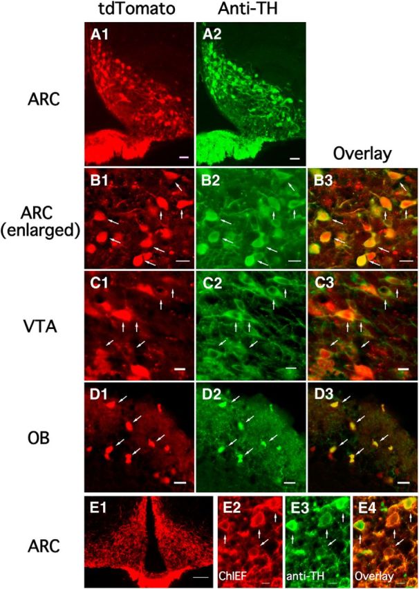
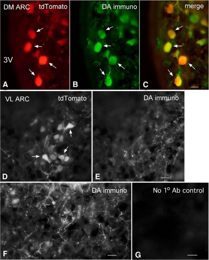
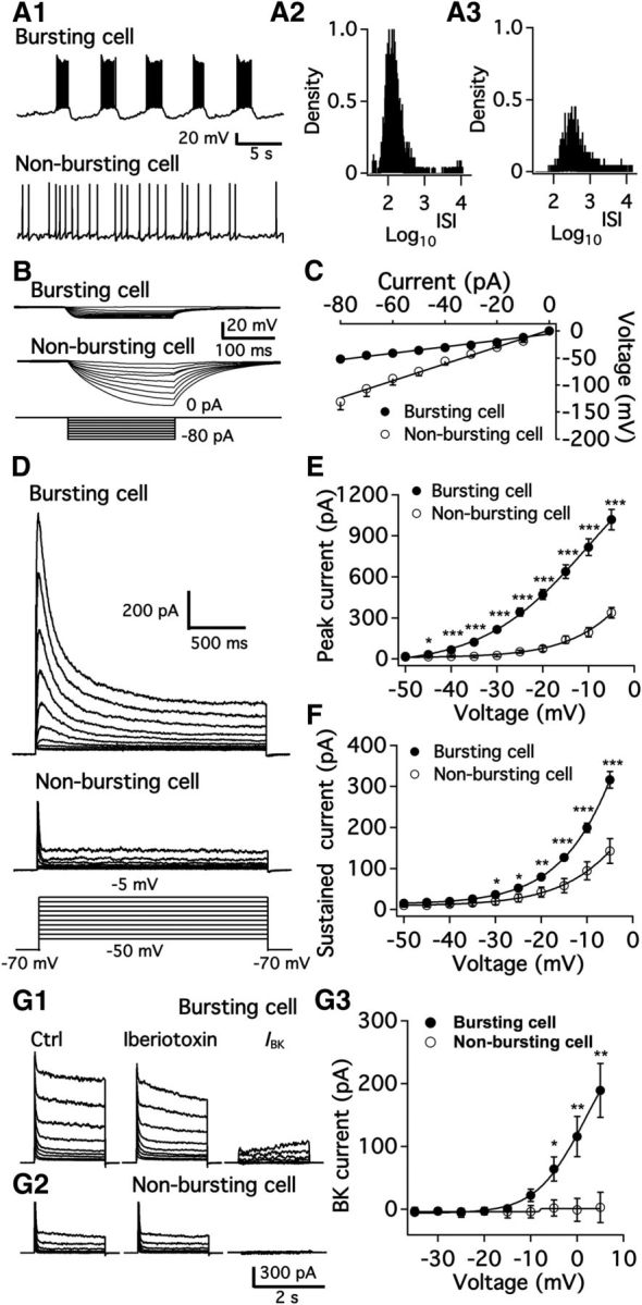
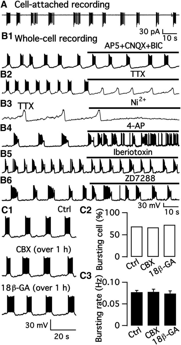
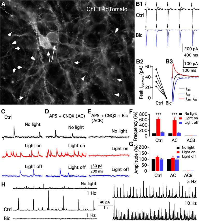
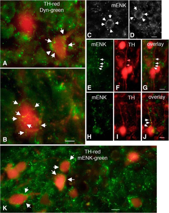
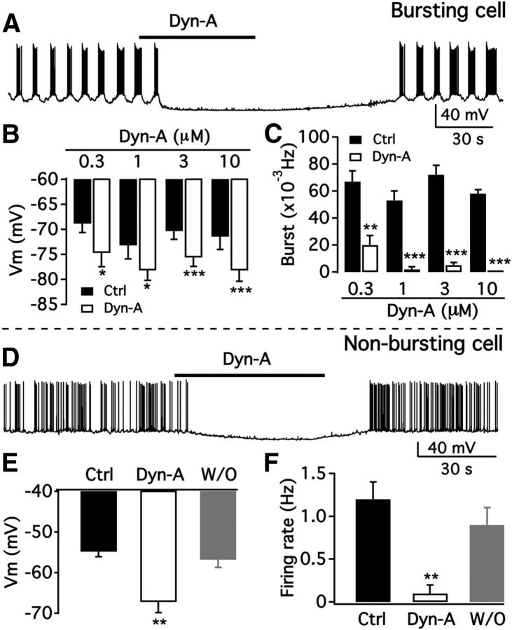
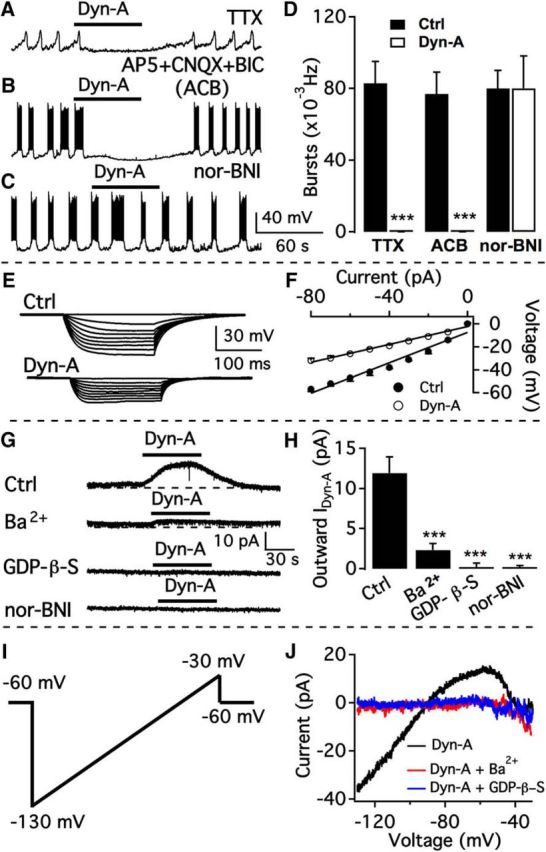
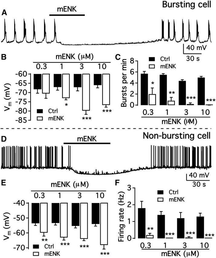
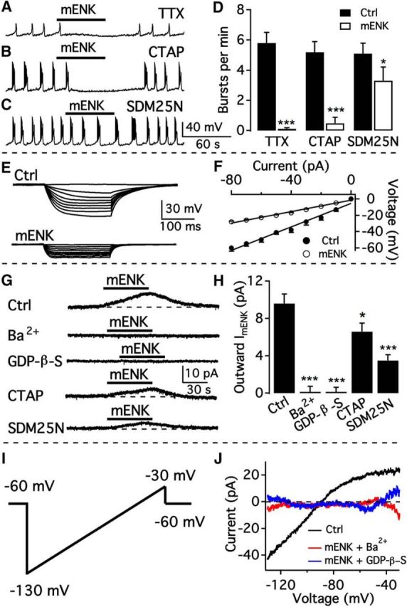
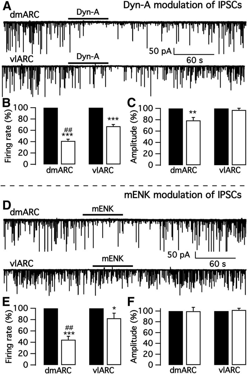
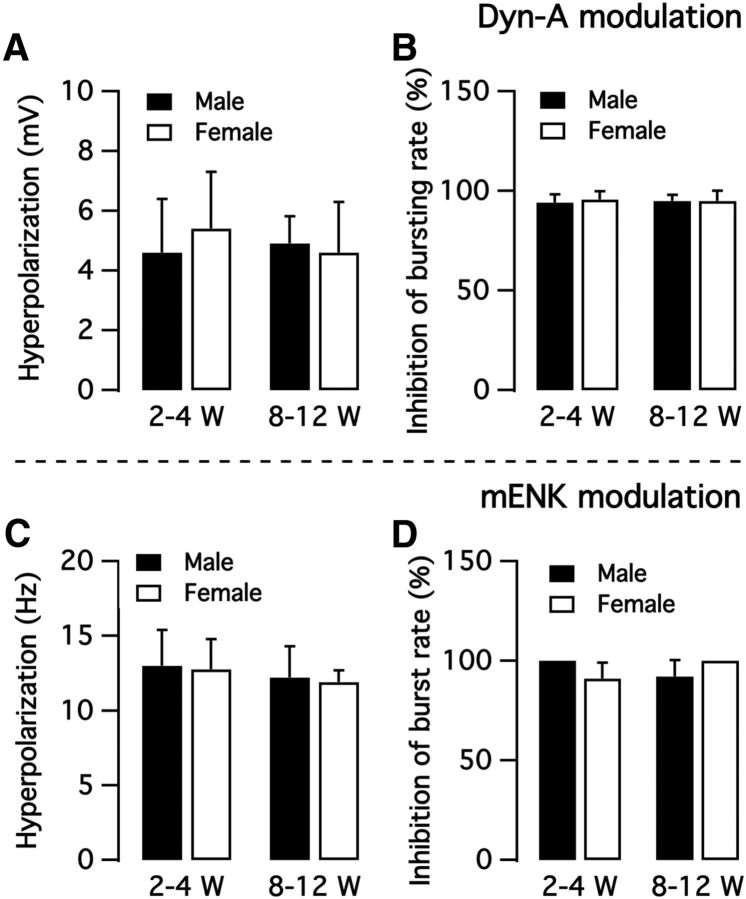
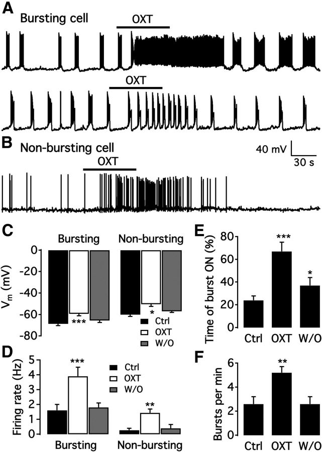
We employ transgenic mice with selective expression of tdTomato or cre recombinase together with optogenetics to investigate whether hypothalamic arcuate (ARC) dopamine/tyrosine hydroxylase (TH) neurons interact with other ARC neurons, how they respond to hypothalamic neuropeptides, and to test whether these cells constitute a single homogeneous population. Immunostaining with dopamine and TH antisera was used to corroborate targeted transgene expression. Using whole-cell recording on a large number of neurons (n = 483), two types of neurons with different electrophysiological properties were identified in the dorsomedial ARC where 94% of TH neurons contained immunoreactive dopamine: bursting and nonbursting neurons. In contrast to rat, the regular oscillations of mouse bursting neurons depend on a mechanism involving both T-type calcium and A-type potassium channel activation, but are independent of gap junction coupling. Optogenetic stimulation using cre recombinase-dependent ChIEF-AAV-DJ expressed in ARC TH neurons evoked postsynaptic GABA currents in the majority of neighboring dopamine and nondopamine neurons, suggesting for the first time substantial synaptic projections from ARC TH cells to other ARC neurons. Numerous met-enkephalin (mENK) and dynorphin-immunoreactive boutons appeared to contact ARC TH neurons. mENK inhibited both types of TH neuron through G-protein coupled inwardly rectifying potassium currents mediated by δ and μ opioid receptors. Dynorphin-A inhibited both bursting and nonbursting TH neurons by activating κ receptors. Oxytocin excited both bursting and nonbursting neurons. These results reveal a complexity of TH neurons that communicate extensively with neurons within the ARC.

Significance statement: Here, we show that the great majority of mouse hypothalamic arcuate nucleus (ARC) neurons that synthesize TH in the dorsomedial ARC also contain immunoreactive dopamine, and show either bursting or nonbursting electrical activity. Unlike rats, the mechanism underlying bursting was not dependent on gap junctions but required T-type calcium and A-type potassium channel activation. Neuropeptides dynorphin and met-enkephalin inhibited dopamine neurons, whereas oxytocin excited them. Most ventrolateral ARC TH cells did not contain dopamine and did not show bursting electrical activity. TH-containing neurons appeared to release synaptic GABA within the ARC onto dopamine neurons and unidentified neurons, suggesting that the cells not only control pituitary hormones but also may modulate nearby neurons.

Keywords: GABA; arcuate nucleus; burst firing; dopamine neuron; neuropeptides; optogenetics.


Figures

Figure 1.
Figure 1.
TH expression in TH-tdTomato transgenic mice. A1, A2, TH-tdTomato fluorescence (A1) and TH immunostaining (A2) showing fluorescence was restricted to the ARC. Scale bar, 20 μm. B1–B3, Enlarged images showing TH-tdTomato fluorescence (B1), TH immunostaining (B2), and the overlay (B3) in the ARC. Scale bar, 10 μm. C1–C3, TH-tdTomato fluorescence (C1), TH immunostaining (C2), and the overlay (C3) in the ventral tegmental area (VTA). Scale bar, 10 μm. D1–D3, TH-tdTomato fluorescence (D1), TH immunostaining (D2), and the overlay (D3) in the olfactory bulb. Scale bar, 10 μm. E, Restricted ChIEF-tdTomato expression was found after AAV Cre-dependent ChIEF-tdTomato was injected into both sides of the ARC of TH-Cre mice (E1). Scale bar, 200 μm. High-magnification image showing cell bodies of ChIEF-tdTomato-expressing neurons (E2), TH-immunostained neurons (E3), and the overlay (E4). Cells expressing ChIEF-tdTomato were immunoreactive for TH. Scale bar, 20 μm.
Figure 2.
Figure 2.
Dopamine immunoreactivity in TH-tdTomato mice. A–C, Dopamine immunoreactivity in tdTomato neurons in dmARC. A, Red tdTomato-positive neurons from transgenic reporter mouse. B, Green neurons immunostained for dopamine. C, Merge of A and B shows that most tdTomato neurons also show dopamine immunoreactivity. Scale bar, 9 μm. D, In vlARC, tdTomato-positive neurons are found (arrows). E, Dopamine immunostaining showed primarily immunoreactive axons with few neurons showing both dopamine-immunoreactive and tdTomato expression. Scale bar, 12 μm. F, Dopamine-immunoreactive axons are seen surrounding unidentified ARC neurons. Scale bar, 9 μm. G, Here, the primary dopamine antibody was omitted, and only the secondary antibody was applied as a control. No immunostaining was found. Scale bar, 10 μm.
Figure 3.
Figure 3.
Electrophysiological characterization of two populations of arcuate TH neurons. A, Representative traces showing burst firing (A1, top) and nonburst firing (A1, bottom) of dmARC TH neurons and histogram of log10ISI of action potentials from bursting (A2) and nonbursting cell (A3). B, Membrane potential changes in a bursting neuron (top) and a nonbursting neuron (middle) induced by a series of negative step current injections (bottom). C, Voltage–current relationship for bursting and nonbursting cells obtained from averaged data with recording as in B. D, Representative traces showing voltage-gated potassium current in bursting and nonbursting neurons at depolarized membrane potentials of −50 to −5 mV from a holding potential of −70 mV. TTX, Cd2+, Bic, AP5, and CNQX were added to the recording solution to block voltage-gated sodium and calcium channels and synaptic responses. E, Summary data showing the peak amplitude of outward potassium current in both types of neuron at various voltages. *p < 0.05 (Student's t test compared with peak current of nonbursting cells at same voltage). ***p < 0.001 (Student's t test compared with peak current of nonbursting cells at same voltage). F, Summary data showing the amplitude of sustained outward potassium current in both types of neuron at various voltages. *p < 0.05 (Student's t test compared with sustained current of nonbursting cells at same voltage). **p < 0.01. ***p < 0.001. G1, Representative traces show voltage-gated potassium current in a bursting neuron using voltage steps from a holding potential of −70 mV to a level of −35 to 5 mV. Data from control (left) and in the presence of iberiotoxin (200 nm, middle) are shown. BK current (right) was obtained by subtraction of potassium current in the presence of iberiotoxin from control. TTX, Bic, AP5, and CNQX were added to the recording solution to block voltage-gated sodium channels and synaptic responses. G2, Representative traces show voltage-gated potassium current in a nonbursting neuron using voltage steps from a holding potential of −70 mV to a level of −35 to 5 mV in a nonbursting neuron in control (left) and in the presence of iberiotoxin (200 nm, middle). No obvious BK current (right) was obtained by subtraction of potassium current in the presence of iberiotoxin from control. G3, Summary data showing the peak amplitude of BK current in both types of neuron at various voltages. *p < 0.05 (Student's t test compared with IBK of nonbursting cells at same voltage). **p < 0.01.
Figure 4.
Figure 4.
Mechanism of bursting activity. A, Regular burst firing recorded in the cell-attached mode from a dmARC TH neuron. B1, Regular burst firing recorded in the whole-cell current-clamp configuration was not obviously affected by a mixture of Bic, CNQX, and AP5. B2, TTX blocked firing but not the regular oscillations of the membrane potential. B3, Membrane potential oscillations were blocked by Ni2+ in the presence of TTX. B4, Regular bursting was changed to irregular bursting with shorter burst duration by 4-AP. B5, Burst firing recorded in normal ACSF and in the presence of the BK channel blocker iberiotoxin. B6, Burst firing recorded in normal ACSF and in the presence of Ih current blocker ZD7288. C1, Representative traces of burst firing recorded in normal ACSF (top) and in the presence of CBX (over 1 h, middle) and 18β-GA (over 1 h, bottom). C2, Percentage of bursting cells in control buffer and in the presence of CBX and 18β-GA. C3, The mean burst rate in control and in the presence of CBX and 18β-GA.
Figure 5.
Figure 5.
Arcuate TH neurons functionally innervate arcuate neurons. A, Photomicrograph shows ChIEF-tdTomato expression in the membrane of dorsomedial ARC of TH-cre mice. Two long arrows indicate positive somata. Short arrowheads indicate very fine positive axonal processes innervating ARC neurons. Scale bar, 7 μm. B, Photostimulation (10 ms) evoked a current with two components in ChIEF-tdTomato-expressing neurons. The membrane potential was held at −40 mV, ensuring an outward current mediated by GABA. B1, Representative traces showing repeated activation using photostimulation (10 ms) of a ChIEF-tdTomato neuron in the absence (Ctrl) and presence of Bic. B2, Mean amplitudes of outward currents evoked by photostimulation in 4 neurons recorded in the absence (Ctrl) and presence of Bic. B3, Mean current traces of five sequential currents induced by photostimulation in the absence (Ctrl) and presence of Bic. The GABAA receptor-mediated synaptic current was obtained by subtracting the current in the presence of Bic from the current in the absence of Bic (Ctrl). C–E, Representative traces showing GABAergic IPSCs before light on (top), during light on (middle), and after light off (bottom), in control (Ctrl) solution (C), in the presence of AP5 and CNQX (D), and in the presence of AP5, CNQX, and Bic (E). F, Summary of data showing that continuous light stimulation increased IPSC frequency in the absence and presence of AP5 and CNQX, but not in the presence of a mixture of AP5, CNQX, and Bic. ***p < 0.001 (one-way ANOVA compared with no light group). G, Bar graph showing that the IPSC amplitude was unaffected by photostimulation. H, Representative traces showing spontaneous (top left) and photostimulation (1 Hz)-evoked IPSCs (middle left) recorded from an unidentified arcuate neuron in control and in the presence of Bic (bottom left). Bic inhibited both spontaneous and photostimulation-evoked IPSCs. The frequency of evoked IPSCs increased with photostimulation frequency (5 Hz, top right, and 10 Hz, bottom right).
Figure 6.
Figure 6.
Dyn and m-ENK-immunoreactive axons contact arcuate TH neurons. A, B, Merged images showing TH neurons (red) surrounded by Dyn axons (green). Scale bar, 10 μm. C, D, Merged images showing TH neuron somata surrounded by mENK axons (white arrows). Scale bar, 10 μm. E–J, Representative images showing mENK axons (E, H), TH-tdTomato neurons (F, I), and the overlay (G, J). Scale bar, 10 μm. K, Merged image showing TH neurons (red) surrounded by mENK axons (green). Scale bar, 10 μm.
Figure 7.
Figure 7.
Selective inhibition of dorsomedial ARC TH neurons by Dyn. A, Dyn-A (3 μm) completely abolished dmARC neuron bursting. B, Bar graph showing dose-dependent effect of Dyn-A on the membrane potential of dmARC bursting neurons. *p < 0.05 (paired t test compared with control without Dyn-A treatment). ***p < 0.001. C, Dose-dependent effect of Dyn-A on the burst frequency of dmARC neurons. **p < 0.01. ***p < 0.001 (paired t test compared with control without Dyn-A treatment). D, Representative trace showing Dyn-A inhibition of a dmARC nonbursting neuron. E, Bar graph showing the mean Dyn-A effect on the membrane potential of nonbursting neurons before Dyn-A treatment (Ctrl), during Dyn-A treatment, and after washout (W/O) of Dyn-A. **p < 0.01 (one-way ANOVA compared with control before Dyn-A treatment). F, Bar graph showing the mean Dyn-A effect on the firing rate of nonbursting neurons before Dyn-A, during Dyn-A treatment, and after washout. **p < 0.01 (one-way ANOVA compared with control before Dyn-A treatment).
Figure 8.
Figure 8.
Dyn-A activates GIRK current mediated by κ opioid receptors. A, Dyn-A (3 μm) inhibited membrane potential oscillations in dmARC TH neurons in the presence of TTX. B, Dyn-A inhibited burst firing in dmARC neurons in the presence of AP5, CNQX, and Bic. C, Lack of effect of Dyn-A on burst firing of dmARC neuron in the presence of the κ opioid receptor antagonist nor-BNI (100 nm). D, Bar graph showing the summary of Dyn-A effects on burst frequency of dmARC neurons in different conditions. ***p < 0.001 (paired t test compared with control before Dyn-A treatment). E, Representative traces showing the response of a bursting neuron to current injection from −80 to 0 pA before (top) and during (bottom) application of Dyn-A in the presence of TTX, AP5, CNQX, and Bic. F, Voltage–current relationship of the averaged responses to current injection before and during Dyn-A application in 6 cells. G, Dyn-A (3 μm) activated current in the presence of TTX, AP5, CNQX, and Bic was blocked by Ba2+, GDP-β-S, or nor-BNI. H, Bar graph showing the average Dyn-A-activated current in the absence (Ctrl) and presence of various blockers. ***p < 0.001 (one-way ANOVA compared with control group). I, Voltage ramp protocol from −130 to −30 mV in 800 msec used to activate GIRK current. J, Dyn-A-activated current at voltages ranging from −130 to −30 mV. The current was blocked by Ba2+ or GDP-β-S.
Figure 9.
Figure 9.
mENK inhibits both types of arcuate TH neurons in a dose-dependent manner. A, Representative trace showing mENK (3 μm) inhibition of a bursting dmARC neuron. B, Bar graph showing dose-dependent effect of mENK on the membrane potential of bursting dmARC neurons. *p < 0.05 (paired t test compared with control without mENK treatment). ***p < 0.001. C, Dose-dependent effect of mENK on burst frequency of dmARC neurons. D, Representative trace showing mENK inhibition of a nonbursting dmARC neuron. E, Bar graph showing the average Dyn-A effect on the membrane potential of nonbursting dmARC neurons. **p < 0.01 (paired t test compared with control without mENK treatment). ***p < 0.001 (paired t test compared with control without mENK treatment). F, Bar graph showing the mean Dyn-A effect on the firing rate of nonbursting dmARC neurons. **p < 0.01 (paired t test compared with control without mENK treatment). ***p < 0.001.
Figure 10.
Figure 10.
mENK is inhibitory by activating GIRK current mainly mediated by δ opioid receptors. A, mENK (3 μm) hyperpolarized bursting dopamine neurons and inhibited burst firing in the presence of TTX. B, Representative trace showing the effect of mENK on the bursting activity of a dmARC TH neuron in the presence of the μ opioid receptor antagonist CTAP. C, Representative trace showing the effect of mENK on bursting in the presence of the δ opioid receptor antagonist SDM25N (1 μm). D, Bar graph summarizing the effects of mENK on the burst frequency of dorsomedial ARC neurons in the absence and presence of various blockers. *p < 0.05 (paired t test compared with control before mENK treatment). ***p < 0.001 (paired t test compared with control before mENK treatment). E, Representative traces showing the response of a bursting dmARC neuron to current injection from −80 to 0 pA before (top) and during (bottom) application of mENK in the presence of TTX, AP5, CNQX and Bic. F, Voltage–current relationship of the averaged responses to current injection before and during mENK application from 5 cells. G, mENK-activated current in the presence of TTX, AP5, CNQX, and Bic and its decrease by Ba2+, GDP-β-S, CTAP, or SDM25N. H, Bar graph showing the average current in the absence and in presence of various blockers. *p < 0.05 (one-way ANOVA compared with control without mENK treatment). ***p < 0.001 (one-way ANOVA compared with control without mENK treatment). I, Voltage ramp protocol from −130 to −30 mV in 800 msec used to activate GIRK current. J, mENK-activated current at voltages from −130 to −30 mV, which was blocked by Ba2+ or GDP-β-S.
Figure 11.
Figure 11.
Dyn-A and mENK inhibit IPSCs in arcuate TH neurons. A, Representative traces showing Dyn-A actions on sIPSCs in a dmARC bursting TH neuron (top) and a vlARC nonbursting TH neuron (bottom). B, Bar graph showing that sIPSCs in both dmARC and vlARC TH neurons were significantly inhibited by Dyn-A. Black bar, control; white bar, Dyn-A. C, Dyn-A attenuated the sIPSC amplitude of dmARC TH neurons but not that of vlARC neurons. D, Representative traces showing mENK inhibition of sIPSCs in a dmARC bursting TH neuron (top) and a vlARC nonbursting TH neuron (bottom). E, Bar graph showing that sIPSC of both dmARC and vlARC TH neurons were significantly inhibited by mENK. Black bar, control; white bar, mENK. F, No obvious effect of mENK on sIPSC amplitude was seen in either dmARC or vlARC TH neurons. *p < 0.05, **p < 0.01, ***p < 0.001 compared to control without neuropeptide application in the same ARC subregion, ##p < 0.01 compared to neuropeptide application in vlARC.
Figure 12.
Figure 12.
Comparison of opioid peptide responses in bursting neurons in male and female mice, and in mature versus immature mice. A, Dyn-A hyperpolarizes membrane potential of bursting neurons from both male and female mice at age 2–4 weeks and 8–12 weeks. B, Dyn-A inhibition of bursting rate. C, mENK hyperpolarizes bursting neuron membrane potential. D, mENK inhibition of bursting rate.
Figure 13.
Figure 13.
Oxytocin excites bursting and nonbursting neurons. A, Representative trace showing oxytocin altered the firing pattern of a bursting neuron from burst firing to rapid tonic firing (top) and excited another dopamine neuron from a low burst rate to a higher burst rate (bottom). B, Example trace showing that oxytocin excited a nonbursting dmARC neuron. C, Bar graph showing that oxytocin depolarized both bursting and nonbursting dmARC neurons. *p < 0.05 (one-way ANOVA compared with control before OXT treatment). ***p < 0.001 (one-way ANOVA compared with control before OXT treatment). D, Summary of results showing oxytocin increased the firing rate of both bursting and nonbursting dmARC neurons. **p < 0.01 (one-way ANOVA compared with control before OXT treatment). ***p < 0.001 (one-way ANOVA compared with control before OXT treatment). E, Summary of results showing oxytocin increased the time of “burst ON.” *p < 0.05 (one-way ANOVA compared with control before OXT treatment). ***p < 0.001. F, Bar graph showing oxytocin increased burst number per minute of bursting neurons. **p < 0.01 (one-way ANOVA compared with control before OXT treatment).

Similar articles

Cited by

References

    1. Acuna-Goycolea C, Tamamaki N, Yanagawa Y, Obata K, van den Pol AN. Mechanisms of neuropeptide Y, peptide YY, and pancreatic polypeptide inhibition of identified green fluorescent protein-expressing GABA neurons in the hypothalamic neuroendocrine arcuate nucleus. J Neurosci. 2005;25:7406–7419. doi: 10.1523/JNEUROSCI.1008-05.2005. - DOI - PMC - PubMed
    1. Andrews ZB, Grattan DR. Opioid receptor subtypes involved in the regulation of prolactin secretion during pregnancy and lactation. J Neuroendocrinol. 2003;15:227–236. doi: 10.1046/j.1365-2826.2003.00975.x. - DOI - PubMed
    1. Arbogast LA, Voogt JL. The responsiveness of tuberoinfundibular dopaminergic neurons to prolactin feedback is diminished between early lactation and midlactation in the rat. Endocrinology. 1996;137:47–54. doi: 10.1210/endo.137.1.8536641. - DOI - PubMed
    1. Bayer VE, Pickel VM. Ultrastructural localization of tyrosine hydroxylase in the rat ventral tegmental area: relationship between immunolabeling density and neuronal associations. J Neurosci. 1990;10:2996–3013. - PMC - PubMed
    1. Ben-Jonathan N. Dopamine: a prolactin-inhibiting hormone. Endocr Rev. 1985;6:564–589. doi: 10.1210/edrv-6-4-564. - DOI - PubMed

Publication types

MeSH terms