. 2015 Nov 11;35(45):15000-14.
doi: 10.1523/JNEUROSCI.0629-15.2015.

Potential Mechanisms Underlying Intercortical Signal Regulation via Cholinergic Neuromodulators

Affiliations

Potential Mechanisms Underlying Intercortical Signal Regulation via Cholinergic Neuromodulators

Jung H Lee et al. J Neurosci. .

Abstract

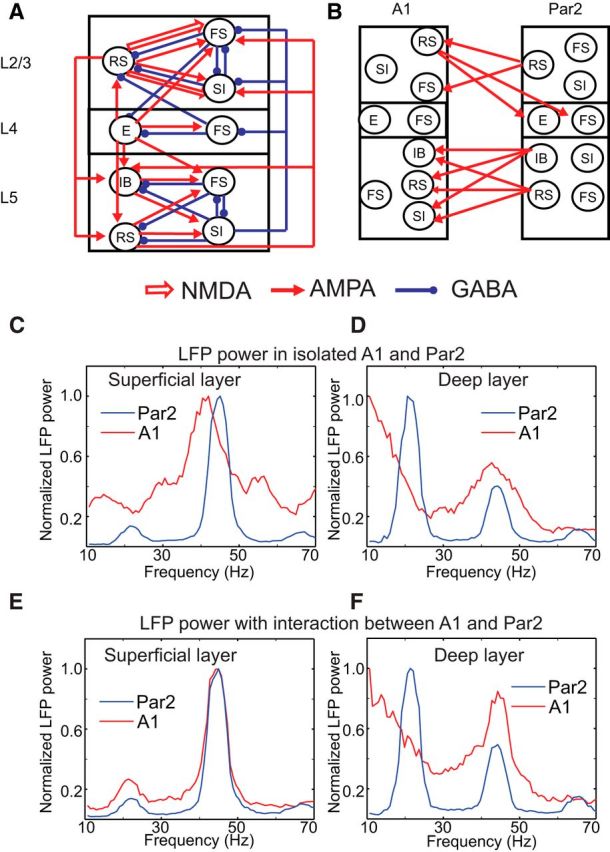
The dynamical behavior of the cortex is extremely complex, with different areas and even different layers of a cortical column displaying different temporal patterns. A major open question is how the signals from different layers and different brain regions are coordinated in a flexible manner to support function. Here, we considered interactions between primary auditory cortex and adjacent association cortex. Using a biophysically based model, we show how top-down signals in the beta and gamma regimes can interact with a bottom-up gamma rhythm to provide regulation of signals between the cortical areas and among layers. The flow of signals depends on cholinergic modulation: with only glutamatergic drive, we show that top-down gamma rhythms may block sensory signals. In the presence of cholinergic drive, top-down beta rhythms can lift this blockade and allow signals to flow reciprocally between primary sensory and parietal cortex.

Significance statement: Flexible coordination of multiple cortical areas is critical for complex cognitive functions, but how this is accomplished is not understood. Using computational models, we studied the interactions between primary auditory cortex (A1) and association cortex (Par2). Our model is capable of replicating interaction patterns observed in vitro and the simulations predict that the coordination between top-down gamma and beta rhythms is central to the gating process regulating bottom-up sensory signaling projected from A1 to Par2 and that cholinergic modulation allows this coordination to occur.

Keywords: cholinergic modulation; computational model; cortical rhythms; dynamical regulation; intercortical communication.


Figures

Figure 1.
Figure 1.
The structure of the model and kainate-induced LFPs. A, Structure of a single column of A1. Each circle is a population of 20 cells. Open and solid arrows represent NMDA and AMPA synapses, respectively. Blue circles are GABA synapses. Par2 was built using the same structure, but we made two changes in the deep layer of Par2: inhibition from L5 SI to L5 IB cells was removed and L5 IB cells were connected via axonal-axonal gap junctions (see text and Kramer et al., 2008). B, Full model with intercortical connections. L2/3 and L5 of Par2 sent top-down signals into L2/3 and L5 of A1, respectively. In contrast, L2/3 A1 sent bottom-up signals into L4 of Par2. We did not display recurrent connections inside the neuron population: all neurons receive recurrent inputs from other others that belong to the same population (see Materials and Methods). C, D, Power spectral density of LFPs in superficial and deep layers of A1 and Par2 without interactions between the two areas. The red and blue represent LFP power from A1 and Par2, respectively. Similarly, spectral power of A1 and Par2 with interaction between them is shown in E and F.
Figure 2.
Figure 2.
Layer-specific cell activity in the presence of kainate and causal interaction between the two areas. All panels show neural responses with synaptic connections between A1 and Par2. A, B, Superficial neural responses of A1 and Par2. C, D and E, F, L4 and L5 responses, respectively. Each dot represents an action potential. x-axis shows simulation time and y-axis displays the cell number. G, GC between superficial layer LFPs. H, GC between deep layer LFPs.
Figure 3.
Figure 3.
Causal relationship dependent on top-down connections. A, GC between superficial layer LFPs from Par2 to A1 as a function of strength of top-down signals to L2/3 RS cells of A1. Numbers in the inset show the maximal conductances of top-down signals to L2/3 RS cells. B, GC between superficial layer LFPs from Par2 to A1 as a function of strength of top-down signals to L2/3 FS cells of A1; numbers in the inset show the maximal conductances of top-down signals to L2/3 RS cells. C, Superficial layer cell activity when the maximal conductance of top-down signals to FS cells is increased to 0.3 μS/cm2.
Figure 4.
Figure 4.
Blockade of bottom-up signals induced by top-down gamma rhythms. A, B, Superficial layer and granular cell activity. In this experiment, although L4 E cells fire continuously, they cannot induce L2/3 RS cells to fire. C, GC between superficial layers, indicating that top-down communication is dominant even with a stronger L4 E cell activity of A1.
Figure 5.
Figure 5.
Effect of cholinergic modulation on cell activity of A1. The two columns compare neural activity of isolated A1 with and without cholinergic modulation. A, B, L2/3 cell activity. C, D, L4 cell activity. E, F, L5 cell activity.
Figure 6.
Figure 6.
Effect of cholinergic modulation on A1 reflected on the spiking activity. All panels compare the firing rate of A1 cells with and without cholinergic modulation. The effect of cholinergic modulation on the spiking activity of L5 SI cells, L4 E cells, and L2/3 SI cells are shown in A, B, and C, respectively.
Figure 7.
Figure 7.
Cell activity of A1 and Par2 with inter-areal connections in the presence of both kainate and cholinergic modulation. A, B, Superficial neural responses of A1 and Par2 with synaptic connections between the two areas. C, D, and E, F, L4 and L5 responses, respectively. G, GC between superficial layer LFPs. H, GC between deep layer LFPs.
Figure 8.
Figure 8.
Inter-areal and inter-laminar communication reflected in the spike-field coherence. A, B, Spike-field coherence between L2/3 RS cell-spiking activity in A1 and granular layer LFPs in A1 with and without ascending synaptic projection from L4 to L2/3. C, D, Spike-field coherence between L2/3 FS cell-spiking activity in A1 and superficial layer LFPs in Par2 with and without the top-down pathway.
Figure 9.
Figure 9.
Cholinergic gating mechanism. A, Spectral power of L2/3 RS cell-spiking activity depending on the connection strengths from FS to RS and from SI to FS cells, respectively. The x-axis shows the connection strength from SI to FS cells. Red, green, and blue lines show the results with 60%, 80%, and 100% connection strength from FS to RS cells, respectively. B, Average spectral power of L4 E cell activity depending on the connection strength from L4 FS to L4 E cells. The numbers in the inset indicate relative strengths of the connection. C, D, Mean and SEs of spectral powers of L2/3 RS cell activity depending on the excitability of SI cells and the amount of M-currents in L2/3 SI cells, respectively. As before, 10 simulations are used for their calculations. The x-axes in C and D represent the applied current (Iapp) and the amount of M-current, respectively.
Figure 10.
Figure 10.
Effect of cholinergic modulation on the sensitivity of L4 E cells in response to synchronous external inputs. A, B, L4 cell activity of A1 in response to 40 Hz synchronous trains of EPSCs to L4 E and FS cells with and without cholinergic modulation, respectively. C, Spiking activity of L4 E cells with and without cholinergic modulation. D, Power spectra of L4 LFPs.

Similar articles

Cited by

References

    1. Ainsworth M, Lee S, Cunningham MO, Roopun AK, Traub RD, Kopell NJ, Whittington MA. Dual gamma rhythm generators control inter-laminar synchrony in auditory cortex. J Neurosci. 2011;31:17040–17051. doi: 10.1523/JNEUROSCI.2209-11.2011. - DOI - PMC - PubMed
    1. Akam TE, Kullmann DM. Efficient “communication through coherence” requires oscillations structured to minimize interference between signals. PLoS Comput Biol. 2012;8:e1002760. doi: 10.1371/journal.pcbi.1002760. - DOI - PMC - PubMed
    1. Arroyo S, Bennett C, Aziz D, Brown SP, Hestrin S. Prolonged disynaptic inhibition in the cortex mediated by slow, Non- 7 nicotinic excitation of a specific subset of cortical interneurons. J Neurosci. 2012;32:3859–3864. doi: 10.1523/JNEUROSCI.0115-12.2012. - DOI - PMC - PubMed
    1. Bastos AM, Vezoli J, Bosman CA, Schoffelen J-M, Oostenveld R, Dowdall JR, … Fries P. Visual areas exert feedforward and feedback influences through distinct frequency channels. Neuron. 2015:390–401. - PubMed
    1. Bauer M, Kluge C, Bach D, Bradbury D, Heinze HJ, Dolan RJ, Driver J. Cholinergic enhancement of visual attention and neural oscillations in the human brain. Curr Biol. 2012;22:397–402. doi: 10.1016/j.cub.2012.01.022. - DOI - PMC - PubMed

Publication types

MeSH terms

LinkOut - more resources